
Case Series
Austin J Forensic Sci Criminol. 2015;2(2): 1016.
Forensic Evaluation of Crania Exhibiting Evidence of Sharp Force Trauma Recovered from Archaeological Excavations
Nikolett Marton1, Boglárka Marcsa2, Ildikó Pap3, Ildikó Szikossy3, Balázs Kovács4, Kinga Karlinger4, Aletta Váradi-T2 and Klára Töro5*
1Department of Genetics, Cell, and Immunobiology, Semmelweis University, Hungary
2Department of Medical Chemistry, Molecular Biology and Pathobiochemistry Semmelweis Universit, Hungary
3Anthropology Department of the Hungarian Natural History Museum, Hungary
4Department of Radiology and Oncotherapy, Semmelweis University, Hungary
5Department of Forensic and Insurance Medicine, Semmelweis University, Hungary
*Corresponding author: Klára Töro, Semmelweis University Department of Forensic and Insurance Medicine, Hungary,
Received: February 24, 2015; Accepted: March 21, 2015; Published: April 01, 2015 2015
Abstract
The authors examined human skulls originating from the eighth to thirteenth centuries AD obtained from the repository of the Anthropology Department of the Hungarian Natural History Museum. The skulls underwent direct visual examination as well as spiral CT imaging provided by the Department of Radiology and Oncotherapy Semmelweis University. Most of the skulls exhibited slash wounds, but there were also stab wounds. We have determined that some of the victims survived their injuries, as evidenced by the CT images in which the bone defects had smoothed edges indicating healing in these cases. The implements which inflicted these wounds on the ancient victims’ skulls were characteristic of the close quarters and long range weapons in use during their respective periods.
Keywords: Cranial trauma; Sharp injury; Archeology; Post mortem examination
Introduction
The category of sharp force injuries includes stabbing, slashing and chopping wounds. Stab wounds occur when a generally long, pointed implement enters the tissue with the force applied parallel to its long axis. Slash wounds occur when a bladed implement enters the tissues tangentially with the force both parallel and perpendicular to its axis. Chop wounds occur when a heavy bladed implement enters the tissue at an angle perpendicular to the axis of the blade with the aid of its own kinetic energy as well as applied force. Chop wounds also occur when the body falls upon a sharp edge, or when a descending or thrown sharp implement’s blade collides with the surface of the body [1-2].
As a result of the mechanical forces applied to the head, bony structures are injured in addition to the covering soft tissues. The resultant skull fractures can be described as open or closed, linear, depressed, spider-web or circular. A skull fracture may result in the exposure of the intracranial cavity to the external environment which could lead to life-threatening complications such as hemorrhage for example in the epidural and subdural space, herniation or infection [3]. Determination of the exact location of the fracture as well, diagnosis and planning of therapy of accompanying complications is most precisely achieved with the aid of computed tomography. In the era prior to the age of modern medical technological advances, in the event of a serious injury resulting from sharp force, the only diagnostic tool available was visual inspection [4-8].
In the eighth to ninth centuries, the Hungarians’ military forces and tactics resembled that of nomadic horsemen of Asia. Generally the use of long range weapons, such as the bow and arrow dominated. These were supplemented by the saber, lance, battle-axe and mace. In the era of the tenth to thirteenth century kings, knights first appeared whose weapons and armor became progressively heavier, making the use of larger swords popular. Long range weapons became the domain of the infantry. The use of full body armor and helmets with visors spread throughout medieval Hungary. Among the close quarter’s weapons in use at that time, the sword was most important, but in addition, lances, battle-axes, halberds and maces were also used, as the first prototypes of rifles also appeared [9-10].
Therefore, in this period of history, during the course of close quarters and long range fighting, combatants were subjected to stabbing, slashing and chopping injuries. In modern times, injuries due to sharp force trauma are largely the result of accidents or murder attempts [11]. Our goal was to determine if and at what rate the victims were able to survive their wounds.
Materials and Methods
We examined five skulls with sharp force trauma injuries provided by the Hungarian Natural History Museum’s Anthropology Department, originating from the historical periods between 900 and 1500 AD ( Age of Conquest, Árpád Era, Middle Ages). The skulls were discovered by archaeologists during the twentieth century in excavations at Nagykorös, Jászdózsa, and Nyársapát (Hungary). recording measures the varying radio densities of the subject and produces cross-sectional and special images in much the same way as traditional roentgenograms. The images were produced by the CT in a continuously rotating, steadily advancing fashion, imaging every aspect of the skulls inclusively, in a “spiral” manner. From this high resolution, large data set a multiplanar, 3D reconstruction, as well as detailed cross-sectional views was created. The MSCT scan (Brilliance 16, Philips Medical Systems, Best, and The Netherlands) had the following parameters: slice thickness 0.8 mm, increment 0.4mm, high resolution, collimation 16 x 0.75m. The image post processing was completed using multiplanar reconstructions (MPR), maximal intensity projections (MIP) and three-dimensional reconstructions. The CT is very appropriate in examining the bony structures in living as well as post-mortem subjects. This technique allows an accurate exploration of the skull structure including the diploe as well, which otherwise could not been investigated. Inner parts of the crania could be examined which otherwise would be invisible to the naked eye.
Signs of healing on computed tomography are rounded bony edges, or fusion of bone fragments to the bony defect edges. Note: reference to orientation is irrelevant as the injuries would vary. Smoothed edges were qualitatively coded as sign of healing and therefore as an indicator of survival. In the case of likely lethal injuries the lamina external and lamina internal edges exhibit no evidence of bone reparation. The bony edges are not rounded, but jagged and sharp [12-17].
Results
The five examined skulls (don’t they have more for a larger sample size? ) provided to us by the Hungarian Natural History Museum’s Anthropological Repository were uncovered from varying archaeological excavations. Archaeological science evaluates past cultures by studying their artifacts. Archaeologists’ most commonly used method is excavation, where they unearth ancient buildings’ remains according to painstaking plans, preparation and cataloging.
Weapons enable attacks and defensive parries to extend beyond the limits of the human body, increasing their destructive force and distance of impact. In this study we studied the effect of sharp-edged fighting implements, therefore we will briefly characterize these stabbing and cutting medieval implements. The most commonly encountered cutting weapon found in Conquest Era tombs is the saber. Sources from this period refer to it as a sword or gladius. Battleaxes were also commonly used. A very commonly used battlefield weapon during the Age of Conquest was the powerfully penetrating reflex bow, normally consisting of a grip from which two flexible, spring-like limbs extend, joined at the ends and brought under great tension by a bow string capable of launching arrows. The arrow was basically a long, thin, straight rod with an arrowhead at the tip, usually made of metal. The long double-edged sword found its way into Hungary in the late tenth century. The lance utilized during the Árpád Era was basically a long wood-handled, metal-tipped hunting or attack weapon [18-19]. The mace was an old crushing weapon, establishing itself in Hungary in the thirteenth century. The battle axe resembles a hatchet with a spike on the opposite aspect of the head, used by infantry for chopping and stabbing [9-11].
Case No 1
On this tenth to thirteenth century, Árpád era skull’s frontal bone we see a stab type of wound, from which emanates an 8cm long fracture line which traverses the frontal bone and the superior wall of the orbit (Figure 1). The 1.5cm arrowhead tip lodged in the stab wound and is still firmly embedded. The wound’s direction is anterior to posterior. The victim did not survive his injury. On the CT images sharp bone edges can be seen in both the horizontal and sagittal views. This is a sign that the bone edges did not heal.
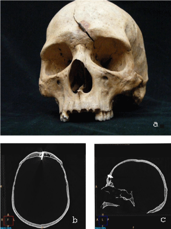
    Figure 1:  A/ Photograph of the skull. A 1.5 cm metal arrowhead tip lodged in
the frontal bone and an 8cm long fracture line are visible.
B/ HRCT image multiplanar reconstruction in the parasagittal plane. The
arrowhead tip appears as a hyper dense object, penetrating the lamina
external, diploe and lamina internal.
C/ HRCT image (axial plane) shows the foreign body penetrating the frontal
sinus.
Case No 2
On the left frontal bone of this Árpád era (A.D. 1000- 1300) skull, above the orbit, is a 1.5cm wide, 6cm long, narrowing defect of the bone. The thickened superciliary arch and rounded bone are visible to the naked eye and on CT imagery, which informs us this individual survived his injury (Figure 2) even though the injury opened into the intracranial cavity. Additionally, a 5cm long indentation can be seen on the right parietal bone which did not enter the cavity, but most certainly the implement used penetrated the overlying soft tissues but was non-lethal.
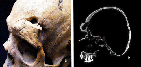
    Figure 2:  A/ Figure A Photograph of the skull shows a 6 cm long and 1.5 cm
wide bony defect with a thickened superciliary arch and rounded bony edges.
B/ HRCT image shows the bone edges along the cut are rounded and the
damaged portion has thickened
C/ HRCT sagittal view shows the frontal bone and superciliary arch have
thickened considerably and the injury opened the intracranial cavity.
Case No 3
This specimen originates from the Árpád Era. A 6cm long, 3cm wide bone defect is seen traversing the right parietal, temporal and frontal bones which was caused by a sharp implement descending from above. The shattered bone fragments are lodged in the inferior edge of the neo-fenestra’ may be the skin and loose connective tissue bound the fragments during the convalescence. On the computed tomographic image the rounded bone edges and bone remodeling is obvious evidence of healing ( Figure 3).
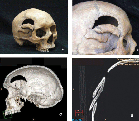
    Figure 3:  A/ The photograph shows a curved, 6cm long, 3cm wide bone
defect in the region of the right temporal region. The window involves the
temporal, parietal and frontal bones. The dislodged bone fragments have
reattached and healed distally to the injury
B/ Closeup photograph of the penetrating chop wound.
C/ HRCT 3D reconstructed image shows the inside view of the fenestrated
bone. The bony edges are clearly rounded.
D/ HRCT image similarly exhibits the rounded edges. (rec. coronal)
Case No 4
This skull from the Middle Ages exhibits a 12cm long chopping wound which extends from a posterior/inferior to anterior/superior direction. The mastoid process has been amputated. The victim did not survive the head injury, since the bony edges along the wound defect are sharp and show no signs of healing. Probably the victim was decapitated ( Figure 4).
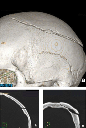
    Figure 4:  A/ HRCT multiplanar reconstructive views show a 12 cm chop
injury of the left parietal bone extending posterior/inferior to anterir/superior
B/ HRCT image shows the jagged edges to the wound.
C/ HRCT image similarly shows the sharp bone edges.
Case No 5
On the medial portion of the superciliary arch on this medieval skull one can see a 4cm long chop injury which resulted in elevation of the bony edges but did not succeed in separating them. Rounded, healed edges are seen on the CT scans also. Several 1-3cm chop wounds caused by a sharp implement are seen on the calvarium, which did not enter the intracranial cavity ( Figure 5).
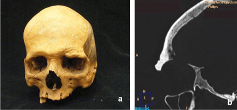
    Figure 5:  A/ On this frontal view photograph of the skull one can see the
4 cm long chop injury on the medial portion of the superciliary arch and the
thickening of the affected bone.
B/ On this photograph are shown the 1-3 cm long, non-penetrating chop
wounds
C/ Close-up view of the chop wounds.
D/ On this parasagittal view of the HRCT multiplanar reconstruction image
the supraorbital bone thickening is evident.
On the medial portion of the superciliary arch on this medieval skull one can see a 4cm long chop injury which resulted in elevation of the bony edges but did not succeed in separating them. Rounded, healed edges are seen on the CT scans also. Several 1-3cm chop wounds caused by a sharp implement are seen on the calvarium, which did not enter the intracranial cavity ( Figure 5).
Discussion
Whenever the force exerted from the impact of a sharp-edged implement striking the cranium overcomes the resistance of the bony structure, disrupting its continuity, the result is a fracture. Several weeks are required for the bone to regenerate, even in cases that are free of complications. In the event that the bone fracture is accompanied by injuries to larger blood vessels, nerves or an organ, the healing period may even extend to six months. In cases beset with complications, the healing process of the bone may still leave behind serious declines in overall health as well as permanent disabilities. Conspicuous facial structural deformities that cause esthetic and dysfunctional problems, such as blindness, are an example. In the examined cases there weren’t any facial bone injuries. Skull fractures may extend to the upper cranium, the base and the facial skeleton. Upper cranial injuries are most often the result of direct trauma. A bone fracture in of itself does not describe the severity of an injury, but nonetheless is an important indicator which indirectly gives evidence of the intensity of the traumatic force exerted, giving an assessment of potential serious complications. Skull fractures may be open or closed but the latter are harder to diagnose. The typical depressed fracture resulting from considerable blunt force delivered to a larger surface area is the spider web fracture. The extent and shape of the fracture are often indicative of the shape of the administering implement and degree of force encountered. If the force is applied tangentially rather than perpendicularly, the bones fracture in a step-wise, or terrace fashion. Force applied perpendicularly by a large, blunt instrument produces fragmented fractures. Facial skeletal fractures are often caused by trauma and may be accompanied by fractures of the cranial base. Currently, the diagnosis of cranial fractures is determined by cranial radiography and CT imagery in conjunction with neurologic, otolaryngologic and ophthalmologic consultation [20-27].
Fighting implements of old were larger in size and heavier, causing larger injuries. Injuries acquired during the course of 21st century hostilities tend to be of the crushing or disruptive type due to explosives or gunshot wounds from firearms. In modern times the majority of cranial injuries affect victims as a result of motor vehicle accidents, mostly pedestrians suffering head trauma [28]. However, they can also be the result of falls from a high elevation and murder or suicide attempts. Today, when sharp force cranial injuries do occur, rarely are they due to battlefield weapons such as explosives or firearms; rather, they are usually caused by everyday objects such as knives, machetes or scissors. Cranial fractures persist as a very serious type of injury to this day, often leading to permanent damage or death. The methods of violence and killing correlate with the societal, social and moral milieu of the time period. The study of historic Hungary’s cranial injuries reveals the mechanisms of injury typical of the given era, refers to the type of implement used, sheds light on the therapeutic methods in use (I didn’t read any of this in the text?) and the odds (you don’ of surviving [29-31].
References
- Ormstad K, Karlsson T, Enkler L, Law B, Rajs J. Patterns in sharp force fatalities-A comprehensive forensic medical study. J Forensic Sci. 1986; 31: 529-542.
- Terazawa K. [Observations on injuries by blunt objects]. Nihon Hoigaku Zasshi. 2010; 64: 103-120.
- Yoganandan N, Pintar FA, Sances A Jr, Walsh PR, Ewing CL, Thomas DJ, et al. Biomechanics of skull fracture. J Neurotrauma. 1995; 12: 659-668.
- Geisler FH. Skull fractures. Wilkins RH, Regachary SS, eds. Neurosurgery, 2nd ed. New York: McGraw-Hill; 1996; p: 2741–2755.
- Rickels E. Diagnosis and treatment of traumatic brain injury. Chirurg. 2009; 80: 153-162.
- Tallon JM, Ackroyd-Stolarz S, Karim SA, Clarke DB. The epidemiology of surgically treated acute subdural and epidural hematomas in patients with head injuries: a population-based study. Can J Surg. 2008; 51: 339-345.
- Ortler M, Langmayr JJ, Stockinger A, Golser K, Russegger L, Resch H. Prognosis of epidural hematoma: is emergency burr hole trepanation in craniocerebral trauma still justified today?. Unfallchirurg. 1993; 96: 628-631.
- Ryan CG, Thompson RE, Temkin NR, Crane PK, Ellenbogen RG, Elmore JG. Acute traumatic subdural hematoma: Current mortality and functional outcomes in adultpatients at a Level I trauma center. J Trauma Acute Care Surg. 2012; 73: 1348-1354.
- Bovill F W. B. Hungary and the Hungarians. Charleston: Nabu Press; 2013.
- De Vries K. Medieval Military Technology Peterborough: Broadview Press; 1992.
- Waldman J. Hafted Weapons in Medieval and Renaissance Europe: The Evolution of European Staff Weapons Between 1200 and 1650. Leiden: Brill Academic Publisher; 2005.
- Wozniak K, Moskala A, Urbanik A, KÅéys M. Value of postmortem CT examinations in cases of extensive mechanical injuries causing considerable corpse destruction. Arch Med Sadowej Kryminol. 2010; 60: 38-47.
- Hildebolt CF, Vannier MW, Knapp RH. Validation study of skull three-dimensional computerized tomography measurements. Am J Phys Anthropol. 1990; 82: 283-294.
- Nakahara K, Shimizu S, Kitahara T, Oka H, Utsuki S, Soma K, et al. Linear fractures invisible on routine axial computed tomography: a pitfall at radiological screening for minor head injury. Neurol Med Chir (Tokyo). 2011; 51: 272-274.
- Scholing M, Saltzherr TP, Fung Kon Jin PH, Ponsen KJ, Reitsma JB, Lameris JS, et al. The value of postmortem computed tomography as an alternative for autopsy in trauma victims: a systematic review. Eur Radiol. 2009; 19: 2333-2341.
- Lorkiewicz-Muszynska D, Kociemba W, Zaba C, Labecka M, Koralewska-Kordel M, Abreu-Glowacka M, et al. The conclusive role of postmortem computed tomography (CT) of the skull and computer-assisted superimposition in identification of an unknown body. Int J Legal Med. 2013;127: 653–660.
- Ampanozi G, Ruder TD, Preiss U, Aschenbroich K, Germerott T, Filograna L, et al. Virtopsy: CT and MR imaging of a fatal head injury caused by a hatchet: A case report. Leg Med (Tokyo). 2010; 12: 238-241.
- Maenchen-Helfen O. The World of the Huns. Berkeley: University of California Press; 1973.
- Hamm J. The traditional bowyer’s bible Connecticut: The Lyons Press; 2008.
- Jennett B. Epidemiology of head injury. J Neurol Neurosurg Psychiatry. 1996; 60: 362-369.
- van den Heever CM, van der Merwe DJ. Management of depressed skull fractures. Selective conservative management of nonmissile injuries. J Neurosurg. 1989; 71: 186-190.
- Jennett B, Miller JD. Infection after depressed fracture of skull. Implications for management of nonmissile injuries. J Neurosurg. 1972; 36: 333-339.
- Sande GM, Galbraith SL, McLatchie G. Infection after depressed fracture in the west of Scotland. Scott Med J. 1980; 25: 227-229.
- Taha JM, Haddad FS, Brown JA. Intracranial infection after missile injuries to the brain: report of 30 cases from the Lebanese conflict. Neurosurgery. 1991; 29: 864-868.
- Plese JP, Humphreys RP. The use of prophylactic systemic antibiotics in compound depressed skull fractures in infancy and childhood. Arq Neuropsiquiatr. 1981; 39: 286-288.
- Marion DW. Complications of head injury and their therapy. Neurosurg Clin N Am. 1991; 2: 411-424.
- Potapov AA, Shahinian GG, Kravtchouk AD. Surgical management of Penetrating brain injury. The Journal of Trauma: Injury, Infection, and Critical Care. 2001; 51:16–25.
- Piantini S, Grassi D, Mangini M, Pierini M, Zagli G, Spina R, et al. Advanced accident research system based on a medical and engineering data in the metropolitan area of Florence.BMC Emerg Med. 2013; 13: 3.
- Bozzeto-Ambrosi P, Costa LF, Azevedo-Filho H. Penetrating screwdriver wound to the head. Arq Neuropsiquiatr. 2008; 66: 93-95.
- Okay O, Da─člio─člu E, Ozdol C, Uckun O, Dalgic A, Ergungor F. Orbitocerebral injury by a knife: case report. Neurocirugia (Astur). 2009; 20: 467-469.
- Hubschmann O, Shapiro K, Baden M, Shulman K. Craniocerebral gunshot injuries in civilian practice--prognostic criteria and surgical management: experience with 82 cases. J Trauma. 1979; 19: 6-12.