Case Series
Austin J Forensic Sci Criminol. 2015;2(2): 1017.
Aortic Intimal Staining In Fresh Water Drowning – A Case Series
Jayanth S Hosahally*, Girish Chandra YP and A Gokulakrishnan
Department of Forensic Medicine, M.S. Ramaiah Medical College, India
*Corresponding author: Jayanth S Hosahally, Department of Forensic Medicine, M.S. Ramaiah Medical College, India
Received: February 04, 2015; Accepted: March 20, 2015; Published: March 30, 2015
Abstract
There is usually a suspicion on the cause of death whenever a body is recovered from water. There are no pathognomonic findings to indicate the diagnosis of drowning at autopsy. The diagnosis is based on the circumstances of the death and various nonspecific anatomical findings. It is one of the most difficult modes of death to prove at postmortem. Fresh water drowning brings about hemodilution and hemolysis; which in turn stains the intima of the aortic root. The present study was undertaken to know the importance of this phenomenon in fresh water drowning. A total of ten cases were studied and the study group was divided into group A and group B. Group A consists of five drowning cases and Group B consists of five control cases other than drowning with similar post mortem interval. Aortic intimal staining was demonstrated in all cases of drowning.
Keywords: Drowning; Autopsy; Fresh water; Aortic root; Intimal staining
Introduction
Drowning is a difficult autopsy diagnosis due to various reasons. Most of the significant autopsy findings start to fade after body is recovered from water. In addition to drowning, injuries, intoxications or natural conditions are all among the potential causes of death in bodies found in water or the factor that may have contributed to the fatal outcome [1]. It has been suggested in the literature, that hemodilution caused by drowning causes red cell lysis which further stains the intima of the aorta. However, this has been based predominantly on anecdotal autopsy observations, and recent texts have not tended to mention this phenomenon resulting in uncertainty as to its significance [2]. Differential hemolytic staining of the intima of the pulmonary artery and aortic root may be useful at autopsies on bodies recovered from water. German literature has recognized this feature for many years as ‘hemoglobin imbibitions’ of the intima due to ‘hypo-osmolar hemeolysis’ and is used as an indicator of freshwater drowning [3]. This observation may have considerable diagnostic significance. This study was taken up to evaluate this in fresh water drowning cases.
Material and Methods
This study was conducted at the Department of Forensic Medicine, M.S. Ramaiah Medical College Bangalore, in 2013, wherein 5 fresh water drowning cases and 5 control cases were evaluated. Pulmonary trunk and aorta were dissected and looked into as part of standard autopsy examinations.
- A total of ten cases were studied and the study group was divided into group A and group B.
- Group A (Table 1) consists of five drowning cases and Group B (Table 2) consists of five control cases other than drowning. In both groups, cases with similar post mortem interval were taken to avoid false positive findings as decomposition also brings about hemolytic changes.
- In all cases heart was dissected along the flow of blood. Both aortic and pulmonary roots were examined for intimal staining by washing them under running tap water and later drying with cotton.
- Tsokos M. In: Lunetta P and Modell JH. Forensic pathology reviews, vol. 3. Totowa (NJ): Humana Press; 2006. pp. 3- 5.
- Saukko P, Knight B. 3rd ed. Knight’s forensic pathology. London: Arnold; 2004. p. 395–411.
- Brinkmann B. Tod im Wasser. In: Brinkmann B, Madea B, editors. Handbuch gerichtliche Medizin, vol.1. Berlin, Heidelberg, New York: Springer; 2004. p. 797–824.
- Modell JH, Bellefleur M, Davis JH. Drowning without aspiration: is this an appropriate diagnosis? J Forensic Sci. 1999; 44: 1119-1123.
- Piette MH, De Letter EA. Drowning: still a difficult autopsy diagnosis. Forensic Sci Int. 2006; 163: 1-9.
- Polson CJ, Gee DJ, Knight B. The essentials of forensic medicine. 4th ed. Oxford: Pergamon Press; 1985. p. 485.
- Tsokos M, Cains G, Byard RW. Hemolytic staining of the intima of the aortic root – A useful pathological marker of freshwater drowning? The American Journal of Forensic Medicine and Pathology. June 2008; 29: 128-130.
    Cases
    Age
    Sex
    PMI


    1
    34
    M
    8-10 hr


    2
    41
    M
    8-10 hr


    3
    27
    F
    12-14 hr


    4
    31
    M
    14-16 hr


    5
    25
    F
    24-28 hr
Table 1: Group A- consists of five drowning cases.
    Control
    Age
    Sex
    PMI


    1
    26
    M
    8-10 hr


    2
    34
    F
    8-10 hr


    3
    39
    M
    12-14 hr


    4
    42
    M
    14-16 hr


    5
    28
    M
    24-28 hr
Table 1: Group B- consists of five control cases.
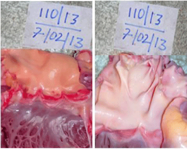
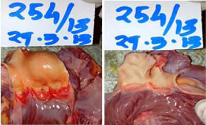
Case 1 and 2 (Figure 1 & 1a) with post mortem interval of 8-10 hr showed staining of aortic intima with lack of pulmonary root staining. Cases 3 (Figure 2 & 2a) and 4 with post mortem interval of 12- 14 hr and 14 – 16 hr respectively showed increased intensity of intimal staining in the aortic root compared to cases 1 and 2. Case 5 with a post mortem interval of 24 to 28 hr showed staining of both aortic intima and pulmonary root. In this case decomposition had set in and pulmonary root is stained with less intensity than aortic root, in addition to flabbiness and reddish brown discoloration of heart.

Figure 1 a: Lack of pulmonary root staining (Case 2).

Figure 2 a: Lack of staining of pulmonary root (Case 3).
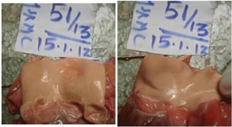
Control 1, a case of road traffic accident, with post mortem interval of 8 to 10 hr showed lack of staining in both aortic and pulmonary root (Figure 3 & 3a).

Figure 3 a: Lack of staining of pulmonary root (Control 1).
Control 2, a case of natural death, with post mortem interval of 8 to 10 hr showed lack of staining in both aortic and pulmonary root.
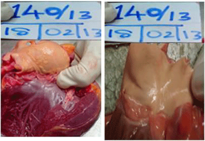
Control 3, a case of natural death, with post mortem interval of 12 -14 hr, showed mild staining of pulmonary root compared to aortic intima (Figure 4 & 4a).

Figure 4 a: Minimal staining pulmonary root (Control 3).
Control 4, a case of hanging with PMI of 14 to 16 hr, showed minimal staining in both aortic and pulmonary root.
Control 5, a case of hanging with decomposition changes with PMI of more than 24 to 28 hr showing marked intimal staining in both aortic and pulmonary root.
Discussion
The medico legal investigation of bodies found in water focuses on determination of the cause and manner of death. Cause of death varies in bodies recovered from water. It could be due to drowning or intoxication or due to injury or natural disease. Modell et al. stated that ‘‘to ascribe drowning as a cause of death to a body found in water without some evidence of the effect of having aspirated water is risky’’ and concluded that ‘‘in this situation, it may be more accurate to list a differential diagnosis rather than a specific cause of death’’ [4].
The macroscopical and microscopical signs in the fresh drowned victim are non-specific and moreover putrefaction will vanish these autopsy findings quite rapidly [5].
It was on the basis of presumed hemodilution of blood returning to the left side of the heart that Gettler’s chloride test was formulated. This test predicted that hemodilution of blood in the left side of the heart in fresh water drowning would significantly lower plasma chloride levels with saltwater drowning having the opposite effect. However, although these changes occur, this test has been shown to be neither useful nor reliable in confirming the diagnosis of drowning as Gettler proposed, due to the marked biochemical fluxes that follow death [2] i.e. individual and circumstantial variability are such that there is no consistency in results. While there may be predictable changes in certain electrolyte levels, such as sodium, the overlap between fresh and saltwater drowning and controls renders it unreliable as a diagnostic marker of drowning in specific cases [5].
Drowning in fresh water is thought to occur more quickly than in salt water, as fresh water is absorbed more rapidly into the pulmonary circulation causing hemodilution and hemolysis. Hyponatraemia, hyperkalemia and circulatory overload ultimately result in lethal cardiac arrhythmias [6].
Red blood cells containing hemoglobin break down over time releasing iron-containing pigment which colors surrounding tissues. In the aorta and pulmonary trunk this takes the form of a characteristic dark red discoloration. Sometimes this may also be seen in badly burnt bodies where heat-induced hemolysis has occurred and in cases of generalized hemolysis due to sepsis. Staining of the vessels was also observed as an artifact, when water from the dissection table mixed with blood causing red cell lysis. To prevent this, grading of vessel wall color should be undertaken within a very short time of separation of the heart from the pulmonary trunk and aorta [7].
However, given that hemodilution occurs following drowning, is it possible that intimal staining may be a more stable and useful marker than electrolyte changes. This is unclear, as studies in the literature have concentrated on biochemical, rather than pathological parameters. The aim of the current study was to evaluate the differential staining of the great vessels in cases of fresh water drowning.
In our study, in all drowning cases intima of aortic root was intensely stained. One case where the body was decomposed, marked intensity of intimal staining was seen in both vessels, but aortic root was stained with intensity than pulmonary root.
Amongst the controls, in the first two which were in the 8- 10 hr PMI, no staining was observed in either vessel. Only mild pulmonary staining was observed in control 3 where in decomposition changes had begun. In the other 2 controls intimal staining was observed in both vessels in same intensity. However in control 5 which was in advanced decomposition, both vessels were markedly stained with equal intensity than control 4.
Here the concept of hemoimbibition arises. Factors influencing the different intensity of intimal staining in the aortic intima are:
I) Hemodilution in the left ventricle - more dilution of blood in the left ventricle leads to excessive destruction of RBCs. Hemodilution depends upon both the struggle period (amount of water swallowed) and the time for which body present in water after death (water enters the lung because of hydrostatic pressure).
II) Post mortem interval- After body is recovered from water the decomposition sets in fast and causes hemolysis. Hence more the PMI, intimal staining will be marked. Thus post mortem interval also plays an important role in the intensity of aortic root intimal staining.
When the staining is seen in pulmonary trunk only or when seen in both vessels but more intensely in pulmonary trunk, this could be attributed to hemolysis associated with decomposition. When the staining is seen in aortic trunk only or when seen in both vessels but more intensely in aortic trunk, this could be attributed to hemolysis associated with fresh water drowning. Thus, this differential staining pattern of the great vessels when demonstrated correctly at autopsy can be used to attribute the cause of death to fresh water drowning.
Conclusion
Aortic root staining is an easy sign to demonstrate at autopsy. It excludes other causes of death in bodies recovered from water and aids in ascertaining the cause of death to drowning. Thus if aortic intimal staining is seen more intensely than in the pulmonary trunk, one could use this finding to supplement other autopsy features to ascertain the cause of death as drowning.
References