Research Article
Austin J Forensic Sci Criminol. 2016; 3(1): 1047.
The Difficult Task of Interpreting Cut Marks, Gunshot Wounds and Ligature Marks on the Skin: A Cautionary Note
Amadasi A, Cerutti E, Spagnoli L, Gibelli D*, Gorio C and Cattaneo C
Dipartimento di Scienze Biomediche per la Salute, Università degli Studi di Milano, Italy
*Corresponding author: Daniele Gibelli, LABANOF, Laboratorio di Antropologia e Odontologia Forense, Dipartimento di Scienze Biomediche per la Salute, Università degli Studi di Milano, Milan, Italy
Received: June 07, 2016; Accepted: June 28, 2016; Published: June 30, 2016
Abstract
The morphological assessment of skin injuries is a stronghold of forensic pathology and relies upon standard “rules” for a correct interpretation. But are these parameters reliable? When is the interpretation dangerously difficult? This study aims at quantifying the difficulties in the assessment of stab wounds, gunshot wounds and ligature marks by the observation of photographs in three questionnaires given to 11 experts (forensic pathologists) and 11 non-experts (trainees in forensic pathology). For stab wounds the overall percentage of correct answers was 47.5%, 56.9% for type of blade and 64.2% for type of edge. Gunshot wounds were correctly assessed only in 41.3% of cases. Finally, only in 47.1% of the cases a correct match ligature mark/object of constraint was found. The results show that wounds on the skin can frequently be misinterpreted if classification is strictly based on only morphological parameters; the judgment should therefore be based on an overall evaluation of all evidences, including also those provided by more advanced technological analyses (for example, SEM-EDS for the search of residues, radiological analyses, etc.).
Keywords: Forensic science; Forensic pathology; Stab wounds; Gunshots wounds; Ligature marks; Pictures; Skin
Introduction
The morphological evaluation of wounds on the skin is one of the strongholds of every autopsy and of the common forensic practice: a correct interpretation of type of wound and of the type of weapon is of crucial importance and usually relies upon macroscopic features whose correct analysis is the basis for every further investigation.
As concerns stab wounds, many studies have been performed to find a relationship between sharp wounds and weapons, and some parameters have been identified as useful: size of the wound, depth, edges, the presence of additional features (notches, bruises, abrasions). However, it is quite evident that many variables can come into play: general features strongly depend on the characteristics of the knife (or other type of sharp weapon), and on body site, strength of the assailant, movements of assailant and victim [1-7]. Many authors consider morphological analyses on skin lesions reliable in the identification of the type of weapon, but the actual reliability in real case scenarios has never been practically tested and cases of atypical presentations and diagnostic difficulties have been frequently reported [8,9].
As concerns gunshot wounds, the distinction between entry and exit wounds and the identification of the range of the shot is crucial. The analysis of the shape, margins, presence or lack of additional elements (abrasion ring, stippling, searing, soot soiling) are the common basics of macroscopic assessment [10-13]; it is therefore evident how errors between entry and exit wounds can lead to misleading evaluations concerning the number of projectiles entering and exiting the body as well as the direction of the shot and the wound track. However, both entries and exits can show peculiar features and several cases of “diagnostic errors” and atypical presentations of gunshot wounds have been previously reported [14-16].
Finally, among blunt force injuries, ligature marks are characteristic and typical of asphyxial deaths but they can be found even in cases of victims of abuse and torture, when some sort of object of constraint is used. These marks usually arise from a combination of bruises and abrasions, as an expression of the mechanical effect on the skin’s surface [17-19]. The morphological analysis of the mark may be an important source of information for the identification of the object of constraint but ligature marks can be variable, depending on the nature of the ligature, strength and location on the body [20].
However, experience teaches that each and every autopsy and especially skin injuries may be misleading. As a matter of fact, several case reports have previously shown how difficult to interpret skin wounds can be, especially when “traditional” parameters (i.e. shape, dimensions, edges) are insufficient or even deceiving [8,9,14-16].
This led to the present study which aims at verifying how frequently such wounds can be misleading or misinterpreted by experts.
Thus, the study consisted in three questionnaires on stab wounds, gunshot wounds and ligature marks submitted to experts and non-experts, which aimed attesting the assessment of wounds on photographs of different types of injury on the skin.
Materials and Methods
A total of 66 questionnaires were submitted to 22 observers (13 females and 9 males) composed of 11 experts (forensic pathologists of 5-10 year experience) and 11 non-experts (trainees in forensic pathology, with at least one year of experience in the field). Assessments were performed on a total of 15 pictures for stab wounds, 15 pictures for gunshot wounds and 10 pictures for ligature marks.
pictures for gunshot wounds and 10 pictures for ligature marks. The results were then analyzed aiming at quantifying the difficulties of the observers in the morphological analysis and the differences among experts and non-experts. The details of every questionnaire are reported in the following paragraphs:
Stab wounds
Among the total 15 pictures, 4 were selected from real autopsy cases with known weapons (single and double-edged knives) and 11 were experimentally produced on pig skin with the wounding weapon kept perpendicular to the surface of the skin in order to simulate a stab wound. Two piglets, who had died of natural death, were used for the study: they were shaved to remove bristles and stabbed several times in different areas (abdomen, chest and thighs) with 9 different sharp weapons: 7 knives (5 single-edged knives, 3 with a smooth blade and 2 with a serrated blade and 2 double-edged knives, both with a smooth blade) and two different pairs of scissors.
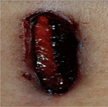
In the related questionnaire (Figure 1) the observers were asked to state:

    Figure 1:  Wound produced by single-edged knife in autopsy case. Correct
assessments: 23%.
1) if the injury they were looking at could have been produced by a single or a double-edged weapon.
2) if the weapon could have been with a smooth or a serrated edge.
If they were not able to reach a decision, they could cross the option “not assessable”.
Gunshot wounds
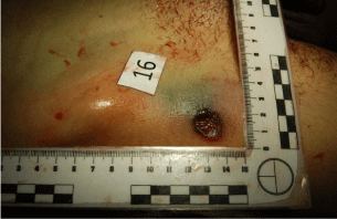
Observers had to assess 15 pictures (9 entrance and 6 exit wounds) taken from real cases. In the questionnaire they were asked:
1) to describe if it was an entry or an exit.
2) to write what they had relied on (Figure 2).

Figure 2: Exit wound. Correct assessments: 18.8%.
Ligature marks
Assessments were performed on 10 pictures of ligature marks produced on the skin of the upper arm of volunteers, after fifteen minutes of tight constriction. Pictures were then taken immediately after the removal of the constriction. Eight different ligatures were used: a white and grey rope of rolling shutters, a red string, a green cord, a beige hemp rope, an orange-yellow-silver cord, a white cord, a grey electric cable and a belt in leather. In the questionnaire they were asked to state:
1) if a ligature mark was detectable in the picture.
2) if the answer positively, they had to try to find a match between the mark and the ligature.
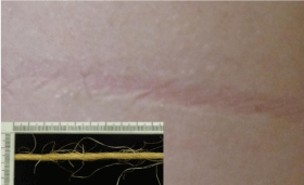
3) To indicate the features they had observed most to reach a decision (Figure 3).

    Figure 3:  Ligature mark produced with an hemp rope (down-left): Correct
assessment: 25%.
Results
Answers were put in a spreadsheet (Microsoft Office Excel™ 2010) with automatic computation of correct/incorrect answers and related percentages. The questionnaires were listed separately, first with the overall results and then divided into the two groups considered, experts (forensic pathologists) and non-experts (trainees in forensic pathology).
Stab wounds
The results are shown in Table 1. In general, a correct identification of the weapon was reached in 47.5% of the cases, and only with slight differences between experts (48%) and non-experts (47.2%). For what concerned the single questions, the type of blade (single/double-edged) was correctly identified in 56. 9% of the cases, whereas the type of edge (linear/serrated) in 64.2%, again with slight differences between the two categories of observers. The percentage of “non assessable” answers was always around 10-15%. For what concerned the identification of a different type of sharp weapon (eg scissors) correct answers fell to 14, 8%, mainly given by experts.
    Stab    wounds


    Overall    results


    Total
    Experts
    Non-experts


    Correct
    Incorrect
    NA*
    Correct
    Incorrect
    NA*
    Correct
    Incorrect
    NA*


    47,5%
    38,4%
    14,1%
    48,0%
    34,7%
    17,3%
    47,2%
    40%
    12,8%


    Type of    blade


    Total
    Experts
    Non-experts


    Correct
    Incorrect
    NA*
    Correct
    Incorrect
    NA*
    Correct
    Incorrect
    NA*


    56,9%
    31,0%
    12,2%
    61,3%
    29,3%
    9,3%
    55,0%
    31,7%
    13,3%


    Type of    edge


    Total
    Experts
    Non-experts


    Correct
    Incorrect
    NA*
    Correct
    Incorrect
    NA*
    Correct
    Incorrect
    NA*


    64,2%
    23,5%
    12,3%
    60,0%
    23,3%
    16,7%
    66,0%
    23,6%
    10,4%


    *NA =    not assessable
Table 1: Results of the assessments on stab wounds.
Gunshot wounds
Table 2 summarizes the results concerning the questionnaires on gunshot wounds: on average, 41, 3% of subjects gave a right answer, with no differences at all between experts and non-experts. “Not assessable” answers were higher than in cases of stab wounds (20.4% on average) and more in non-experts (22.4%).
    Gunshot    wounds


    Total
    Experts
    Non-experts


    Correct
    Incorrect
    NA*
    Correct
    Incorrect
    NA*
    Correct
    Incorrect
    NA*


    41,3%
    38,3%
    20,4%
    41,3%
    42,7%
    16,0%
    41,2%
    36,4%
    22,4%


    *NA =    not assessable
Table 2: Results of the assessments on gunshot wounds.
Entry wounds were more easily assessed, with an average of 49.3% of correct answers. On the other hand, the assessment on exit Entry wounds were more easily assessed, with an average of 49.3% of correct answers. On the other hand, the assessment on exit The main features that were observed for most of the evaluations, as reported by the observers in the questionnaire, were (in order of frequency): presence/lack of abrasion ring, flaring of the edges, shape/ general appearance of the wound, burning and soot deposition.
Ligature marks
Table 3 shows the results concerning ligature marks. Firstly, the presence of a furrow, or at least of a mark on the skin, was detected only in 69.4% of the cases and among this percentage, only in 41.7% of the cases a correct match between the mark and the ligature was reached. Moreover, non-experts gave surprisingly more correct answers (50.7%) than the experts (39.2%). Some marks were easily linked to the specific ligature, like in the case of woven fabric ropes, even with 100% correct identification. Some cases, even in front of clear marks in the pictures, were scarcely associated to the right ligature, like in the case of the hemp rope (Figure 3) which was correctly assessed only in 25% of the cases. The features on which the evaluation most frequently relied upon were the general pattern of the ligature and the shape/appearance of the mark on the skin (i.e. weave and dimensions).
    Ligature    marks


    Identification    of the presence of a furrow


    Total
    Experts
    Non-experts


    Yes
    No
    NA*
    Yes
    No
    NA*
    Yes
    No
    NA*


    69,4%
    28,8%
    1,9%
    64,0%
    34,0%
    2,0%
    71,8%
    26,4%
    1,8%


    Identification    of the right ligature when a furrow was detected


    Total
    Experts
    Non-experts


    Correct
    Incorrect
    Correct
    Incorrect
    Correct
    Incorrect


    47,1%
    52,9%
    39,2%
    60,8%
    50,7%
    49,3%


    *NA =    not assessable
Table 3: Results of the assessments on ligature marks.
Discussion
The results of the study clearly show the difficulties of the interpretation of wounds on skin in the common forensic practice: one of the strongholds of autopsies and of scene of crime investigations seems to be filled of risks of misinterpretation. As a matter of fact, all the types of lesions analyzed showed a significant percentage of error, and moreover, a considerable “indecision” rate (14.1% of “not assessable” answers for stab wounds, 20.4% for gunshot wounds). The high amount of incorrect assessments is clearly shown in gunshot wounds with an average of only four out of ten correct assessments; even in cases of stab wounds interpretations were correct in less than half of the cases.
Observers usually relied on the common and widely accepted criteria of macroscopic interpretation of different lesions [1-7,10- 13,17-19], but whose reliability decreases when atypical features are present. For stab wounds, in fact, different parts of the same singleedged knife can produce different injuries, so that, for example, the handle can cause bruises or abrasions around the wound’s edges.
The same can be said for gunshot wounds: when additional features (i.e. abrasion ring, soot soiling, stippling) are lacking, the evaluation usually relies upon the general shape and appearance of the wound, but the high variability of the wound’s features in different body parts has always to be kept in mind: i.e. an entry wound to the skull can be stellate or irregular, or a gunshot wound to the abdomen can be regular both in entry and in exit, even with a sort of pseudo-abrasion ring in the exit when the skin is pressed against a firm surface [10-13].
The analysis of ligature marks has been scarcely investigated in literature yet, but a correct match between marks/furrows and ligatures could be crucial not only in cases of strangulation but even in cases of maltreatment, when observed soon after the events [17- 19]. The presence of a distinguishable mark on the skin and the chance of recognizing a specific ligature seem to be strictly dependent on the characteristics of the ligature itself, obviously in addition to the time and strength of the constriction.
The study is affected by the limitation of being performed on pictures. It is clear that the best way to assess injuries on the skin is directly at the moment of the autopsy, but the importance of pictures must not to be underestimated, since one could be called even years later to re-evaluate a forensic case of which only photographs are available, since it is the only way to “fix” the characteristics of injuries on the skin over time. For what concerns pictures gained from autopsy cases, in many of them some sort of uncertainty was present even at the moment of the autopsy. Moreover, the reliability of photographs has already been verified in several studies in literature in clinical forensic medicine, especially with child abuse [21-23], and the importance of photographs in the forensic context has been highlighted in several articles [24-26]. However, to our knowledge, the reliability of wound interpretation in blind tests has never been tested. The study arose from these assumptions, considering the evaluation of photographs as a good “testing ground” for the reliability of the macroscopic features of skin wounds. The study also showed that there are curiously no significant differences between experts between trainees and older pathologists. This may be because the non-experts were trainees usually attending the dissection room hence with some experience.
The results of the present study have to be taken into account, as they concern the morphological diagnosis, one of the most applied tools by forensic pathologists to real cases. Generally forensic pathologist learns to recognize lesions from standard images, usually representing ideal conditions and the most typical characteristics of each type of trauma. However, lesions may acquire different features and render difficult the diagnosis as they are distant from the ideal didactic models. One should therefore be aware that in several cases the judgment concerning lesion becomes often a subjective opinion, which may be different according the observer. This suggests that more research needs to be performed on technical analyses of lesions in order to reach a more objective conclusion about their origin.
In conclusion the study highlights the fact that in such a crucial part of the forensic field there may be important flaws in the interpretation and thus some cases need to be thoroughly investigated with further analysis. This means that experts should rely on other techniques to confirm their suspicions, such as microscopic or chemical analyses. There are in fact several techniques and further investigations one can rely upon in the evaluation of injuries on the skin: in case of stab wounds, a valuable help can come from SEM-EDX testing [27], simple scanning electron microscopy [6,28,29] or radiological investigation [30,31]; for gunshot wounds many studies have been already reported concerning the analysis of gunshot residues with chemical methods [32,33], scanning electron microscopy [34,35], sodium-rhodizonate [36], or with radiological investigations like micro-CTs [37]; finally, when one has to deal with ligature marks, aid for a correct identification of the ligature can come from further examination with casts, inking or searching for fibers [20].
This implies stepping up sampling and procedures during autopsies, which may be time-consuming but crucial.
This implies stepping up sampling and procedures during autopsies, which may be time-consuming but crucial.
References
- DiMaio VJM, DiMaio DW. Wounds caused by pointed and sharp-edged weapons. In: Forensic Pathology. CRC Press, Boca Raton, 2001; 187-228.
- Shkrum MJ, Ramsay DA. Penetrating trauma-Sharp force injuries. In: Forensic Pathology of Trauma. Humana Press Inc. 2007; 356-403.
- Houck MM. Skeletal trauma and the individualization of knife marks in bones. In: Reichs K: Forensic Osteology: Advances in the Identification of Human Remains. Second edition. Springfield: C.C. Thomas: 410-424.
- Saville PA, Hainsworth SV, Rutty GN. Cutting crime: the analysis of the “uniqueness” of saw marks on bone. Int J Legal Med. 2007; 121: 349-357.
- Thompson TJ, Inglis J. Differentiation of serrated and non-serrated blades from stab marks in bone. Int J Legal Med. 2009; 123: 129-135.
- Pounder DJ, Cormack L. An experimental model of tool mark striations in soft tissues produced by serrated blades. Am J Forensic Med Pathol. 2011; 32: 90-92.
- Pounder DJ, Bhatt S, Cormack L, Hunt BA. Tool mark striations in pig skin produced by stabs from a serrated blade. Am J Forensic Med Pathol. 2011; 32: 93-95.
- Menon A, Kanchan T, Monteiro FN, Rao NG. A typical wound of entry and unusual presentation in a fatal stab injury. J Forensic Leg Med. 2008; 15: 524-526.
- Rothschild MA, Karger B, Schneider V. Puncture wounds caused by glass mistaken for with stab wounds with a knife. Forensic Sci Int. 2001; 121: 161- 165.
- Ramsay DA, Shkrum MJ. Penetrating Trauma – Close-Range Firearm Wounds. In Ramsay DA, Shkrum MJ: The Forensic Pathology of Trauma: Common Problems for the Pathologist. New Jersey: Humana Press: 2007; 295-356.
- Vincent J.M, Di Maio VJ. GunshotWounds – Practical aspect of firearms, ballistics, and forensic Techniques. Second edition. New York: CRC Press LLC: 1999.
- Denton JS, Segovia A, Filkins JA. Practical pathology of gunshot wounds. Arch Pathol Lab Med. 2006; 130: 1283-1289.
- Dodd MJ. Terminal ballistics. Boca Raton: CRC Press: 2006.
- Hiss J, Kahana T. Confusing exit gunshot wound--”two for the price of one”. Int J Legal Med. 2002; 116: 47-49.
- Ersoy G, Gurler AS, Ozbay M. Upon a failure to equal entry and exit wounds: a possible case of tandem bullets in view of the literature. J Forensic Sci. 2012; 57: 1129-1133.
- Molina K, Rulon JJ, Wallace EI. The Atypical Entrance Wound – Differential Diagnosis and Discussion of an Unusual Cause. Am J Forensic Med Pathol. 2012; 33: 250-252.
- DiMaio VJM, DiMaio DJ. Asphyxia. In: Forensic Pathology. Second edition. Boca Raton: CRC Press: 2001; 229-277.
- Dolinak D, Matshes E, Lew E. Asphyxia. In: Forensic Pathology – Principles and Practice. Elsevier: 2005; 201-226.
- Sharma BR, Harish D, Singh P. Ligature mark on neck: how informative? Journal of Indian Academy of Forensic Medicine. 2005; 27:1.
- Spagnoli L, Mazzarelli D, Porta D, Gibelli D, Grandi M, Kustermann A, et al. The persistence of ligature marks: towards a new protocol for victims of abuse and torture. Int J Legal Med. 2014; 128: 243-249.
- Cooper SW. The medical analysis of child sexual abuse images. J Child Sex Abus. 2011; 20: 631-642.
- Muram D, Arheart KL, Jennings SG. Diagnostic accuracy of colposcopic photographs in child sexual abuse evaluations. J Pediatr Adolesc Gynecol. 1999; 12: 58-61.
- Starling SP, Frasier LD, Jarvis K, McDonald A. Inter-rater reliability in child sexual abuse diagnosis among expert reviewers. Child Abuse Negl. 2013; 37: 456-464.
- Wright FD. Photography in bite mark and patterned injury documentation-- Part 1. J Forensic Sci. 1998; 43: 877-880.
- Pilin A, Pudil F, Bencko V. Changes in colour of different human tissues as a marker of age. Int J Legal Med. 2007; 121: 158-162.
- Ernst EJ, Speck PM, Fitzpatrick JJ. Usefulness: forensic photo documentation after sexual assault. Adv Emerg Nurs J. 2011; 33: 29-38.
- Ferllini R. Macroscopic and microscopic analysis of knife stab wounds on fleshed and clothed ribs. J Forensic Sci. 2012; 57: 683-690.
- Luna A, Solano C, Gomez M, Bañon R. Incised wound margins caused by steel blades. Scanning electron microscopy to determine wound direction. Forensic Sci Int. 1989; 43: 21-26.
- Vermeij EJ, Zoon PD, Chang SB, Keereweer I, Pieterman R, Gerretsen RR. Analysis of microtraces in invasive traumas using SEM/EDS. Forensic Sci Int. 2012; 214: 96-104.
- Bolliger SA, Ruder TD, Ketterer T, Gläser N, Thali MJ, Ampanozi G. Comparison of stab wound probing versus radiological stab wound channel depiction with contrast medium. Forensic Sci Int. 2014; 234: 45-49.
- Pounder DJ, Sim LJ. Virtual casting of stab wounds in cartilage using microcomputed tomography. Am J Forensic Med Pathol. 2011; 32: 97-99.
- Amadasi A, Merli D, Brandone A, Poppa P, Gibelli D, Cattaneo C. The survival of gunshot residues in cremated bone: an inductively coupled plasma optical emission spectrometry study. J Forensic Sci. 2013; 58: 964-966.
- Turillazzi E, Di Peri GP, Nieddu A, Bello S, Monaci F, Neri M, et al. Analytical and quantitative concentration of gunshot residues (Pb, Sb, Ba) to estimate entrance hole and shooting-distance using confocal laser microscopy and inductively coupled plasma atomic emission spectrometer analysis: an experimental study. Forensic Sci Int. 2013; 231: 142-149.
- Saverio Romolo F, Margot P. Identification of gunshot residue: a critical review. Forensic Sci Int. 2001; 119: 195-211.
- Molina DK, Martinez M, Garcia J, DiMaio VJ. Gunshot residue testing in suicides: Part I: Analysis by scanning electron microscopy with energydispersive X-ray. Am J Forensic Med Pathol. 2007; 28:187-90.
- Andreola S, Gentile G, Battistini A, Cattaneo C, Zoja R. Forensic applications of sodium rhodizonate and hydrochloric acid: a new histological technique for detection of gunshot residues. J Forensic Sci. 2011; 56: 771-774.
- Cecchetto G, Giraudo C, Amagliani A, Viel G, Fais P, Cavarzeran F, et al. Estimation of the firing distance through micro-CT analysis of gunshot wounds. Int J Legal Med. 2011; 125: 245-251.