
Case Report
Austin J Forensic Sci Criminol. 2016; 3(1): 1049.
Identification of Suspect by Bite Mark Analysis in a Dead Woman: A Case Report
Costa ST¹, Carvalho GP¹, Matoso RI¹, Freire AR¹, Junior ED², Prado FB¹ and Rossi AC¹*
¹Department of Morphology, University of Campinas, Brazil
²Department of Social Odontology, University of Campinas, Brazil
*Corresponding author: Rossi AC, Department of Morphology, Anatomy Division, Piracicaba Dental School, University of Campinas, FOP-UNICAMP, Av. Limeira, 901- Vila Rezende, Piracicaba - SP - Brazil
Received: June 07, 2016; Accepted: July 08, 2016; Published: July 11, 2016
Abstract
Human bite marks are unique and can provide precise biter identification. When a dead woman was found with a thoraco abdominal bite mark, the first step was tried to recognize the owner of the guilty teeth. A suspect was found and comparisons between bite mark and plaster models and wax impression was made. After investigations, the suspect was identified as the author of dental impression found on the dead victim. Plaster models of teeth and dental wax impression fully supported this statement. In consequence, he was condemned for qualified homicide, because it was committed with cruelty and feature that impeded or made impossible the offended defense. He was sentenced to nineteen years imprisonment in fully closed regime.
Keywords: Forensic science; Forensic dentistry; Bite marks; Identification; Teeth; Plaster models
Introduction
Bite marks may be present in police investigations, involving certain specific crimes, such as homicides, rapes, children abuse and domestic violence [1]. Typically, a bite mark is round or ovoid, followed by a profuse bruising [2]. Petechial bruising is due to suction, made by humans. They are caused by loose skin being sucked into the mouth and then pressed against the palate by the tongue [1]. It may be possible to detect individual tooth marks, especially in more aggressive biting [1,2]. Upper and lower incisors teeth mark are expected to be rectangular and canine’s teeth marks are circular, triangular or diamond-shaped in their normal relations to one another [3].
Initial bite mark screening is generally conducted by local polices and coroners, but the specific analysis it is in charge of a forensic dental expert, because the investigation final aims to identify the owner of the guilty teeth. In order to achieve the purpose, the first step is to recognize 4 to 5 marks that resembled teeth, before a given mark can be defined as a human bite mark. Then, such injuries can eventually allow identification of the originator [4]. Biter identification is based on uniqueness teeth features. Human teeth and their related oral structures, like fingerprints are unique for each individual, even including identical twins [2,5]. Identification based on bite mark impression is made based on the shapes and arrangements of the bite mark impressions left behind and the degree of match to the teeth of the human who might have left these impressions [6].
Bite mark analysis can make criminals be sentenced to prison [7], since it can reveal unique individual features. Therefore, because teeth relations and characteristics are unique for each individual, the present study proposes to report a case, which a human bite marks, was the essential element to convict the principal author.
Case Presentation
In 2001, a woman was found dead with several bruises. In the complaint, it was stated that the defendant, freely and consciously, with intent to kill, assaulted the victim with punches, kicks, bites, and blunt instrument, causing the numerous injuries. Author was arrested but denied the murder because he lived cohabiting with the victim and that he acted in self-defense of his honor. Finally, it maintains that the intent of killing remains unproven, because when he departed from the crime scene, the victim was still alive. Thus, faced with allegations imposed by the defense and the statement of not being the author of several injuries, there was a need to prove the authorship of the attacks. One of the lesions called attention, because it seemed to be a typical bite mark (Figure 1). In the left anterior thoraco abdominal region there was a well-defined lesion that clearly resembled a human bite mark, with no dilacerations. Thus, the bite mark was a chance to define the suspect as the author of all injuries.
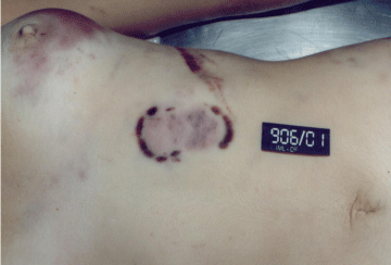
Figure 1: General aspect of bite mark.
Description of the observed injuries on the victim
Singular tooth marks could be recognized from the details normally left by cusps from upper and lower teeth. Bite mark showed all anterior superior and inferior teeth, from canine to canine, numbered according FDI (World Dental Federation) as 23, 22, 21, 11, 12, 13, 43, 42, 41, 31, 32 and 33. It was also noted the upper left first premolar, 24, upper right second premolar, 15, and lower right first premolar, 44.
Some particularities were distinguished such as: palatine cusp from tooth 24 is more printed than the vestibular cusp; only palatine cusp is imprinted on tooth 15 and impression of the tooth 44 is smoother than the other teeth. Differing from what is usual as common print in bite marks, the tooth 23 was printed as an incisive, showing a rectangular form. There were diastemas between the teeth 22 and 23, between the teeth 12 and 13 and teeth 43 and 42, measuring respectively 1.94 mm, 2.93mm and 2.00 mm. Upper lateral incisors seemed to be buccally inclined when compared to adjacent. Its mesial face was also displaced to vestibular (Figure 2).
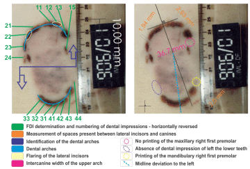
Figure 2: Description of characteristics of the bite mark.
The partition between upper and lower teeth was safely established and the midline could also be indicated. However, there was a deviation on the inferior midline, when compared to the superior one. The upper arch transverse width from canine to canine was 36.7mm (Figure 2).
Examination of author dental arch
Author dental arches had elliptical format, with great amplitude. Dental elements were well fixed with higid periodontal aspects, presenting cervical decreases in upper posterior teeth (Figure 3). The transverse width from 13 to 23 was 40.1 mm. Teeth 16, 26, 27, 37, 36, 35, 46 and 47 were absent with gingival tissue healed without recent surgery signals. The subject reported not using prosthesis. The upper and lower dental elements adjacent to the prosthetic spaces did not show any detritions consistent with prosthetic dental treatment.
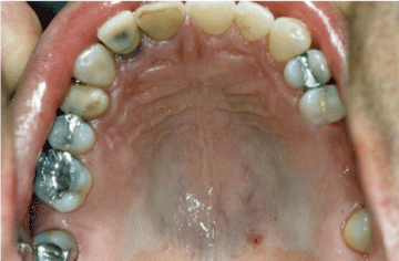
Figure 3: Maxillar arch of author.
The anterior superior and inferior tooth presented dental substance loss by detritions in the incisal border, reaching dentin. Tooth 13 showed detritions, but not physiological, on its mesial third, feature not observed in its upper counterpart. There was a diastema between 42 and 43 (Figure 4).
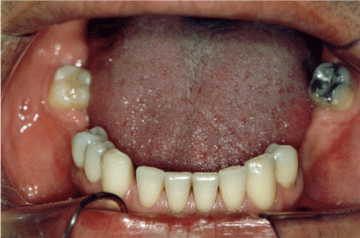
Figure 4: Mandibular arch of author.
The occlusal plane of the superior right hemi-arch showed a depression in the tooth 14, explained by the dental substance loss throughout the occlusal surface. There was also vestibular and palatal cusp height loss, when compared to the tooth 15 and the absence of physiological contact points, both on its mesial and distal faces. The tooth 12 had a total crown restoration buccal inclined when compared to adjacent teeth. The upper arch midline showed deviation, when compared to the lower arch midline. In occlusion, overbite on the right side is more accentuated (Figure 5).
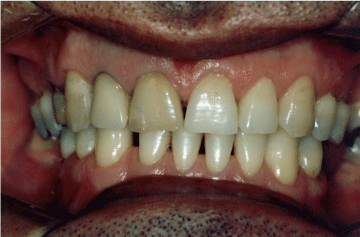
Figure 5: Arches of author in frontal occlusion.
Author dental impression in wax, dental casts and photographs
Suspect dental casts were produced in die stone (Herodent, Vigodent, Rio de Janeiro, Brazil). Author made a bite on a sample of bite registration wax sheet (Size 10 x 6 x 0.5 cm). Bites were made with an incisive action to get impression of the incisal edges and a portion of the labial and lingual surfaces of upper and lower tooth.
Dental impression wax revealed dental elements 28, 25, 24, 23, 22, 21, 11, 12, 13, 14, 15, 17, 18 from upper arch, and 48, 45, 44, 43, 42, 41, 31, 32, 33, 34, 38 from lower arch. Teeth 25, 14, 17 and 45 disclosed less deep impressions. The impression wax had interstice of 2.80 mm between teeth 23 and 22 as well an interstice of 3.90 mm between teeth 12 and 13. There was also a diastema of 3.00 mm between dental elements 43 and 42. The dental impression of the upper arch shows the tooth 22 and 12 buccally from adjacent with its mesial face displaced buccal (Figure 6).
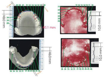
    Figure 6:  (A) Dental casts. Yellow circles represent lateral incisors buccally
inclined. Dental substance loss marked in pink circle. Absence of inferior
dental impression, marked in a purple circle. (B) Wax impressions.
Bite marks, determined on the victim, caused by the aggressor, were analyzed. The photographs that belong to the victim exposed to the attack were taken. Intraoral photographs included frontal view, two lateral views, right and left sides and an occlusal view of each arch. Dental casts were also photographed. Identification of criminal based on bite mark was performed by comparing unique attributes and patterns in the author dental impression wax, photographs and dental casts with similar characteristics in the injury.
Discussion
The present case report displays the great significance of bite mark in forensic investigations. Bite mark value in forensic dentistry relies mainly on the uniqueness of human dentition and this asserted oneness is reproduced and recorded in the injury [5]. When analyzing a bite mark, a very important step is to compare dental features between a subject dentition and the bite injury [3].
Bite marks are not so easily recognized, and it may appear in any part of the body, particularly the protruding ones [2]. Besides prominent areas, bite marks anatomical locations are related to different crime type, age and sex [8,9,10]. Although literature relates one bite mark to another similar bite on the body [1], the present case involves only one bite mark.
The absence of a tooth may be regarded as a distinctive feature in a bite mark that could prove to be of great discriminative value in excluding an author or concluding that they are a probable or possible biter [3]. Missing or present posterior teeth that are not relevant in bite mark injuries but useful data can still be extracted from their findings for forensic purposes [3]. In this case, premolars are posterior teeth found in the bite mark and were valuable for author confirmation. Confrontation between the victim and dental impression in the author dental arches demonstrate that the bite mark shows the teeth 24, 23, 22, 21, 11, 12, 13, 15, 44, 43, 42, 41, 31, 32 and 33, which are also present in the author. The presence of premolar teeth in bite mark occurs by force deduced by the bite, which after closing the mandible, it follows that the suction skin suffers distortions by its elasticity and shrinkage. However, teeth impressions of 14, 25, 45 and 34, also premolars, are absent.
Teeth 28, 16, 18, 48 and 38, present in the author, are absent in the dental impression, not participating in the hold and cut action performed by anterior teeth. Nevertheless, since they all are third molars and a first molar, and rarely appear in bite marks [9], their absence could not be considered as a factor of exclusion.
Besides teeth absence, other important occlusal parameters should be taken into account. When a tooth fails to register in a bite mark it does not necessarily mean that it is missing; it may be fractured, displaced or infra-occluded [3]. In the present case, teeth 25 and 14 exhibit different impression patterns. Tooth 14 has no effective contact with its lower counterpart, lying outside the occlusal plane, due to dental substance loss of all occlusal surfaces and tiny height of its cusps (Figure 7). Tooth 25 has no lower counterpart for an effective occlusal action. Actually, the particularities were very helpful in identifying the author as the biter.
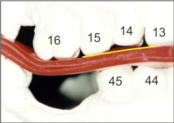
Figure 7: Dental casts demonstrating 14 teeth infra-occluded.
Missing teeth, when considered in combination with other dental characteristics such as rotated teeth or distinctive crowding patterns they have the potential to act as a useful discriminator in bite mark analysis [3]. (Figure 8).
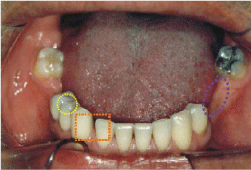
    Figure 8:  Diastemas between teeth 42 and 43, marked by orange rectangle.
Absence of inferior posterior teeth, in purple circle, and right first pre molar,
in a yellow circle.
Bite mark evidence in court presentation has a current hurdle: deformations left by the author’s dentition. Irrespective of the techniques used to analyze bite marks, there will always be warping, shrinkage, and distortion. These deformations are inherent by variations in tissue structure, dehydration, and photographic technique. Despite measurements made in bite marks will present variations, the relationship to the adjacent teeth are the same. Since tooth position remains constant, identifications based on these features are allowed. This concept can be applied to diastema, rotated teeth, missing teeth, teeth out of the arch, intercanine widths, incisal grooves, or any other recognizable tooth features [11]. In our case, comparing the widths measured in the dental impression and author plaster model, it was found a difference of 3.4 mm more in the last one, probably owing to human skin distortion and shrinkage by the elasticity of the tissues. Therefore, the upper canine widths of the dental impression found on the victim and in plaster model were considered compatible. Interstices of 2.0 mm between impressions of the teeth 43 and 42 were also found in wax impressions, measuring 3.0 mm. It was also observed the presence of diastema in the plaster model, as well as in author dental arch photograph. So, despite the discrepancy, it was found another coincidence point.
Teeth 22 and 12 buccally inclined when compared to its adjacent, with its mesial face buccal displaced was found in dental impression, wax impression and plaster model. Thus, it is pointed out another matching feature. Lower midline arch deviation relative to higher, observed in dental impression can also be seen in plaster models and dental arch photography. It was noticed a greater deviation in the bite mark, probably caused by the elasticity and shrinkage of the tissues and the dynamics of the biting action [12] (Figure 9).
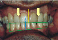
    Figure 9:  Overbite on the right side more accentuated, marked by the green
rectangle. Upper lateral incisors buccally inclined, highlighted by yellow
arrows. Midline deviation pronounced by yellow and blues lines.
Spaces present in dental impression among canines and upper lateral incisors come from the anatomical difference of canines presenting blade conformation, different from the anatomy of the incisors side having a trapezoidal shape, thus creating a gap between the distal edge of the upper lateral incisor and canine cusp. However, differences of 0.99 mm and 1.10 mm more between the teeth 12 and 13 in dental and wax impression come from probably the non physiological detritions on the third mesio incisal tooth 13 and, therefore, coincident characteristics of impressions (Figure 10).
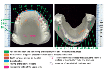
Figure 10: Description of cast models of the author and found coincidences.
The comparison of bite marks and teeth model can bed one by two methods: odontometric triangle method (Objective) and superimposition method (Subjective). In Objective method a triangle is made on the tracing of bite marks and teeth models by making three points-A,B,C. Points A and B are plotted on the outermost convex point on the canine teeth. Center of upper two central incisors is selected as point C. All the three points are joined to form a triangle ABC. Lines AB, BC, CA are measured and angles a, b, c were calculated. It is done for both upper and lower jaw [13]. Geometric morphometric analysis has been applied to bite marks. Shapes are quantitatively analyzed by capturing the geometry of morphological structures of interest [14]. 3D imaging technique also allow investigate bite marks [15]. In our study, we choose to analyze bite marks using comparisons between photographs from victim and author because is a cheap, fast and trustworthy method. Missing teeth, malformed teeth, fractures, crowding of the teeth, diastema and other peculiar characteristics of the teeth are helpful in the comparison process on these individualistic characters [16].
Author produced the mark in question. Plaster models of teeth and dental wax impression fully supported this statement (Table 1). In consequence, he was condemned for qualified homicide, because, according to Brazilian laws, it was committed with cruelty and feature that impeded or made impossible the offended defense. He was sentenced to nineteen years imprisonment in fully closed regime.
    Characteristics
    Dental    Impression
    Author


    1
    Arch
    Large    and elliptical
    Large    and elliptical


    2
    Upper    canine width
    36.7 mm
    40.1 mm


    3
    Tooth 14
    Missing    print
    Unevenness    of the occlusal plane


    4
    Tooth 25
    Missing    print
    Absence    of antagonist


    5
    Tooth 34
    Missing    print
    Neighbor    to the prosthetic space


    6
    Tooth 45
    Missing    print
    Neighbor    to the prosthetic space


    7
    Space    between teeth 12 and 13
    Present
    Atypical    13 tooth detrition


    8
    Space    between teeth 42 and 43
    Present
    Present


    9
    Tooth 12    buccally inclined
    Present
    Present


    10
    Tooth 22    buccally inclined
    Present
    Present


    11
    Lower    midline deviation
    Present
    Present
Table 1:  Overlapping points between the dental impression and the author dental
arch.
Conclusion
- Lesion observed in victim is a human dental impression produced intra-vitae.
- The points matched presented in dental impression and the author; identify him as the author of dental impression found on the dead victim.
- Bite marks analysis was the most important research tool utilized to author condemning.
References
- Crane J. Interpretation of non-genital injuries in sexual assault. Best Pract Res Clin Obstet Gynaecol. 2013; 27: 103-111.
- Sperber N. Identification of children and adults through federal and state dental identification systems: recognition of human bite marks. Forensic Sci Int. 1986; 30: 187-193.
- Kouble RF, Craig GT. A survey of the incidence of missing anterior teeth: Potential value in bite mark analysis. Sci Justice. 2007; 47: 19-23.
- Jakobsen JR, Kieser-Nilsen S. Bite mark lesions in human skin. Forensic Sci Int. 1981; 18: 41-55.
- Pretty IA, Sweet D. The scientific basis for human bite mark analyses - a critical review. Sci Justice. 2001; 41: 85-92.
- Tuceryan M, Fang L, Blitzer HL, Parks ET, Platt JA. A Framework for Estimating Probability of a Match in Forensic Bite Mark Identification. J Forensic Sci. 2011; 56: S83-S89.
- Clement JG, Blackwell SA. Is current bite mark analysis a misnomer?. Forensic Sci Int. 2010; 201: 33-37.
- Afsin H, Karadayi B, Cagdir SA, OzaslanA. Role of bite mark characteristics and localizations in finding an assailant. J Forensic Dent Sci. 2002; 6: 202- 206.
- Lessig R, Wenzel V, Weber M. Bite mark analysis in forensic routine case work. EXCLI Journal. 2006; 5: 93-102.
- Page M, Taylor J, Blenkin M. Reality bites - A ten-year retrospective analysis of bite mark casework in Australia. Forensic Sc Int. 2012; 216: 82-87.
- Stols G, Bernitz H. Reconstruction of Deformed Bite Marks Using Affine Transformations. J Forensic Sci. 2010; 55: 784-787.
- Sheasby DR, MacDonald DG. A forensic classification of distortion in human bite marks. Forensic Sci Int. 2001; 122: 75-78.
- Verma AK, Kumar S, Bhattacharya S. Identification of a person with the help of bite mark analysis. J Oral Biol Craniofac Res. 2013; 3: 88-91.
- Heras SM, Tafur D, Bravo M. A quantitative method for comparing human dentition with tooth marks using three dimensional technology and geometric morphometric analysis. Acta Odontol Scand. 2014; 72: 331-336.
- Sainte Croix MM, Gauld D, Forgie AH, Lowe R. Three dimensional imaging of human cutaneous forearm bite marks in human volunteers over a 4 day period. J Forensic Leg Med. 2016; 40: 34-39.
- Krishan K, Kachan K, Garg AK. Dental evidence in Forensic Identification - An Overview, Methodology and Present status. Open Dent J. 2015; 9: 250-256.