Special Article - Forensic Case Studies
Austin J Forensic Sci Criminol. 2016; 3(2): 1050.
Forensic Age Estimation According to Fusion of Proximal Humeral Epiphysis in 1367 Living Turkish Subjects’ Radiographs; A Preliminary Study
Erol OB¹, Bayramoglu Z¹*, Ertem F², Sharifov R³, Yılmaz R¹ and Yekeler E¹
¹Department of Radiology, Istanbul University, Turkey
²Istanbul School of Medicine, Istanbul University, Turkey
³Department of Radiology, Bezmialem Vakif University School of Medicine, Turkey
*Corresponding author: Bayramoglu Z, Department of Radiology, Istanbul Medical Faculty, Fatih, 34093, Istanbul, Turkey
Received: May 21, 2016; Accepted: July 22, 2016; Published: July 25, 2016
Abstract
Purpose: Precisely evaluation of bone age around and after 18years old has distinctive importance for legal issues in Turkey. Closure degree of proximal humeral epiphysis has significant value in this critical age group. We aimed to determine the age groups around this critical age among the male and females.
Methods: Shoulder radiographs of 1367 living Turkish individuals including 590 males and 777 females between 14-20 ages were evaluated according to closure degree of epiphysis divided into four stages; fusion less than one-third of the proximal humeral epiphysis (stage 1), fusion more than one-third to less two-third (stage 2), fusion more than two-third (stage 3), and recently closed (stage 4).
Results: Mean ages in females and males were 14.8 and 16.1 years for stage 1; 15.2 and 16.5 for stage 2; 16.2 and 17.5 for stage 3; 17.3 and 18.6 for stage 4, respectively. Minimum age for stage 4 was 15.6 in females and 17.0 in males. Maximum age for stage 4 was 18.5 in females and 19.6 in males. The mean ages between the age groups and gender were highly significant.
Conclusion: Evaluation of closure degree of proximal humeral epiphysis has a crucial role to differentiate the gender and radiological bone age around legal age of 18 years.
Keywords: Forensic age estimation; Proximal humeral epiphysis; Shoulder radiograph
Introduction
Age estimation for criminal proceedings has a significant importance in forensic cases. Radiological bone age determination is commonly used in forensic medicine and various forensic cases regarding marriage, social rights and liberty, pediatric judgment, employment, legal age alteration and gender determination.
Age thresholds concerning criminal responsibility varies between countries from 13 to 18. Teenagers may be referred to the forensic medicine for age estimation. Hand and wrist radiographs are used up to 17 years old in girls and 18 years old in males for bone age estimation. Precise evaluation around and after 18 years old has distinctive importance for legal issues in Turkey. Therefore, the next step for bone age determination in this critical age period will be evaluation of proximal humeral epiphysis after closure of radius and ulna epiphyses.
The aim of the present study was to document the age groups according to the fusion degree of proximal humerus epiphysis.
Methods
Conventional shoulder radiographs of the living Turkish subjects aged between 14 and 20 which were selected from the PACS (Picture Archiving Communication Systems) between the years 2012 and 2015 were evaluated retrospectively. A total of 1367 left shoulder radiographs with anteroposterior projection were enrolled into this study. The patients had been referred to the Emergency Department and discharged without hospitalization. The age and gender of the patients were confirmed from the hospital registrations. Individuals with a previous disease affecting skeletal development and evolution of the epiphysis such as a chronic disease, long bone fracture, previous radiotherapy, and chronic steroid usage were excluded in addition to the individuals of other nations. We selected the radiographs obtained with anteroposterior projection of glenohumeral joint in addition to humeral external rotation and performed with approximately 60 mA and 15 kVp radiation exposure and optimal magnification demonstrating the proximal humeral epiphysis clearly. Retrospective evaluation of selected appropriate radiographs was performed after the approval of the ethics committee.
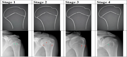
The left shoulder radiographs with DICOM (Digital Imaging and Communications in Medicine) format were evaluated on a workstation by consensus of two radiologists (E. OB., Y. E.) blinded to the subjects’ chronological age. A cartilage of proximal humeral epiphysis on radiographs is seen as a thin, radiolucent line between proximal humeral epiphysis and proximal humeral metaphysis. Closure degrees of proximal humeral epiphysis were divided into four groups from stage 1 to 4. Closure of proximal humeral epiphysis less than one-third is labeled as stage 1, more than one-third to less than two-third is labeled as stage 2, and more than two-third is labeled as stage 3. Recently closed proximal humeral epiphysis determined from a coarse indistinct radiopaque contour around the epiphysis and signed as stage 4. Examples for each stage on left shoulder radiographs as DICOM images and schematic drawings are shown in Figure 1. Stages of closure and chronological ages of individuals were recorded and adequate number of male and female subjects for each year from age 15 to 19 included in this study. The data of both males and females is compared among stage groups and gender. Cases causing disagreement between radiologists were excluded. Statistical analysis was performed using SPSS (Statistical Package for the Social Sciences) 21.1 statistical software. Based on the data, the age parameters between all stages and both genders were expressed as minimum, maximum, mean ± standard deviation, standard error, median values. Distribution of the data was evaluated with Kolmogorov-Smirnov test. Differences between the stages and gender were analyzed using Mann-Whitney U test for two independent groups in order to avoid skew distributions. Significance was assessed at p< 0.05. Differences of each stage in males and females were analyzed using one-way ANOVA.

    Figure 1:  Continuous line shows closed epiphysis, dotted line shows not ossified proximal humeral epiphysis. Schematic drawings of all stages correspondence to
the shoulder radiograph are seen on same column. Green arrows show closed part of epiphysis and red arrows designate open portion of epiphysis.
Results
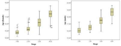
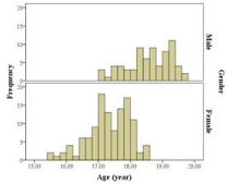
The numbers of the subjects aged from 14 to 20 are shown in Table 1. All subjects staged according to the closure degree (n=916) except those with totally open or old-closed epiphysis (n=451). The data was tabulated as the age parameters according to the four stages in both genders (Table 2). Since the data was not normally distributed, comparison of mean age values between each stages in both males and females was obtained from one-way ANOVA (Analysis of Variance) test and differences were highly significant (p< 0.001) (Table 3). The stages in both males and females showed positive correlation with the ages (Graph 1). The mean age of each stage in males compared with the girls was significantly different (p< 0.001) (Table 4). Girls were at more advanced stages than boys among all stages of proximal humeral epiphysis closure. Based on the data obtained from our sample (17/71) 24% of male patients in stage 4 are older than 18 years while the ratio is (17/118) 14% in females (Graph 2).
    Age (years)
    Female
    Male


    14
    60
    -


    15
    152
    90


    16
    114
    153


    17
    156
    135


    18
    207
    95


    19
    60
    86


    20
    28
    31


    Total
    777
    590
Table 1: Number of participants for each age according to gender.
    Male
    N
    Mean
    Std. Deviation
    Std. Error
    95% Confidence Interval for    Mean
    Minimum
    Maximum


    Lower Bound
    Upper Bound


    Stage 1
    113
    16.07
    0.43
    0.04
    15.99
    16.15
    15.47
    17.24


    Stage 2
    82
    16.49
    0.42
    0.04
    16.4
    16.58
    15.75
    17.37


    Stage 3
    166
    17.47
    0.65
    0.05
    17.37
    17.57
    15.93
    19


    Stage 4
    71
    18.57
    0.68
    0.08
    18.41
    18.73
    17.01
    19.62


    Total
    432
    17.1
    1.03
    0.04
    17
    17.2
    15.47
    19.62


    Male
    N
    Mean
    Std. Deviation
    Std. Error
    95% Confidence Interval for Mean
    Minimum
    Maximum


    Lower Bound
    Upper Bound


    Stage 1
    31

    14.82
    0.42
    0.07
    14.67
    14.98
    14.19
    15.83


    Stage 2
    95

    15.24
    0.42
    0.04
    15.16
    15.33
    14.49
    16.64


    Stage 3
    170

    16.2
    0.74
    0.05
    16.09
    16.31
    14.44
    17.8


    Stage 4
    118

    17.33
    0.64
    0.05
    17.21
    17.44
    15.56
    18.54


    Total
    414

    16.2
    1.05
    0.05
    16.1
    16.3
    14.19
    18.54
Table 2: Descriptive statistics of age parameters in each stages.
    Female
    Sum of Squares
    df
    Mean Square
    F
    Sig.


    Between Groups
    295.44
    3
    98.48
    244.83
    0


    Within Groups
    164.92
    410
    0.4


    Total
    460.36
    413


    Male
    Sum of Squares
    df
    Mean Square
    F
    Sig.


    Between Groups
    325.74
    3
    108.58
    333.36
    0


    Within Groups
    139.4
    428
    0.32


    Total
    465.15
    431
Table 3: Comparison of the mean ages of all stages in males and females.
    Table    4
    Mann-Whitney    U
    Wilcoxon    W
    Z
    Asymp.    Sig. (2-tailed)
    Exact    Sig. (2-tailed)
    Exact    Sig. (1-tailed)
    Point    Probability


    Stage 1
    59
    555
    -8.22
    0
    0
    0
    0


    Stage 2
    848
    7869
    -9.17
    0
    0
    0
    0


    Stage 3
    2961
    17496
    -12.52
    0
    0
    0
    0


    Stage 4
    848
    7869
    -9.17
    0
    0
    0
    0
Table 4: Statistical analysis of the differences between males and females by
using Mann-Whitney U test.

Graph 1: Mean ages for each stage in males and females.

    Graph 2:  Histogram comparing the age distribution of the participants in
stage 4.
Discussion
The study of union of bones epiphysis is considered a reasonable scientific and accepted method for estimation of age by the courts of law all over the world [1]. Changes in bones especially time related appearance and fusion of different ossification centres in growing periods are valuable indices for assessing the age. It can be evaluated up to 25 years of age by addition evaluation of medial clavicular epiphysis [2,3]. The medial clavicular epiphysis has been proved to be a valuable physical marker for age assessment especially around the age of 18 and 21 years [3]. At birth, there is no calcification in the carpal bones. Approximate ossification times are as follow; capitate 1-3 months, hamate 2-4 months, triquetral 2-3 years, lunate 2-4 years, scaphoid 4-6 years, trapezium 4-6 years, trapezoid 4-6 years, pisiform 8-12 years [4,5]. Fusion of epiphysis of distal end of radius occurs at 21 years in male and at 20 years in female. Distal epiphyseal fusion of ulna is observed at 21 years in male and 19 years in female. Thus in female, the ossification centers of distal end of radius and ulna occurs one to three years earlier than males [6]. Gilsanz, et al. divided skeletal development into six major categories to facilitate bone age assessments. At infancy (from birth to 14 months of age) the carpal bones and radial epiphyses, at toddlers (from 10 months to 3 years of age) the number of epiphyses visible in the long bones of the hand, at pre-puberty ( from 2 years to 9 years of age) the size of the phalangeal epiphyses, at early and mid-puberty (from 7 years to 14 years of age) the size of the phalangeal epiphyses, at late puberty (from 13 years to 16 years of age) the degree of epiphyseal fusion of distal phalanges, metacarpals, proximal phalanges and middle phalanges, and at postpuberty (from 15 years to 19 years of age) the degree of epiphyseal fusion of the radius and ulna are valuable for bone age assessments [7].
Poor knowledge concerning age estimation around the critical age of 18 makes the usability fusion of proximal humeral epiphysis more considerable. American literature presents anthropological methods of estimating chronological age on the basis of the morphology of the upper ends of the humerus. Objective morphometric method assessment of the upper end of the humerus was projected by Zydek, et al. and they found out lack of statistical correlation between atrophy of the spongy structure within the upper end of the humerus and the chronological age and the assessment of humerus structure should be omitted in the forensic medical age estimation [8]. However, a recent study have been published about reliability and usability of bone age estimation based on radiographic evaluation of proximal humeral epiphyseal closure [9,10]. Visually seriated radiographs of the proximal femur, proximal humerus, clavicle, and calcaneus from 130 individuals by Hamann-Todd collection were reported to be examined as indicators of skeletal age at death [10]. Radiograms of the humerus were reported to be analyzed using visual seriation qualitative assessment method and objective bone structure relative radiolucency method.
Low resolution of computed tomography (CT) than radiograph, increased susceptibility artifacts besides signal reduction in MRG (magnetic resonance imaging) and artifacts caused by the subcutaneous fat and intense fibrous tissue around glenohumeral joint makes the conventional roentgenogram optimal diagnostic tool for age estimation. Also, to refer an individual to CT for age estimation is controversial because of excess radiation exposure in absence of any previous disease [3].
Our observations showed that the proximal humeral epiphysis does not follow a standard arrangement during closure. Scattered closed segments in epiphysis were the most common observation especially stage 2 and 3. Therefore, whole epiphysis must be evaluated from beginning to the end attentively in order to decide current stage. Totally open epiphysis may be seen as a double or single thin radiolucent line between medial to lateral cortex of humerus. Recently closed epiphysis is seen as a coarse and thick radiopaque line concerning the epiphysis while old closed epiphysis is seen as thin slightly radiopaque line.
The proximal humeral epiphysis is mentioned to start ossification by the age of 14-20 years in females and 14-21 years in males while the complete closure is mentioned to be observed by the age of 18-25 in both genders. However, we excluded this hypothesis with reliable statistical analysis and suggested that the complete closure is earlier than this. Memon, et al. reported that the proximal humeral epiphysis has distinctive importance for bone age assessment in females between 16-17 years and in males between 17-18 ages [11]. Based on our data most of the recently closed epiphysis found between 17-18 ages in females and 18-19 ages in males.
The Gök Atlas is the most commonly used method in Turkey by forensic specialists in determining skeletal maturity from X-rays [12]. By using the Gök Atlas, the skeletal ages of boys between the ages of 11 and 22 were determined by examining the degree of epiphyseal fusion of shoulder, elbow, hand and wrist, and pelvic bones [13].
It is suggested that if the humeral epiphysis is fused the bone age will be 19. However, we obtained that the age of fusion of proximal humeral epiphysis is at least one year earlier especially in females. Thus, in addition to review the published data, we provided further information to the radiologists, forensic physicians, orthopedic surgeons and anthropologists. 15-18 age interval has significant importance for criminal liability cases and should be determined according to fusion of humerus epiphysis.
The algorithm of at least one year earlier epiphyseal fusion in females than males is acceptable for proximal humeral epiphysis too. Also each stage takes average one year period to progress in both males and females. Including adequate number of cases and obtaining mean values with low standard deviations in each stage strengthen our study to be a reference in evaluation of closure of proximal humeral epiphysis for radiological bone age estimation based on anteroposterior shoulder radiographs. This is the first study which evaluates the applicability of radiographs in assessing the closure of epiphysis of the proximal humeri for the purposes of forensic age diagnostics in living people. Comparable data based on statistical analysis is not existent in the literature about bone age estimation concerning closure of proximal humeral epiphysis.
Limitations of our study are that; it is not a longitudinal study giving permission to achieve birth records of the subjects documenting whether they borne in a hospital or not, and percentile values of the subjects during growing-up. However, including large number of subjects facilitates to diminish the standard deviation.
Conclusion
The evaluation of proximal humeral epiphysis is appropriate for both genders and a valuable method in bone age estimation in critical age. In order to determine whether an individual has already reached the criminal responsibility age (18 years) or not; evaluation of proximal humeral epiphysis closure is important after closure of hand and wrist bone epiphyses just before evaluation of iliac crest and ischion apophyses.
Acknowledgement
Any financial support was taken during the materials were preparing. A part or total of this study has not been published elsewhere. We (all authors) declare that there is no conflict of interests regarding the publication of this paper.
References
- Banerjee KK, Aggarwal BB. Estimation of Age from Epiphyseal Union at the wrist and ankle joints in the capital city of India. Forensic Sci Int. 1998; 31-39.
- Reddy KSN. The essentials of Forensic Medicine and Toxicology. 28th ed. Hyderabad: K.Suguna Devi; 2009.
- Schemling, 2013. Forensic age estimation in JA Siegel, PJ Saukko. Ecyclopedia of Forensic Sciences. Academic Press, Waltham.
- Butler P, Mitchell A, Healy JC, editors. Applied Radiological Anatomy. 2nd ed. Cambridge University Press. 2012.
- Garn SM, Rohmann CG. Variability in the order of ossification of the bony centers of the hand and wrist. Am J Phys Anthropol. 1960; 18: 219-30.
- Rajdev BM, Gajera CN, Rajdev SB, Govekar GP, Tailor CI, Chandegara PV. Age of fusion of epiphysis at distal end of radius and ulna. Int J Int Med Res. 2014; 1; 5-11.
- Gilsanz V, Ratib O. Hand Bone Age, a Digital Atlas of Skeletal Maturity. Berlin: Springer-Verlag Heidelberg, 2005; 1-92.
- Zydek L, Barzdo M, Meissner E, Berent J. Assessment of Bone Age Based on Morphometric Study of the Upper End of the Humerus. J Forensic Sci. 2011; 56: 1416-1423.
- Wachholz L. O oznaczaniu wieku. Poland: Akademia Umiejetnos´ci. Krak w. 1894.
- Walker R, Lovejoy O. Radiographic changes in the clavicle and proximal femur and their use in the determination of skeletal age at death. Am J Phys Anthropol 1985; 68: 67-68.
- Memon N, Memon M U, Memon K, Junejo H, Memon J. Radiological Indicators for Determination of Age of Consent and Criminal Responsibility. Journal of Liaquat University of Medical & Health Sciences. 2012; 11: 64-70.
- Büken B, Büken E, Yazıcı B, Şafak A, Mayda A. Is the Use of the Gök Atlas Sufficiently Reliable for Age Determination of Turkish Children ?. Fifth Annual Anatolian Forensic Science Congress. 2006; 38: 319-327.
- Gök Ş, Erölçer N, Özen C. Age determination in forensic medicine. 2nd ed. Istanbul: Turkish Republic Ministry of Justice, Council of Forensic Medicine Press; 1985.