Clinical Image
Gastrointest Cancer Res Ther. 2016; 1(2): 1008.
Primary Malignant Melanoma of Esophagus
Liu L, Lu X, Zhao Y and Cao D*
Department of Radiology, The First Hospital of Jilin University, China
*Corresponding author: Dr Dianbo Cao, Department of Radiology, The First Hospital of Jilin University, 71 Xin Min Zhu Street, Chang Chun, China
Received: September 02, 2016; Accepted: September 07, 2016; Published: September 09, 2016
Keywords
Esophageal tumors; Melanoma; MSCT; Endoscopy
Clinical Image
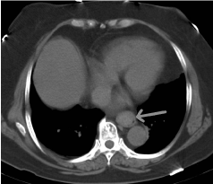
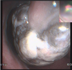
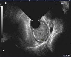
A 63-year-old woman presented with progressive dysphagia for 1 month, and had lost approximately 2kg in weight since the onset of his illness. Physical examination showed no abnormality. MSCT scan revealed a solid mass in the distal third of the esophagus, where local fat space around the esophagus still existed (Figure 1). Endoscopy revealed an irregular dark endoluminal and lobulated mass suited 30-35cm from the incisors (Figure 2). Endosonography showed esophageal mass to be solid, heterogenous in echo-architecture and arising from the deep mucosa (Figure 3). The biopsy specimen suggested suspected malignant melanoma. Extensive detailed examination revealed no other skin, anal, facial or rectal lesions. The patient underwent an Ivor-Lewis esophagogastrectomy and lymph node dissection. The surgical specimen showed a 5.0cm ×4.0cm ×4.0cm, polypoid and pigmented lesion located in the distal esophagus. The tumor was composed of plump spindle-shaped cells with pigmented cytoplasm, prominent nuclei and numerous mitosis. Immunohistochemical stains were positive for S-100 protein and HMB-45. Therefore, a diagnosis of primary malinant melanoma of esophagus (PMME) was made. The patient had an uneventful recovery and was in good condition on the follow-up of one year.
Malignant melanoma is a typical cutaneous tumor, originating from melanocytes. PMME is exceedingly rare, varying its incidence from 0.1% to 0.2% of all esophageal malignant tumors. Imaging studies such as barium meal and CT only reveal esophageal occupying lesion. The characteristic appearances is a polypoid irregular pigmented and friable lesion at endoscopy, but only 54.7% of cases are diagnosed preoperatively as malignant melanoma. Endoscopic ultrasound is seldom applied and common a hypo echoic or a mixed echogenicity mass, as described in our case.