Case Report
Gastrointest Cancer Res Ther. 2017; 2(1): 1016.
An Unusual Cause of Melena: Brunner’s Gland Hyperplasia
Loganadane G1,2*, Honoré C³, Dartigues P4, Marty O5, De La Lande P5 and Levy A1,6,7
¹Department of Radiation Oncology, Gustave Roussy, Université Paris-Saclay, F-94805, Villejuif, France
²Department of Radiation Oncology, Henri Mondor, University of Paris-Est, F-94010 Créteil, France
³Department of Surgical Oncology, Gustave Roussy, Université Paris-Saclay, F-94805, Villejuif, France
4Department of Gastroenterology, St Joseph Hospital, Paris, France
5Department of biology and medical pathology, Gustave Roussy, Université Paris-Saclay, France
6Univ Paris Sud, Université Paris-Saclay, F-94270, Le Kremlin-Bicêtre, France
7INSERM U1030, Molecular Radiotherapy, Gustave Roussy, Université Paris-Saclay, France
*Corresponding author: Gokoulakrichenane Loganadane, Department of Radiation Oncology Henri Mondor, University of Paris-Est, F-94010 Créteil, France
Received: March 30, 2017; Accepted: April 26, 2017; Published: May 03, 2017
Abstract
A 43 year old woman presented melena with severe anemia without particular medical history or medication intake. Esophagogastroduodenoscopy showed a large ulcerative mass located from the first to second portion of the duodenum. Endoscopic ultrasonography and the abdominal computed tomography described a 5cm mass with a tumoral aspect. A gastrointestinal stromal tumor (GIST) was suspected and a partial duodenectomy was performed. Finally the pathological concluded to the diagnosis of Brunner-gland hyperplasia.
Introduction
Brunner’s gland hyperplasia is a rare and benign lesion of the duodenum, which generally has a good prognosis although some cases of association with adenocarcinoma have been described. Its clinical presentation is quite variable, ranging from an asymptomatic condition to abdominal pain, intestinal obstruction, gastrointestinal hemorrhage, and occasionally the mimicking of a duodenal malignancy.
Case Presentation
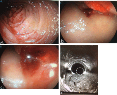
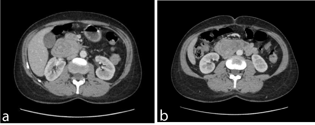
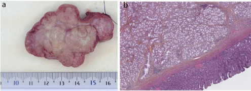
A 43 year-old-woman was admitted at the emergency ward for melena. Her history was unremarkable, and she was not taking any medication. The physical examination was not remarkable and the blood count found a haemoglobin level of 6.9g/dl. Esophagogastroduodenoscopy showed a large ulcerative mass located from the first to second portion of the duodenum (Figures 1a). Endoscopic ultrasonography displayed a 48mm isoechoic tumor with cystic change arising from the submucosal layer (Figure 1b). Abdominal computed tomography confirmed the round mass with internal multifocal low densities in the proximal duodenum without distant lesion (Figures 1c). Repeated endoscopic biopsies were not contributive. A gastrointestinal stromal tumor (GIST) was suspected and a partial duodenectomy was performed. A cross-section of the specimen showed a well-circumscribed, nodular mass including fibrous septa (Figure 2a). Figure 2b shows a histologic section of the mass stained with hematoxylin and eosin. Pathologic analysis retrieved a nodular proliferation of normal Brunner’s glands separated by fibrous septa at the junction of the duodenum, confirming the diagnosis of Brunner-gland hyperplasia (Figures 3a, 3b). There was no surgical complication and the patient had a complete resolution of her symptoms.

    Figure 1: Esophagogastroduodenoscopy (1a to 1c) and endoscopic
ultrasonography (1d) showing the duodenal mass.

    Figure 2: Abdominal computed tomography showing the mass
without distant lesion.

    Figure 3: Cross and histologic section of the surgical mass stained
with hematoxylin and eosin.
Discussion
Brunner’s gland is an alkaline-secreting gland that is usually located in the deep mucosa or submucosal layer of the duodenum. Brunner’s gland hyperplasia/adenoma is a benign neoplasm with an unknown exact pathogenesis. Malignant transformation was exceptionally reported, with overexpression of P53 and MIB1 demonstrated in a single patient [1]. Clinical presentation involves a large spectrum of digestive symptoms and rare complications include gastrointestinal bleeding and obstruction. Misdiagnosis may be frequent given the pseudo-tumour presentation, and this may be reinforced by the tumour’s high avidity for 18F-fluorodeoxyglucose [2,3]. The great majority of duodenal mesenchymal tumors are GISTs and local resection is an adequate management [4]. Only a deep endoscopic or a surgical biopsy provides adequate tissue for diagnosis, as Brunner’s glands are located in the submucosa. Endoscopic ultrasound with fine needle aspiration may help to get a correct diagnosis and avoid overtreatment [5]. In symptomatic patients with large lesions, limited surgery is the main therapeutic modality but endoscopic resection most likely represents a reasonable alternative when the tumour is small or pedunculated [6].
Acknowledgements
GL, CH, PD, OM, PdL, and AL participated to the acquisition, analysis and interpretation of data, critical revision of the manuscript, and approved the manuscript.
GL and AL drafted the manuscript.
References
- Fujimaki E, Nakamura S, Sugai T, et al. Brunner’s gland adenoma with a focus of p53-positive atypical glands. J. Gastroenterol. 2000; 35: 155–158.
- Lin C-C, Chang W-H, Wang T-E, et al. Why is this abdomen swollen? Lancet Lond. Engl. 2008; 371: 274.
- Park SH, Park K-M, Kim JS. 18F-FDG-avid brunner gland hyperplasia. Clin. Nucl. Med. 2014; 39: 728–730.
- Johnston FM, Kneuertz PJ, Cameron JL, et al. Presentation and management of gastrointestinal stromal tumors of the duodenum: a multi-institutional analysis. Ann. Surg. Oncol. 2012; 19: 3351–3360.
- Arena M, Rossi S, Morandi E, et al. Brunner’s glands hyperplasia: diagnosis with EUS-FNA for suspected pancreatic tumor involving the duodenum. JOP J. Pancreas. 2012; 13: 684–686.
- Ohba R, Otaka M, Jin M, et al. Large Brunner’s gland hyperplasia treated with modified endoscopic submucosal dissection. Dig. Dis. Sci. 2007; 52: 170–172.