Clinical Image
Gastrointest Cancer Res Ther. 2022; 6(1): 1035.
Adenomyomatosis Looks Like a Pearl Necklace
Raissa Albertine KN¹*, Fatimzahra L² and Jerondi L³
1Resident Doctor in Radiology Emergency Department of Radiology Hospital Avicenne Rabat Morocco
2Assistant Professor Specialist in Radiology, Radiology Emergency Department at Avicenne Rabat Morocco Hospital, Morocco
3Professor Head of the Radiology Department Radiology Emergency Department Avicenne Hospital Rabat Morocco
*Corresponding author: Kaukone Nyare Raissa Albertine, Resident Doctor in Radiology Emergency Department of Radiology Hospital Avicenne Rabat Morocco
Received: May 23, 2022; Accepted: June 16, 2022; Published: June 23, 2022
Keywords
Adenomyomatosis; MRI; Pearl Necklace Sign.
Clinical Image
Focal and diffuse thickening of the gallbladder, Benign and noninflammatory pathology.
Gallbladder adenomyomatosis is a benign alteration of the gallbladder wall characterized by excessive epithelial proliferation associated with muscle hyperplasia, resulting in thickening of the gallbladder wall. Excessive epithelial proliferation leads to epithelial folding in the underlying muscle layer with the subsequent formation of diverticular pockets lined with epithelium, the Rokitansky-Aschoff sinuses (SRA).
MRI
Thickening of the gallbladder wall can be clearly represented on both T1 and T2 weighted images, and is not a specific finding.

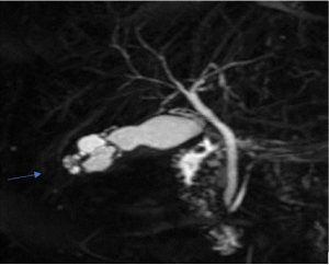
    T1: Coronal MRI biliséquences showing rounded hyper-intense intramural

cavities.

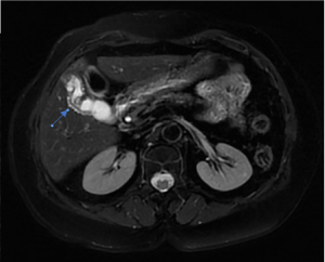
T2: Axial T2 rounded hyper-intense intramural cavities.
MRI guarantees high specificity in the diagnosis of GA by precisely excluding extra-parietal infiltration, which is indicative of gallbladder carcinoma.
ARS generally appear clearly in hyperintensity on T2-weighted images [1-3], hypointense on T1-weighted images and do not show any enhancement of contrast.
However, the progressive concentration of bile and the development of calcifications can modify the MRI appearance of the RAS which can become increasingly hyperintense on T1-weighted images and relatively hypointense on T2-weighted images.
The pearl necklace sign refers to the characteristic curvilinear arrangement of multiple rounded hyperintense intramural cavities seen on T2-weighted MRI.
References
- Matteo Bonatti, NorbertoVezzali, Fabio Lombardo, Federica Ferro, Giulia Zamboni, et al. Gallbladder adenomyomatosis: imaging findings, tricks and pitfalls. Insights into Imaging. 2017; 8(2): 243–253. doi: 10.1007/s13244-017- 0544-7.
- Haradome H, Ichikawa T, Sou H, Yoshikawa T, Nakamura A, et al. The pearl necklace sign: an imaging sign of adenomyomatosis of the gallbladder in MR cholangiopancreatography. Radiology. 2003; 227(1): 80-88. doi: 10.1148/ radiol.2271011378.
- Teelucksingh S, Welch T, Chan A, Jason D, Fidel S.R. Gallbladder adenomyomatosis presenting with abdominal pain. Cureus. 2020; 12(9): e10485. doi:10.7759/cureus.10485.