
Clinical Image
Ann Hematol Oncol. 2015;2(3): 1026.
Idiopathic Pericardial Cyst
Calin S1, Sepesi B2, Swisher S3, Yusuf S.W4 and Ferrajoli A5*
1Department of Hematopathology, USA
2Department, Thoracic & Cardiovascular Surgery, USA
3Department, Surgery, USA
4Department of Surgery, USA
5Cardiology and Leukemia, USA
*Corresponding author: Ferrajoli A, Department of Leukemia, UT MD Anderson Cancer Center, USA
Received: December 08, 2014; Accepted: January 27, 2015; Published: January 29, 2015
Clinical Image
Patient is a 52 years old man referred for treatment of chronic lymphocytic leukemia. At time of referral a routine chest radiograph showed a mass projected over the right lower chest. Computerized tomography images were obtained that demonstrated a pericardial cyst measuring 10.9 cm in the largest diameter with some compressive atelectasis of the right middle lobe (Figure A). The patient had no complains of chest pain, shortness of breath or cough. An ultrasoundguided needle aspiration yielded the diagnosis of idiopathic pericardial cyst based on fluid characteristics (transudate, with no evidence of malignant cells, acid fast bacilli, fungi or anaerobic organisms). Four years later, the patient developed recurrent episodes of retro-sternal discomfort. A surgical intervention was performed and a 13.5 cm pericardial cyst was removed by median sternotomy (Figure B, C) with resolution of the patient symptoms. Pericardial cysts are rare (1 per 100,000 individuals) congenital abnormalities.
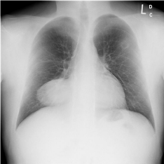
    Figure 1:  Chest radiograph image showing a mass projected over the right
lower chest.
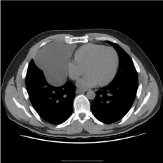
    Figure 1:  Computerized tomography image of chest showing a large
pericardial cyst and compressive atelectasis of the right middle lobe.
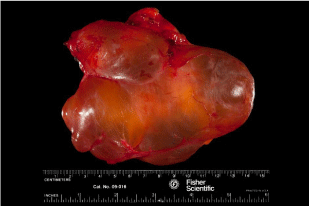
Figure 1: Resected pericardial cyst (13.5 x 10.5 x 5.8 cm).