Clinical Image
Austin J Musculoskelet Disord. 2015;2(3): 1023.
Os Trigonum Syndrome: A Rare Cause of Posterior Ankle Pain
Mustafa Ozsahin¹*, Besir Erdogmus² and Safinaz Ataoglu¹
¹Department of Physical Medicine and Rehabilitation, Duzce University, Turkey
²Medical School of Duzce University, Department of Radiology, Turkey
*Corresponding author: Mustafa Ozsahin, Department of Physical Medicine and Rehabilitation, Duzce University, Turkey
Received: June 30, 2015; Accepted: July 01, 2015; Published: July 04, 2015
Keywords
Os trigonum syndrome, posterior foot pain, posterior tibiotalar impingement syndrome
Musculoskeletal Imaging
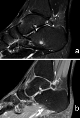
A 43-year-old male presented to our outpatient clinic complaining of left foot pain for the last 6 months. He had been competing in orienteering for 7 years, but had no history of trauma or injury to the ankle. In the physical examination, plantar flexion of the ankle and great toe was painful. There was mild tenderness to palpation at the posterior aspect of the subtler joint. Os trigonum syndrome was diagnosed based on the clinical examination and magnetic resonance imaging (MRI) findings (Figure 1). Physical therapy consisting of cold packs and foot and ankle range of motion and strengthening exercises was started. His symptoms improved with rest, activity modification, and physical therapy. The os trigonum is an accessory bone of the foot located just behind the talus. An os trigonum is seen in up to 14% of the normal, asymptomatic population [1]. It is generally asymptomatic, but occasionally presents as chronic posterior ankle pain. It occurs commonly as a result of overuse of the posterior ankle caused by repetitive plantar flexion stress [2]. Although most often seen in ballet dancers, os trigonum syndrome has also been reported in football and volleyball players, downhill runners, and even non-athletes [2,3]. This painful condition is usually called os trigonum syndrome, but is also called a symptomatic os trigonum, posterior ankle impingement syndrome, posterior tibiotalar impingement syndrome, and talar compression syndrome [2, 3].The initial treatment for this syndrome is conservative. If conservative treatment fails to provide relief, poster lateral arthroscopic resection with or without a capsulectomy or debridement of the os trigonum may resolve symptoms [3]. In many cases, however, the evaluation of posterior ankle pain is complex, making diagnosis difficult. A painful os trigonum is a common cause of posterior ankle pain, resulting from the impingement of posterior ankle structures. Os trigonum syndrome should be considered in patients with posterior foot pain that is made worse by plantar flexion of the ankle. The clinical presentation and radiographic findings can suggest the diagnosis of os trigonum syndrome, but MRI should be performed to confirm the diagnosis.

    Figure 1:  (a, b) Sagittal fat-saturated T2 images show marrow edema of the
os trigonum and talus at the synchondrosis, along with fluid in the fatty tissue
surrounding the os trigonum.
References
- Donovan A, Rosenberg ZS. MRI of ankle and lateral hind foot impingement syndromes. AJR Am J Roentgenol. 2010; 195: 595-604.
- Nault ML, Kocher MS, Micheli LJ. Os Trigonum Syndrome. See comment in PubMed Commons below J Am Acad Orthop Surg. 2014; 22: 545-553.
- Song AJ, Del Giudice M, Lazarus ML, Lomasney LM, Demos TC, Dux K. Radiologic case study. Os trigonum syndrome. Orthopedics. 2013; 36: 5, 63-68.