
Case Report
Austin J Musculoskelet Disord. 2016; 3(1): 1029.
Salvage of an Existing Construct With Stacked Plating
Hsi-Ting Lin¹ and Andy Li-Jen Liu²*
1Department of Orthopedic Surgery, Cathay General Hospital, Taipei, 10689, Taiwan, ROC
2Department of Orthopedic Surgery, Hope Doctors Hospital, Taiwan, R.R.China
*Corresponding author: Andy Li-Jen Liu, Department of Orthopedic Surgery, Hope Doctors Hospital, No. 125, Xindong Street, Miaoli City, 36052, Taiwan, R.R.China
Received: December 01, 2015; Accepted: March 11, 2016; Published: March 14, 2016
Abstract
Fracture fixation with double plating has been well documented. We report an 82-year-old female who suffered a proximal humerus fracture who was treated surgically with locking plate and screws. Unfortunately, patient fell seven weeks later and suffered a peri-implant humeral shaft fracture at the junction of the proximal and middle third just around the most distal screw from the initial surgery. Because the previous fracture had not healed completely and removing the prosthesis would probably cause a collapse of the previous reduction, a decision was made to stack an exact but longer proximal humeral locking plate for fixation. After three months of follow up, patient has achieved functional recovery with satisfactory ranges of motion in her right shoulder. This outcome demonstrates that salvage of an existing construct with stacked plating can work as an alternative method of fixation when other options are less appropriate.
Keywords: Overlap plates; Proximal humerus fracture; Stacked plates; Veterinary cuttable plates
Introduction
Most salvage procedures involving a pre-existing construct usually requires the removal of the original prosthesis followed by the application of a new one after proper reduction and fixation have been performed. However, when the removal of the original construct is not suitable, an alternative method must be available. Depending on the type of fracture, orthogonal double plates and overlapping connecting plates have proven effective [1-6]. we present an unusual case report describing the salvage of an existing construct with stacked plating. Even though it has been commonly used for fracture repair in small animals, its application in human fracture fixation has been limited and does provide worthwhile and novel information for the orthopedic surgeon seeking different treatment options. A search of literature shows that only a handful of similar articles have ever been published [4-11].
Case Report
Patient is an 82-year-old woman who fell while strolling through the park. She entered the emergency department (ER) complaining of extreme pain on her right upper arm. Physical examination revealed abundant ecchymosis around the arm and chest, tenderness upon palpation, and limited Range of Shoulder Motion (ROM). No neurovascular abnormalities were noted. Initial radiograph of the right upper arm revealed a proximal humerus fracture with severe comminution of the humeral head (Figure 1). Considering the age of the patient, the severity of the fracture, and the osteoporotic quality of her bone, a hemiarthroplasty would be the treatment of choice. However, after informing the patient of the benefits and risks of such procedure, she decided that restoring shoulder motion, function and strength as close to her pre-injury levels as possible would suit her active lifestyle better. Therefore, Open Reduction and Internal Fixation (ORIF) were performed. During surgery, with the help of both allogenic and artificial bone grafts, the fracture was reduced and fixated with a proximal humerus internal locking system (PHILOS) plate (Synthes, Switzerland). Postoperatively, the radiograph showed good alignment and the arm was immobilized with a shoulder sling (Figure 2).
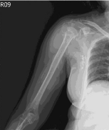
    Figure 1: Radiograph of the right upper arm showing a four-part proximal
humerus fracture. Note the surgical clips in the axillary region that remain
from her previous breast surgery.
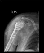
    Figure 2: Postoperative radiograph showing fixation with a locking proximal
humeral plate and bone grafts in the treatment of the aforementioned fracture.
Unfortunately, about seven weeks after the surgery, patient fell again and landed on the injured arm. She complained of similar right upper arm pain and was brought to ER for help, where swelling and bruising of the upper arm were also noted. Precautionary radiograph showed a humeral shaft fracture at the junction of the proximal and middle third just around the most distal screw from the previous surgery (Figure 3). At this point, a second surgical procedure would be necessary not only to allow continuous bone healing of the previous fracture but also to reduce and fixate the new peri-implant humeral shaft fracture. After searching through literature and carefully weighing the advantages and disadvantages of various options such as hemiarthroplasty, Reverse Total Shoulder Arthroplasty (RTSA), and Intramedullary (IM) nailing, a salvage procedure of the existing construct with stacked plating was decided.
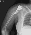
    Figure 3: Radiograph of the right upper arm showing a peri-implant humeral
shaft fracture at the junction of the proximal and middle third. Note the
loosening of the most distal screw but retention of the overall prosthesis from
the initial surgery.
Over the previous operation scar, a vertical incision was made and extended further distally to expose the new fracture site. Since the initial ORIF occurred only seven weeks ago, removing the prosthesis would certainly cause a collapse of the ongoing healing fracture, not to mention a waste of the bone grafts. In addition, because the proximal portion of the prosthesis appeared radiographically stable, a decision was made not to remove the previous construct. After removing four screws from the proximal portion and three screws from the distal portion, an exact but longer (twice the length) PHILOS plate was stacked on top of the existing prosthesis after proper reduction was performed. By using and stacking identical plates of different lengths, it was assured that the screw holes would also be stacked on top of each other and that the same locking screws can be used to fixate and lock both plates to the bone firmly. Overall, a total of six screws were exchanged and locked through the overlapping portion of the stacked plates while six screws were locked distally through the non-overlapping portion of the longer plate for secure fixation. Even though the secondary procedure had extended the exposure to the middle third of the humeral shaft, care was taken during placement of the plate and screws as not to injure the radial nerve.
Postoperatively, the arm was immobilized in a sugar tong splint due to the osteoporotic quality of the bone and because the same bone had suffered two fractures in less than two months (Figure 4). At one month post-op, the splint was removed as gentle mobilization of the right upper arm and shoulder began without resistance. Neither skin necrosis nor wound dehiscence was observed. During the latest follow up at three-months (almost five months after the initial surgery), the patient was asymptomatic, did not complain of reduced shoulder strength, and had good functional recovery with the following ROM: 115 degrees of flexion, 30 degrees of extension, 110 degrees of abduction, 40 degrees of external rotation, and 45 degrees of internal rotation. She was able to perform daily activities without pain or limitations and did not report any discomfort from the bulkiness of the prostheses (Figure 5).
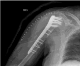
    Figure 4: Postoperative radiograph after the salvage procedure showing
good reduction and fixation with the stacked plates and screws.
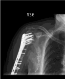
    Figure 5: Radiograph at three months after the salvage procedure. Note the
slight bone resorption near the greater tuberosity but overall good alignment
is maintained.
Discussion
Surgical treatment of fractures has always obeyed the principles first proposed by Lambotte and later modified by the Arbeitsgemeinschaft fur Osteosynthesefragen group [12,13]. Proper plates and screws application, specifically their positioning, provides a solid framework in which the fractured bones may be attached and their reductions facilitated. For example, placing the plates orthogonally in a 90o configuration has proven success both biomechanically and clinically [1]. On the other hand, in situations where placement of plating in such a pattern is not possible, alternatives must be available.
Hemiarthroplasty is usually the preferred treatment option for unreconstructible proximal humerus fractures; or for those fractures with malunion, nonunion, or hardware failure following osteosynthesis fixation [14]. Even though it does provide adequate pain relief, the functional outcomes (i.e. range of motion) of the treated shoulder are frequently affected and disappointing [15,16]. For our patient who has and wants to maintain an active lifestyle, hemiarthroplasty is not a viable option. While adequate clinical results can be obtained with RTSA in patients with comminuted humeral head fractures, functional scores and range of motion are frequently unpredictable [17]. In addition, some authors have found abundant radiographic evidence of glenoid loosening and scapular notching in their studies, which would caution its use [18,19]. Since the ideal candidate for RTSA seems to be elderly with low functional demand [17], it does not seem to be a good option for our patient. Last but not least, even though IM nailing achieves stabilization with minimal surgical invasion, poor outcomes have been reported in comminuted humeral head fractures [20,21]. Since all of the aforementioned options have been shown to be inadequate, another option that can reconstruct the proximal humerus and peri-implant humeral shaft fractures with emphasis on achieving anatomical reduction and stable fixation must be found.
The concept of using double plates for osteosynthesis was previously described in the 1970s [2,3]. However, using overlapping plates as an alternative to treat complicated fractures represents a fairly novel concept. Christodoulou et al [4] used two overlapping plates (a small fragment cloverleaf plate and a narrow 4.5-mm dynamic compression plate with at least two common screws) for the treatment of Maale and Seligson [5] type IV distal tibial intraarticular fractures with proximal spiral extensions. The coupling of the two plates by at least two screws provided adequate rigidity as no loosening or prosthesis breakage occurred. All 11 of their patients returned to work within six months of the surgery and only three reported moderate ankle joint pain after intensive activities. In addition, Adulkasem [6] used overlap connecting plates for the treatment of 15 segmental and comminuted femoral, tibial and radial fractures. The connection between two plates by at least three or four mutual screws allowed adequate fracture healing without infection, shortening and decreased ROM in all patients. Even though these overlapping plates shared only a minimum of two screws and were not fully stacked on top of each other, they were able to achieve successful fracture fixation because basic osteosynthesis principles were followed: minimum of six cortices in each fracture fragment and the plates sat well covering the bone.
Through literature review, the most successful example of using stacked or overlapping plates for fracture repair is the Veterinary Cuttable Plates (VCP) used in small cats and dogs [7-9]. These plates are manufactured as 7.0 mm wide and 300 mm long, 50-hole prostheses. They come in two sizes: 1.5/2.0 mm and 2.0/2.7 mm plates that are 1.0 mm and 1.5 mm thick, respectively [9,10] Since all these thin plates have the same hole-to-hole distance, they can be cut to the desired length, stack on top of one another, and allow for screw placement in any hole. Consequently, stacked VCP result in increased stiffness, strength, and fatigue life of the construct when compared to using one single plate [8,11,22].
Even though it has been reported that the stiffness of two stacked VCP equals the sum of the stiffness of the individual plate [22], stacking two VCP may result in a bulky construct that causes soft tissue morbidity [7]. In addition, using an implant that is too rigid causes stress shielding on the repaired bone and stress concentration at the metal to bone interface; [23] the result of which may appear clinically as osteopenia, delayed bone healing, screw pullout, and implant-related fractures [23-25]. To solve this problem, Bichot et al. [10] performed a study on the effects of the length of the stacked plates and made the following observations: 1) shortening the superficial plate affects the strength of the construct, specifically at the fracture gap; 2) shortening the superficial plate reduces the stress concentration by evenly distributing the strain along the implantbone interface; and 3) lengthening the superficial plate increases the friction between the stacked plates, which in turn results in a stiffer and stronger construct. Therefore, use of partially stacked plates may represent a viable option between a single plate and fully stacked VCP when looking for a balance between strength and stress concentration in a construct.
By combing the aforementioned concepts of overlapping and stacked plates, we placed a locking proximal humeral plate on top of an existing construct to accomplish anatomic reduction and stable fixation. A plate that was double the length of the original construct was chosen to make sure that at least six cortices were firmly secured on each side of the fracture fragment. Fortunately, adequate soft tissue and skin coverage were available and patient does not feel discomfort from the bulkiness of the plates when moving her upper arm and shoulder. In addition, no skin slough was observed during the healing process. However, disadvantages of this procedure do include excessive soft-tissue dissection and periosteal stripping, and bone weakening from multiple screw holes, all of which can cause complications such as nonunion, delayed union, or implant failure.
In gauging performance of any plates used in fracture repair, the goal should be to reduce and evenly distribute the applied stress among the entire construct [26]. Which in turn will help limit stress risers and extend the fatigue life and strength of the prostheses [27]. Despite the lack of concrete biomechanical evidence, stacked plating does provide a sound alternative to other treatment options. It represents a simple and effective technique based on basic osteosynthesis principles that can be used when other options are not suitable, does not require extra learning curve, and provides a fixation that is stable and rigid enough for early physiotherapy and bone healing. Obviously, future studies with a larger treatment group and longer follow up are needed to establish more accurate conclusions about the proposed method of fixation. But so far, the salvage procedure of an existing construct with stacked plating looks promising.
References
- Kosmopoulos V, Nana AD. Dual plating of humeral shaft fractures: orthogonal plates biomechanically outperform side-by-side plates. Clin Orthop Relat Res. 2014; 472: 1310-1317.
- Ruoff AC, Biddulph EC. Dual plating of selected femoral fractures. J Trauma. 1972; 12: 233-241.
- Jergesen F. Double plating of tibial fractures. Clin Orthop Relat Res. 1974; 240-252.
- Christodoulou A, Ploumis A, Terzidis I, Givissis P, Hatzokos I, Pournaras J. Fixation of distal tibial fractures with intraarticular extension using double overlapping plates. Orthopedics. 2004; 27: 1155-1158.
- Maale G, Seligson D. Fractures through the distal weight-bearing surface of the tibia. Orthopedics. 1980; 3: 517-521.
- Adulkasem W. Overlap connecting plate for treating fractures. Thai J of Ortho Surg 2006; 3: 28-31.
- Bruse S, Dee J, Prieur WD. Internal fixation with a veterinary cuttable plate in small animals. Vet Comp Orthop Traumatol. 1989; 1: 40-46.
- Fruchter AM, Holmberg DL. Mechanical analysis of the veterinary cuttable plate. Vet Comp Orthop Traumatol. 1991; 4: 116-119.
- Théoret MC, Moens NM. The use of veterinary cuttable plates for carpal and tarsal arthrodesis in small dogs and cats. Can Vet J. 2007; 48: 165-168.
- Bichot S. Gibson TWG, Moens NM, Runciman RJ, Allen DG, Monteith GM. Effect of the length of the superficial plate on bending stiffness, bending strength and strain distribution in stacked 2.0-2.7 veterinary cuttable plate constructs. An in vitro study. Vet Comp Orthop Traumatol. 2011; 24: 426-434.
- Rose BW, Pluhar GE, Novo RE, Lunos S. Biomechanical analysis of stacked plating techniques to stabilize distal radial fractures in small dogs. Vet Surg. 2009; 38: 954-960.
- Wood II GW. General principles of fracture treatment. In: Canale SJ, Beaty JH, editors. Campbell’s Operative Orthopaedics 12ed. Philadelphia: Mosby, 2013; 2560-2617.
- Harasen G. Orthopedic hardware and equipment for the beginner. Part 2: plates and screws. Can Vet J. 2011; 52: 1359-1360.
- Cadet ER, Ahmad CS. Hemiarthroplasty for three- and four-part proximal humerus fractures. J Am Acad Orthop Surg. 2012; 20: 17-27.
- Antuña SA, Sperling JW, Cofield RH. Shoulder hemiarthroplasty for acute fractures of the proximal humerus: a minimum five-year follow-up. J Shoulder Elbow Surg. 2008; 17: 202-209.
- Boileau P, Krishnan SG, Tinsi L, Walch G, Coste JS, Molé D. Tuberosity malposition and migration: reasons for poor outcomes after hemiarthroplasty for displaced fractures of the proximal humerus. J Shoulder Elbow Surg. 2002; 11: 401-412.
- Lenarz C, Shishani Y, McCrum C, Nowinski RJ, Edwards TB, Gobezie R. Is reverse shoulder arthroplasty appropriate for the treatment of fractures in the older patient? Early observations. Clin Orthop Relat Res. 2011; 469: 3324-3331.
- Cazeneuve JF, Cristofari DJ. The reverse shoulder prosthesis in the treatment of fractures of the proximal humerus in the elderly. J Bone Joint Surg Br. 2010; 92: 535-539.
- Gallinet D, Clappaz P, Garbuio P, Tropet Y, Obert L. Three or four parts complex proximal humerus fractures: hemiarthroplasty versus reverse prosthesis: a comparative study of 40 cases. Orthop Traumatol Surg Res. 2009; 95: 48-55.
- Adedapo AO, Ikpeme JO. The results of internal fixation of three- and four-part proximal humeral fractures with the Polarus nail. Injury. 2001; 32: 115-121.
- Gradl G, Dietze A, Arndt D, Beck M, Gierer P, Börsch T. Angular and sliding stable antegrade nailing (Targon PH) for the treatment of proximal humeral fractures. Arch Orthop Trauma Surg. 2007; 127: 937-944.
- Hammel SP, Elizabeth Pluhar G, Novo RE, Bourgeault CA, Wallace LJ. Fatigue analysis of plates used for fracture stabilization in small dogs and cats. Vet Surg. 2006; 35: 573-578.
- Zahn K, Frei R, Wunderle D, Linke B, Schwieger K, Guerguiev B. Mechanical properties of 18 different AO bone plates and the clamp-rod internal fixation system tested on a gap model construct. Vet Comp Orthop Traumatol. 2008; 21: 185-194.
- Stoffel K, Klaue K, Perren SM. Functional load of plates in fracture fixation in vivo and its correlate in bone healing. Injury. 2000; 37-50.
- Stoffel K, Dieter U, Stachowiak G, Gächter A, Kuster MS. Biomechanical testing of the LCP--how can stability in locked internal fixators be controlled? Injury. 2003; 11-19.
- Strauss EJ, Schwarzkopf R, Kummer F, Egol KA. The current status of locked plating: the good, the bad, and the ugly. J Orthop Trauma. 2008; 22: 479-486.
- O'Toole RV, Andersen RC, Vesnovsky O, Alexander M, Topoleski LD, Nascone JW. Are locking screws advantageous with plate fixation of humeral shaft fractures? A biomechanical analysis of synthetic and cadaveric bone. J Orthop Trauma. 2008; 22: 709-715.