
Review Article
Austin J Nanomed Nanotechnol. 2014;2(4): 1025.
Osteoarticular Regenerative Nanomedicine: Advances and Drawbacks in Articular Cartilage Regeneration Implants
Pascale Schwinté1, Laetitia Keller1, Sandy Eap1, Didier Mainard3 and Nadia Benkirane-Jessel1,2,3*
1INSERM (French National Institute of Health and Medical Research), "Osteoarticular and Dental Regenerative Nanomedicine" laboratory, France
2Department of Dental Surgery, University of Strasbourg, France
3Department of Orthopaedic Surgery, Central Hôspital, France
*Corresponding author: Nadia Benkirane-Jessel, Department of Dental and Orthopaedic surgery, University of Strasbourg, INSERM (French National Institute of Health and Medical Research), "Osteoarticular and Dental Regenerative Nanomedicine" laboratory, Central Hôspital, Strasbourg, France
Received: May 20, 2014; Accepted: June 20, 2014; Published: June 23, 2014
Abstract
Important advances have been made in the last decade in the development of biologically active scaffolds for osteochondral repair, as can be seen from the exponentially growing number of research studies. Articular cartilage lesions are quite common and constitute a significant financial issue.
Multi-tissue regeneration, through the combination of biomimetic scaffold design, and localized active therapeutics delivery system and living cells, represents a promising strategy for the development of complex tissue such as the osteochondral unit.
In this regard there is suitable expectation that such strategies could apply in the future to the repair of large defects or even resurfacing of a whole joint. Obviously, some new challenges will have to be faced, in particular in cell population needed and the controlled release of the active therapeutics.
Introduction
Articular cartilage lesions are quite common and constitute a significant financial issue. For example, on the basis of knee arthroscopy results, articular cartilage lesions represent 60 to 70% of pathologic cases, about half of these cartilage lesions resulting from trauma. According to various sources, up to 60% of these articular cartilage lesions are of grade 3 on the ICRS gradation system (International Cartilage Repair Society), which comprises 5 grades, from 0 (normal cartilage) to 4 (abnormal cartilage, thick osteochondral lesion) [1].
Cartilage lesions are problematic due to the unique biomechanical properties of this tissue. Articular cartilage is relatively avascular, and has a very little ability to self-repair. Articular cartilage is composed of hyaline cartilage. Its function is to bear loads in various joint movements, while minimizing frictions on articular surfaces. The major component of cartilage is the extracellular matrix of chondrocytes, composed of type II collagen fibers which give this tissue its shape, strength and tensile force, and proteoglycans which are responsible for resistance to compression. Cartilage displays three main different specialized layers of differing fiber orientation and chondrocyte population, and each with particular load-bearing properties. These piled layers rest on top of subchondral bone (Figure 1) [2].
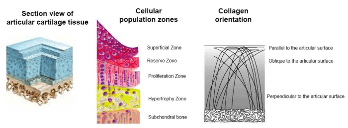
Figure 1: Cellular population and collagen orientation zones in the articular cartilage (reproduction from labrha.com).
The auricular cartilage is composed of different layers defined by the cell specificity: the superficial zone containing small and flat chondrocytes, the reserve zone containing spherical and bigger chondrocytes, the proliferation zone where chondrocytes are disposed in axial isogenic group and where they show an important mitotic activity, the mineralized hypertrophic zone containing hypertrophic chondrocytes, and the sub chondral bone. The collagen fibers are parallel to the articular surface in the superficial zone, oblique in the reserve zone and perpendicular to the articular surface in the proliferation and hypertrophic zones. This specific orientation gives cartilage its resistance to the compression forces.
Asymptomatic lesions in cartilage can degenerate into painful symptomatic chondral disease like osteoarthritis. Osteochondral lesions, which involve both cartilage and sub chondral bone, lead to fibro cartilage, which does not have the same biomechanical properties of hyaline cartilage, and cannot protect the sub chondral bone for further deterioration. Gaining functional repair is a big challenge, and the aim of cartilage repair is to restore the functional properties of the osteochondral unit [3].
Several surgical techniques are commonly used for the treatment of osteochondral lesions. We can cite joint debridement [4], micro fracture (alias chondroplasty) which is a marrow stimulation technique [5], and mosaicplasty (or osteochondral transplantation, or autologous osteochondral graft) which is a resurfacing technique [6]. Joint debridement only consists in eliminating lesion debris from the joint, to reduce pain. It is usually associated with either of the two other techniques. Bone marrow stimulation technique consists in penetrating the sub chondral bone to release progenitor cells from the bone marrow into the defect. These progenitor cells will transform into chondrocyte-like cells and produce some fibrocartilagenous repair tissue. In mosaicplasty, one removes cylindrical plugs of sane cartilage with its sub chondral bone, and implants them into the lesion like a mosaic pattern. These first generation treatments are aimed to relieve pain, recover function and inhibit cartilage lesion progress, but are not fully satisfactory, especially in the long term. Most of these surgical techniques mainly produce repair fibrocartilage, which will not last and does not resist compression and load demands as hyaline cartilage does.
Articular cartilage viability depends on chondrocytes ability in Synthesizing Extracellular Matrix (ECM) and restoring the different zones of hyaline cartilage. This is the aim of Autologous Chondrocyte Implantation (ACI) introduced more recently. ACI is a two-step procedure: first, collection by arthroscopy of a little piece of autologous cartilage, for chondrocyte culture; second, about one month later, surgical procedure of implantation of cultured chondrocytes. ACI has been the first cell engineering application in orthopedic surgery. The procedure was introduced by Peterson et al. in 1987 [7] and the first clinical use of this procedure was reported by Brittberg et al. in 1994 [8] and consisted in injecting autologous chondrocytes under a periosteal patch. ACI is indicated for large symptomatic lesions surrounded by non-osteoarthritic cartilage [9], whereas marrow-stimulating techniques or mosaicplasty are used for small lesions [3,10,11]. Good histological results on implantation sites after treatment were reported by various authors and lesion repair lasted several years [8,12-14]. An estimation of 10 000 patients worldwide having undergone an ACI was given by Brittberg in 2003 [15].
In order to obtain new tissue formation from the implanted cells, there is a need for a suitable environment at the lesion site, which might not be the case if there is extensive cartilage loss. This is why the early Brittberg procedure has been abandoned in favor of implanting cells in biodegradable three dimensional matrices, like collagen membranes, as provisional supportive ECM-like scaffolds. Therefore, new techniques implying tissue engineering have been developing for the last ten years [16-18].
Research to improve cartilage repair is focused on tissue engineering combining three axes: the presence of cells, tridimensional scaffolds mimicking ECM and various environmental factors (growth factors etc.). Tuning these parameters is really the challenge in cartilage repair.
The first point of interest is the development of tridimensional matrices, natural or synthetic, biocompatible and biodegradable, which serve as filling material for the lesion itself, as scaffolds for new cellular growth, and as reservoirs for the release of chondrogenic factors.
Then, one needs to define which are the best cell candidates to concentrate on, either chondrocytes, which are the native cartilage cell types, or stem cells for example Mesenchymal Stem Cells (MSC) from bone marrow. This choice is concerned with cell source requirements, cell adhesion and proliferation and cell efficiency.
The third topic of interest concerns the identification and implementation of specific adequate growth factors or signaling molecules, enabling both chondrocyte cell differentiation and phenotype preservation.
Combinations of these three parameters, as well as optimal implementation conditions are currently under investigation for tissue regeneration in cartilage lesions, both in animal models and clinic studies.
Analysis and Interpretation
Materials used in osteoarticular tissue engineering
Cell adhesion, growth and resulting tissue regeneration will depend on the first place on the scaffold properties. To mimic extracellular matrix, the scaffold must be biodegradable, biocompatible, favor cell adhesion, regulate cell expression, and be a suitable reservoir for bioactive molecules such as growth factors [19]. There have been extensive studies on potential biomaterials, natural, synthetic, ceramics or composite.
Matrices of natural origin
A few natural matrices have been investigated to date, in vitro and in vivo, mainly of protein or carbohydrate origin: collagen, fibrin, agarose, alginate, Hyaluronic Acid (HA), chitosan, cellulose [20].
Early interest has focused on collagen: type I gels and sponges [21,22] and type II sponges [23]. In second generation ACI, a collagen membrane has been used (for example, Chondro-Gide®, from Geistlich, Switzerland) to replace perioste [24], to facilitate the second part of the treatment, which is the implantation of cultured autologous chondrocytes. In third generation ACI, the focus is made on the use of tridimensional scaffolds optimized for chondrocyte implantation, the so-called MACI (Matrix-assisted Autologous Chondrocyte Implantation) for example with Verigen (Leverkusen, Germany) or Genzyme (Boston, USA) [25].
The main drawback encountered with these materials concerns the type of repair tissue obtained: the best tissue obtained was hyaline-like, but still not identical to articular hyaline cartilage in terms of morphology or histo-chemistry, and could only be obtained in some cases. Often, only fibrocartilage was formed [26,27].
After collagen membranes [28], research has focused on HA derivatives as potential scaffold [29], see for example Hyalograft® C (Fidia Advanced Biopolymers, Abano Terme, Italy), an esterified derivative of HA, which showed good results [30-32], namely cartilage function improvement among 91.5% of patients. This graft enables chondrocyte growth together with phenotype conservation [33] and resorbs without inflammatory reaction [34].
Films and sponges of chitosan, chitosan/HA and chitosan/ chondroitin sulfate were prepared by film deposition (films) or lyophilisation (sponges) and were shown to constitute good cell supports [35].
Hydrogels, such as alginate, also constitute a suitable scaffold for cell development and differentiation [36-38], however they display mechanical weaknesses. Agarose has been used as a matrix [39] and more recently in a layered manner, to produce depth-dependent in homogeneity in the scaffold [40]. Some hybrid agarose-alginate gel, Cartipatch® (Lyon, France), has been used in vivo in an ACI case study on man. After two years, all patients showed clinical improvement and eight out of thirteen patients displayed hyaline cartilage restoration [41]. HA-based injectable hydrogels have been widely studied [42], often in combination with chitosan [43-45]. They enable chondrocyte survival and these retain their morphology [46]. New chitosan-based hydrogels have also been shown to enable chondrogenic differentiation of encapsulated MSCs [47]. GAG-augmented polysaccharide hydrogels have also been reported as suitable supports for chondrogenesis, based on chondroitin-sulfate and chitosan, a GAG-analog [48]. Chitosan, which is a polycationic repeating monosaccharide of β-1,4-linked glucosamine monomers with randomly located N-acetyl glucosamine units, may be combined with the polyanionic CSA resulting in hydrogel formation by ionic cross-linking.
Despite their ability for cell adhesion, proliferation, differentiation and subsequent ECM production, these natural gel matrices have several disadvantages, such as potential immunogenicity, possible transmission of animal pathogens, difficulty of processing, mechanical weakness often needing chemical modification, like cross-linking for stabilization and improvement of mechanical properties [49-51]. However, these cross-linking agents, like glutaraldehyde, are often toxic and to avoid possible complications due to these components, various groups have developed composite hydrogels which combine the hydrogel compound and structural proteins. For example, the composite hydrogel matrices fibrin/HA and HA/collagen type I display improved mechanical properties, promote cell development and ECM production [38,52].
Another way of approaching cartilage structure is based on the electro spinning technique to produce collagen fibers [53-57], fibrinogen fibers [58], or other protein fibers like elastin which support the growth of MSCs [59], or gelatin [60]. Some authors combined collagen for the fibrous scaffold and chitosan gel to model ECM proteoglycans [61], or electrospan collagen together with HA [62] or with chitosan [63]. Kim et al. have reported on fibrous electrospun HA hydrogels that direct MSCs chondrogenesis through mechanical (cross linking density) and adhesive (RGD motives density) characteristics [64].
To stabilize collagen based electrospun nanofibers, other groups have focused on the development of safer cross-linking processes, like photopolymerization based on the use of methacrylates [65,66] or rose Bengal [67]. These photo-cross linked matrices successfully encapsulate chondrocytes or MSCs [68,69]. Another approach consisted in inserting some thermo sensitive elements, like poly (N-isopropylacrylamide) in the structure. Upon a certain temperature modified HA chains undergo conformational changes which lead to self-assembly and stabilization of the hydrogel [70]. Self-assembly processes are currently widely explored, with a variety of peptidic building blocks (see next chapter). Jiang et al. have recently reported on the electro spinning of collagen fibers from a non-toxic solvent (ethanol-water) and gentle cross-linking system (citric acid with glycerol) [57]. Native collagen conformation was retained after electro spinning and water stability was enhanced after the cross-linking. Furthermore, cells showed better adhesion and growth than on glutaraldehyde cross-linked scaffolds.
Synthetic matrices
Synthetic materials have been widely used in tissue engineering due to their controllable properties. Various artificial biodegradable scaffolds are being investigated, based on Poly-Lactic Acid (PLA) [71], Poly-glycolic Acid (PGA) [72], and Copolymers (PLGA), Polycaprolactone (PCL), nanocarbon, Dacron®, Teflon® fibers or polymer hydrogels [20].
Based on the characteristics of hydrogels (biocompatibility, hydration and bioactive molecules reservoir capacity), ECM-mimicking matrices have been developed for example with designed peptides amphiphiles [73-75], elastin-like polymers [76,77]. A large number of studies are currently devoted to self-assembling peptides, for instance those developed by O'Leary et al., combining both high water content and structural robustness [78,79]. Amino-acid self-assembling β-sheet interaction has been used by Liu et al. to promote chondrocyte growth and hyaline cartilage formation [80], or chondrogenesis from bone marrow stem cells [81]. Some self-assembled nanofibers of peptides amphiphiles have been shown to display a large number of binding domains for TGF-β1, allowing chondrogenic differentiation of MSCs and cartilage repair in a rabbit chondral defect [82]. Although biocompatible, these polymers do not enable cell adhesion on their own, and sites for cell adhesion have to be added. Moreover, they can induce some local pH lowering upon hydrolysis, with possible inflammatory reaction [19].
Hydrogels based on PEG (polyethylene glycol) have attracted much attention [83,84], and were shown to promote cell adhesion [85], and serve as reservoirs with possible multiphase composition for bioactive molecules, like chondroitin sulfate and specific peptides [86-88].
Moutos et al. have reported on a tridimensional scaffold of specially woven microfibers of PGA impregnated with hydrogel (agarose or fibrin) containing chondrocytes, with tensile and compressive mechanical properties close to that of native cartilage [89]. This study was the first to use composite biomaterials to specifically target these biomechanical properties [90].
Bio seed® (Bio Tissue Technologies, Freiburg, Germany) based on PGA derived matrix, in combination with fibrin gel, used in MACI, has shown promising results [91].
Synthetic electrospun nanofibers have also been studied for MACI: chondrocytes were associated with electrospun poly-lactic acid nanofibers [92], or for example co-electrospun fibers of poly-caprolactone (slow degrading polyester) and poly-ethylene glycol (hydro soluble polymer) forming an architecture with controlled porosity [93]. In general, chondrocytes as well as MSCs grow well on this type of nanofiber scaffold, and produce ECM components like collagen and proteoglycans, as shown for example by Li et al. with PCL scaffolds [94] in a mini-pig model [95] or Foroni et al. with electrospun poly(L-lactic) acid [96]. Recently, some authors have successfully developed specific zones in such nanofibrous scaffolds; based on different fiber organization, in a way to mimic the different cartilage layers [97]. Some others have introduced sacrificial polymer fibers in the scaffold to improve later cell colonization [98]. Wright et al. have developed scaffolds based on electrospun poly(D,L-lactide)/ poly(L-lactide) or poly (D,L-lactide)/polycaprolactone, with salt leached pores and embedded chitosan hydrogel [99] which enabled growth and ECM production by chondrocytes. The increase in the pore sizes to enhance cell infiltration has been investigated by Phipps et al. on a bone-mimetic electrospun scaffold of PCL, collagen I and hydroxyapatite, using three different techniques: limited protease digestion, decrease of fiber packing density during electro spinning, and inclusion of sacrificial fibers (water soluble PEO) [100]. The sacrificial fibers approach appeared to be the most effective. Schneider et al. have studied the influence of fiber orientation (random versus aligned) in electrospun synthetic polymer scaffolds (PDC, PPDO) on adhesion and differentiation of chondrocytes. SEM microscopy revealed a flattened chondrocyte shape on scaffolds with random fiber orientation and growth mainly restricted to the scaffold surface. On aligned fibers the chondrocytes exhibited a more spindle-shaped morphology with rougher cell surfaces but only a minority of the cells aligned according to the fibers [101].
Among ceramic materials, hydroxyapatite and tricalcium phosphate are known to induce the formation of a bony apatite layer when implanted. They have been widely investigated in the last decades in bone regeneration systems [102-105]. Bone and cartilage have very different properties and it is a real challenge to tune systems aiming at osteochondral lesion treatment. For sub chondral bone we are looking for stiffness, porosity and vascularization, to promote cell growth and the production of a bone matrix rich in type I collagen and hydroxyapatite. On the other hand, the cartilage is not vascularized, presents mainly a type II collagen matrix with an embedded proteoglycan hydrogel, allowing altogether resistance and elasticity. Some attempts have been made to mimic more closely this complex multi zone architecture, but few show good results in vivo. For example, I'm et al. have elaborated a multiphase scaffold combining HA and atelocollagen for chondral regeneration, and hydroxyapatite and tricalcium phosphate for the bone layer, and obtained good results in osteochondral regeneration upon implantation in the knee joint of a pig [106]. Promising results have been obtained with a ditopic combination of collagen and glycosaminoglycans on the one side, associated with calcium phosphate on the other side, with a soft interface between them. The physical properties achieved with this architecture are quite good, but further in vivo investigation is needed [107]. Jiang et al. have elaborated a multi-phase scaffold composed of agarose hydrogel and sintered microspheres of PLGA-bioactive glass, which successfully resulted in both osteoblasts and chondrocytes in the appropriate region of the scaffold, leading to the production of three tissues: cartilage, calcified cartilage and bone [108]. Stanishevsky et al. have studied the micro architecture of hydroxyapatite nanoparticle loaded collagen fiber composites [109]. Catledge et al. have elaborated an electrospun triphasic nanofibrous scaffold by electro spinning a mixture of PCL, type I collagen and hydroxyapatite nanoparticles [110]. Qu et al. have recently studied some composite bilayer scaffold of PVA/gelatin, nano-hydroxyapatite and polyamide-6, seeded with marrow MSCs, and have observed in rabbit neocartilage formation in the PVA layer, and sub chondral bone regeneration within the HA-PA6 layer [111].
Osteochondral differentiation factors
Many bioactive molecules intervene in the physiological process of maturation and differentiation of immature bone and cartilage cells. Most of these molecules are proteins (growth factors and cytokines). A major family is the Bone Morphogenetic Proteins (BMPs) [112,113], but there are other potential candidates for osteochondral induction. The differentiation factors tested in various in vitro or in vivo models are among various protein growth factor families: Epidermal Growth Factor (EGF), Fibroblast Growth Factor (FGF) [114], Transforming Growth Factor Beta (TGF-β) [115], Insulin-like Growth Factor (IGF), Vascular Endothelial Growth Factor (VEGF), and also among signaling and regulatory molecules such as Wnt ligands (wingless family) and Hh proteins (hedgehog family) [19,20]. The exploration of their use in MACI is expanding. Different ways are being explored for the controlled release of these growth factors. Limitations encountered concern problems of dose, factor efficiency and over-time delivery, and suitable spatial delivery.
The most common way of delivering these factors consists in direct injection at the lesion site or in direct contact with the implant scaffold, however due to the short half-life of these active protein factors, these methods require high doses for therapeutic effect and do not permit a controlled-time delivery [51].
In many studies the growth factors are delivered via the scaffold itself, by mixing them with the scaffold components during fabrication. In these cases the matrix characteristics such as porosity or cross-linking degree will modulate the protein delivery by diffusion. For example; growth factors like BMP-2 have been incorporated into chitosan and hyaluronan hydrogels, and induced bone formation in the quadriceps muscle of rats [116]. Kopesky et al. have shown sustained delivery of TGF-β1 from self-assembling peptide hydrogels, which induced chondrogenesis by encapsulated bone marrow stromal cells [117].
In other studies reporting microsphere-based scaffolds, TGF-β1, BMP-2 or IGF-1 were loaded in the microspheres (of PLGA, or PEG), resulting in good osteochondral regeneration [118-120]. These systems provide spatial controlled delivery of various growth factors [121], or even co-delivery of adipose derived stem cells and growth factors, as in the study of Sukarto et al. using loaded microspheres in RGD-grafted N-methacrylate glycol chitosan gels for focal chondral repair [122].
Simple Ionic bonding was used to load Insulin-like Growth Factor IGF-1 in a porous collagen-glycosaminoglycan scaffold and the adsorption and release characteristics were examined by the authors, which confirmed the bioactivity profile of the growth factor by the ECM component production from seeded chondrocytes [123]. Lee et al. exploited weak interactions to coat an electrospun poly(lactide-co-glycolic acid) PLGA nanofiber scaffold with polydopamine by immersion of the fibers in a dopamine solution under weakly basic conditions, and further immobilized Bone-forming Peptide 1 (BPF-1) derived from the immature region of Bone Morphogenetic Protein-7 (BMP-7) on the polydopamine-coated fibers, by similar immersion in the peptide solution [124]. These peptide-coated scaffolds acknowledged positive results in bone regeneration, and the same approach could be applied for cartilage tissue. Although this kind of material can control the spatial release of factor, the release in time cannot be controlled, leading to a massive release in the body.
Another way to improve the biodisponibility of the growth factor can be used. In another approach, growth factors have been successfully delivered through innovative nanoreservoirs based on the layer-by-layer technology, on electrospun PCL nanofibrous scaffolds or collagen membranes, resulting in efficient cell response for bone regeneration, process which may easily apply to cartilage tissue regeneration [125,126].
A recent study by Lim et al. [127] has described the development of a new bio functionalized electrospun Poly (L-lactide) scaffold for cartilage differentiation: latent transforming growth factor LTGF-β1 was anchored to the scaffold via surface chemical modification. Both random and orientated bio functionalized scaffolds were tested in vitro and in vivo in rats, and proved chondrocyte differentiation and collagen II production. Jeong et al. have also performed some chemical modification to attach BMP-2 on a 3D PCL scaffold [128]. The authors found that these chemically conjugated BMP-2 PCL scaffolds promote significantly greater cartilage regeneration from seeded chondrocytes, in vitro and in vivo, compared to untreated scaffolds.
Recently magnetic scaffolds have been elaborated, based on biocompatible magnetic nanoparticles, which enable continuous and controlled loading of growth factors by the means of an external magnetic field [129-131].
Gene therapy is an alternative to the direct delivery of proteins, as the delivery of genes encoding for specific factors leads to the synthesis of these factors directly by the cells [132-135]. Among various vectors, the non-pathogenic human Adeno-associated Virus (AAV) is most promising, as recombinant AAV vectors allow the transduction of most relevant tissues and cells involved in cartilage repair, and it has been successfully tested in vivo in a rabbit osteochondral defect, using the FGF-2 gene sequence [133,136]. Moreover, although viral gene vectors are subject to safety considerations, rAAV has recently been recommended for clinical use in the treatment of pancreatitis. The group of Lu et al has recently described porous chitosan scaffolds with embedded HA/chitosan/plasmid-DNA nanoparticles encoding for TGF-β1 which induces DNA controlled release, transfect chondrocytes and promote cell proliferation [137]. Chen et al. have produced simultaneous regeneration of articular cartilage and sub chondral bone in vivo using MSCs by the use of a spatially controlled gene delivery system in bilayered osteochondral scaffolds, consisting of plasmid TGF-β1-activated chitosan-gelatin scaffold for chondrogenic layer and plasmid BMP-2-activated hydroxyapatite/ chitosan-gelatin scaffold for osteogenic layer. The results showed that spatially controlled and localized gene delivery system in the bilayered integrated scaffolds could induce the mesenchymal stem cells in different layers to differentiate into chondrocytes and osteoblasts in vitro, respectively, and simultaneously support the articular cartilage and sub chondral bone regeneration in the rabbit knee osteochondral defect model [138] (Figure: 2).
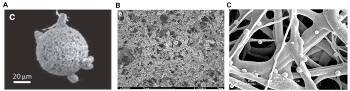
Figure 2:  Examples of scaffolds potentially used for osteoarticular regeneration.
(A) Solorio et al.[121] reported a polymeric 3-D nanofibrous microspheres scaffold used in cartilage engineering to improve the naturally round form of the
chondrogenic cells.
(B) SEM micrograph of hydroxyapatite mineralized collagen-I synthetic scaffold, representing structure and composition close to extracellular bone matrix by
Bernhardt et al.[104] for bone regeneration.
(C) SEM micrograph showing PCL electrospun nanofibers containing nanoreservoirs of BSA added by the layer-by-layer technology. This kind of nanofunctionalized
scaffold can also contain osteogenic or chondrogenic growth factors for osteochondral repair (Eap et al).
Cell candidates for implantation
Two criteria will be determinant for the selection of good cell candidates for osteochondral repair: their easy access and their efficiency to produce specific matrix elements.
Regarding performance, chondrocytes are choice candidates, as they provide a high level of matrix synthesis, and are the only cell source currently approved for clinical use. Several ACI and MACI using chondrocytes have given promising results in clinical applications; however the use of cultured chondrocytes has some disadvantages, like the dedifferentiation of cultured chondrocytes, the need for large numbers of cells to fill large lesions, and the necessity of a two-step surgical procedure (cartilage harvesting and implantation) with the risk of donor site morbidity [139]. Some attempts are being made with allogeneic chondrocytes, as they have immunologic characteristics which limit immune reaction in the host. Thus, allogeneic juvenile chondrocytes have been tested for clinical use [140]. Allogeneic chondrocytes from adults are also under investigation [141,142]. Some examples of non-articular cartilage cell sources, such as ear or nose, can also be used to produce new cartilaginous tissue but its characteristics and potential for defect repair remain to be established [143].
Stem cells, in particular multi potent adult stem cells as Mesenchymal Stem Cells (MSCs), are expected to be good candidates for the treatment of osteochondral lesions, as they can differentiate into various lineages and present immunosuppressive properties [144]. The implementation of these cells requires isolation [145] and chondrogenic differentiation, typically by the means of TGF-β growth factor and dexamethasone [146-148]. Increasing numbers of studies are devoted to explore the in vitro and in vivo chondrogenesis process using MSCs and growth factors in grafts, as MSCs can be injected at a graft site or combined with graft components [149-151]. Few case studies have been made on ACI or MACI using stem cells as candidates for implantation on human cartilage lesion, but these reveal promising results [152-154]. However, progress has to be made on the production of hyaline cartilage. As the cell yield from bone marrow is quite low, other sources of stem cells are investigated, like adipose tissue [155], or the Synovial Membrane (SM) [156]. Stem cells from SM have great potential for chondrogenic differentiation: when cultured in monolayer's they differentiate into fibroblasts, but when seeded in a 3D alginate medium, they readily turn into chondrocytes even in the absence of growth factors [157]. Sampat et al. have shown that seeding SM stem cells in a clinical grade agarose hydrogel scaffold in the presence of TGF-β3, resulted in new tissue with properties comparable to native cartilage [158]. Adipose tissue is another promising source for stem cells, abundant and accessible. Adipose tissue Stem Cells (ASCs) differentiate into different lineages, including bone and cartilage, and are more stable than MSCs for long-term culture [159-161]. ASCs are already widely used in osteochondral tissue regeneration studies, but some drawbacks remain today: chondrogenesis and osteogenesis are slower than adipogenesis with quite low yields despite the use of growth factors. These progenitor cells will gain in attractivity with the improvement of the differentiation performance [162-165]. Umbilical Cord Blood (UCB) is also a promising source for mesenchymal stem cells, and UCB stem cells have also been shown more chondrogenic potential than bone marrow MSCs: they can differentiate and produce cartilaginous ECM in two-three weeks [166,167]. Moreover, when seeded in different matrices, they can form cartilage and/or bone, as shown by Kogler et al. [168,169], in calcium phosphate they produce bone after 12 weeks in a rat bone defect; they produce chondrocytes in gelatin after 3 weeks implantation in mice; in other PGA scaffolds they produce native cartilage after 12 weeks in the presence of TGF-β1.
Pluripotent stem cells as Embryonic Stem Cells (ESCs) are being increasingly explored for chondrogenesis [170] in the literature but due to limitations brought by ethical and regulations considerations, it is unlikely that these would be of practical use in the clinic. Their interest is mainly in the fundamental understanding of biological processes. Finally, there is an increasing interest in Induced Pluripotent Stem Cells (IPSCs), which can be produced from the patient's cells [171]. They have high differentiation potential and can induce chondrogenesis via a multi-stage process, involving micro-mass culture [172]. Like for ESCs, further studies need to be done to evaluate their real efficiency, and to control their production and differentiation, and these will not be suitable for clinical application until long time.
Genetically modified cells, both chondrocytes and MSCs, are being considered as interesting vehicles to introduce in osteochondral implant scaffolds: on the one side they can proliferate and produce new ECM, and on the other side they can generate the secretion of over expressed protein to further stimulate cartilage repair [136,173]. Zhang et al. have used a mixed co-culture of MSCs and transgenic chondrocytes in alginate hydrogel for cartilage engineering [174]. Chondrocytes, pre-transduced with adenoviral vectors carrying the transforming growth factor TGF-β3 gene, were selected and co-cultured side-by-side with MSCs in a 3D environment to provide chondrogenic growth factors in situ. In vitro and in vivo results showed that the growth factor was successfully released from the transgenic chondrocytes, and not only induced MSCs differentiation, but also preserved the chondrocyte phenotype.
Finally, some groups reported some interesting in vitro cell culture modification to improve the colonization of biomedical scaffolds. For instance, Nerurkar et al. investigated the effect of dynamic cell culture on stem cell infiltration and behaviour in an aligned electrospun nanofibrous scaffold [175]. After seeding and pre-culture of MSCs in an electrospun PCL scaffold, dynamic culture was initiated by incubating the construct on an orbital shaker. This dramatically improved cell infiltration into the scaffold and uniform production of collagen.
Clinically used Scaffolds and Clinical Reports
Some scaffolds are available for clinical use, but there still is a general lack in technical reports and full clinical trials on arthroscopic ACI and MACI compared to the exponential development of research studies [176-178].
For the moment, the most common scaffolds clinically used are based on collagen I/III (Chondrogide®) or HA (Hyaff®-11) [49,179]. Recent clinical studies on ACI-MACI can be found for type I/III collagen matrices like ACI-Maix [180], atelocollagen gel [181], Type I collagen scaffold Neocart® [182], esterified hyaluronan Hyalograft-C® [183-186]. Table 1 presents a summary of products commercially available or in clinical trial. Clinical data for many other combinations or synthetic polymer-based scaffolds are scarce and not yet satisfactory [187,188], the various MACI present comparable results. We can cite for example clinical data for the mixed gel-polymer scaffold BioSeed-C® [189,190] or the composite type I collagen- hydroxyapatite scaffold [191].
![]()
Product
Company
Composition
Website
BST-CarGel
Biosyntech Inc., Laval, QC, Canada
Chitosan-Beta glycerolphosphate-based medical device
ChonDux
Biomet Inc., Warsaw, IN, USA
Hydrogel made of polyethylene glycol and a bioadhesive to keep the hydrogel in place after injection
Gelrin C
Regentis, Haifa, Israel
Cellular implant made of polyethyleneglycol diacrylate
(PEG-DA) covalently conjugated with a structural backbone of denatured fibrinogen chains. The
device comes in a liquid form, injected into the lesion site and polymerizes in situ into a stable hydrogel solid matrix.
Salucartilage
SaluMedica, Smyrna, GA, USA
Cylindrical implant based on polyvinyl-alcohol� hydrogel
Chondromimetic
TiGenix NV (Leuven, Belgium)
Bi-layer Collagen-based implant (upper layer: collagen/GAG; bottom layer: collagen/GAG/calcium phosphate
TrueFit Plug
OsteoBiologics/Smith & Newphew, Andover, MA, USA
Synthetic mosaicplasty plugs
OrthoGlide
Advanced Biosurfaces, Minnetonka, MN, USA
Interposition arthroplasty for the knee:� comprises a dished, disc-shaped cobalt chrome component which is inserted into the medial compartment of the knee in a minimally invasive fashion. The device has a lip which locks over the posterior aspect of the tibial plateau.
Carticel, MACI
Genzyme Inc, Cambridge, MA, USA
Autologous cultured chondrocytes on bovine collagen membrane
ChondroGide
Geistlich Biomaterials, Wolhausen, Switzerland
Bilayer collagen membrane
CaReS
Arthro Kinetics, Esslingen, Germany
Autologous chondrocytes embedded in a collagen matrix
Hyalograft-C
Fidia Advanced Biopolymers, Abano Terma, Italy
Autologous chondrocytes seeded on a hyaluronan-based scaffold
www.?diapharma.com
NeoCart, VeriCart
Histogenics, Waltham, MA, USA
NeoCart™ : autologous chondrocytes embedded in proprietary type I collagen scaffold
VeriCart™ :a single-step, cell-free collagen scaffold uniquely designed to be used in conjunction with the patient's own stem cells to repair small cartilage defects
ACT 3D/ARTROcell 3D
Co.don AG/OrmedGmbH, Teltow, Germany
Autologous chondrocytes transplantation using chondrospheres (tissue engineered chondrocytes without matrix, 3D culture)
Chondrotissue
Bioseed-C
BioTissue Technologies, Freiburg, Germany
BioTissue Technologies, Freiburg, Germany
1-step, cell-free implant used to treat traumatic or degenerative cartilage defects in combination with marrow stimulating techniques. Patented 3D matrix of synthetic polymer (PGLA) and a hyaluronic acid.
Autologous 3D chondrocyte graft based on polymer matrix and fibrin
Novocart 3D
TETEC AG/B.Braun-Aescalap, Tuttlingen, Germany
A combination of autologous cartilage cells in a biphasic, three-dimensional collagen-based matrix
Table 1: Commercial products in cartilage engineering [196].
In a review of clinical trials on cell-based therapies for chondral lesions, from 1994 to 2009, Nakamura et al. found no difference between those and other interventions [192]. Later on, Benthien et al. have provided a systematic review of clinical trials from 2002 to 2007 on the treatment of chondral defects in the knee [193], and compared the results for micro fracturing, Osteochondral Autograft Transplantation System (OATS), ACI and MACI (altogether 133 relevant studies, with an average of 32 patients per study, and 24 months follow-up). The conclusion was that no evidence based results could be clearly defined and no technique of choice could be pinpointed. Comparison is very problematic due to the variety of clinical scores applied. From a few studies comparing costs, Derrett et al. concluded that ACI costs were lower than for mosaicplasty [194] but this result needed more prospective studies to be confirmed. On the other hand, MACI still have a poor data base. More randomized prospective trials are needed.
Interestingly, some clinical studies realized on groups of juvenile patients [195] have shown significantly positive results of these ACI (with Geistlich collagen membrane) and MACI (Genzyme collagen), on pain reduction and functional motricity recovery, which leads the authors to believe that these techniques are particularly suitable for this type of patients, a major population for this kind of lesions, as osteochondral lesions are more common in adolescents than in adults.
The biggest problem when considering the transfer of a new chondral or osteochondral implant from the laboratory to the clinic concerns the regulatory aspects. This is both a time consuming and expensive process. Indeed, the functionalized scaffolds that we are discussing have to be considered as combinations of scaffolds, which are devices, and bioactive agents (like growth factors) which are biological material and fall in the drug definition. Both feasibility and safety has to be tested for each component individually and in combination, in early phase of clinical trial, and only later on, the efficiency of the implant in comparison with reference implantation techniques such as mosaicplasty.
Conclusion
Important advances have been made in the last decade in the development of biologically active scaffolds for osteochondral repair, as can be seen from the exponentially growing number of research studies.
Multi-tissue regeneration, through the combination of biomimetic and multi-phasic scaffold design, spatially controlled and localized bioactive molecules delivery system and even multi-lineage differentiation of a single stem cell population, represents a promising strategy for facilitating the development of complex tissue such as the osteochondral unit.
However, the main drawback in this field of research is the lack of reference conditions, for the purpose of reliable comparison. Little progress is being made in the establishment of standard screening conditions, like in pharmaceutical industry [197]. Furthermore, there is no consensus in the choice of animal model for the in vivo studies, whether rabbit, mini-pig, dog, sheep, goat or horse [198-200]. There is no consensus either, whether to study chondral or osteochondral lesions, and whether to implant mature matrices or to leave it mature in the defect.
Primarily aimed at the repair of small defects, all the above discussed technologies are more and more focused on the improvement of the functional biomechanical requirements of the osteochondral tissue. In this respect there is good hope that such techniques could apply in the future to the repair of large defects or even resurfacing of a whole joint, for example in the treatment of osteoarthritis. Of course, some new challenges will have to be faced, in particular in cell population, a large number of cells being required, and in the handling of particular conditions related to this pathology, namely an inflammatory environment [201].
Acknowledgment
We are indebted to The French national Institute of Health and Medical Research (INSERM) and the University of Strasbourg.
References
- Logerstedt DS, Snyder-Mackler L, Ritter RC, Axe MJ, Orthopedic Section of the American Physical Therapy Association. Knee pain and mobility impairments: meniscal and articular cartilage lesions. J Orthop Sports Phys Ther. 2010; 40: A1-1A35.
- Poole AR, Kojima T, Yasuda T, Mwale F, Kobayashi M, Laverty S. Composition and structure of articular cartilage: a template for tissue repair. Clin Orthop Relat Res. 2001; S26-33.
- Ho YY, Stanley AJ, Hui JH, Wang SC. Postoperative evaluation of the knee after autologous chondrocyte implantation: what radiologists need to know. Radiographics. 2007; 27: 207-220.
- Jackson RW, Dieterichs C. The results of arthroscopic lavage and debridement of osteoarthritic knees based on the severity of degeneration: a 4- to 6-year symptomatic follow-up. Arthroscopy. 2003; 19: 13-20.
- Steadman JR, Rodkey WG, Rodrigo JJ. Microfracture: surgical technique and rehabilitation to treat chondral defects. Clin Orthop Relat Res. 2001; S362-369.
- Hangody L, Füles P. Autologous osteochondral mosaicplasty for the treatment of full-thickness defects of weight-bearing joints: ten years of experimental and clinical experience. J Bone Joint Surg Am. 2003; 85: 25-32.
- Peterson L, Minas T, Brittberg M, Lindahl A. Treatment of osteochondritis dissecans of the knee with autologous chondrocyte transplantation: results at two to ten years. J Bone Joint Surg Am. 2003; 85: 17-24.
- Brittberg M, Lindahl A, Nilsson A, Ohlsson C, Isaksson O, Peterson L. Treatment of deep cartilage defects in the knee with autologous chondrocyte transplantation. N Engl J Med. 1994; 331: 889-895.
- Gomoll AH, Filardo G, de Girolamo L, Espregueira-Mendes J, Marcacci M, Rodkey WG, et al. Surgical treatment for early osteoarthritis. Part I: cartilage repair procedures. Knee Surg Sports Traumatol Arthrosc. 2012; 20: 450-466.
- Unziker EB. Articular cartilage repair: basic science and clinical progress. A review of the current status and prospects. Osteoarthritis and Cartilage. 2002; 10: 432-463.
- Magnussen RA, Dunn WR, Carey JL, Spindler KP. Treatment of focal articular cartilage defects in the knee: a systematic review. Clin Orthop Relat Res. 2008; 466: 952-962.
- Micheli LJ, Browne JE, Erggelet C, Fu F, Mandelbaum B, Moseley JB, et al. Autologous chondrocyte implantation of the knee: multicenter experience and minimum 3-year follow-up. Clin J Sport Med. 2001; 11: 223-228.
- Minas T. Autologous chondrocyte implantation for focal chondral defects of the knee. Clin Orthop Relat Res. 2001; S349-361.
- Peterson L, Brittberg M, Kiviranta I, Akerlund EL, Lindahl A. Autologous chondrocyte transplantation. Biomechanics and long-term durability. Am J Sports Med. 2002; 30: 2-12.
- Brittberg M, Peterson L, Sjögren-Jansson E, Tallheden T, Lindahl A. Articular cartilage engineering with autologous chondrocyte transplantation. A review of recent developments. J Bone Joint Surg Am. 2003; 85: 109-15.
- Daher RJ, Chahine NO, Greenberg AS, Sgaglione NA, Grande DA. New methods to diagnose and treat cartilage degeneration. Nat Rev Rheumatol. 2009; 5: 599-607.
- Johnstone B, Alini M, Cucchiarini M, Dodge GR, Eglin D, Guilak F, et al. Tissue engineering for articular cartilage repair--the state of the art. Eur Cell Mater. 2013; 25: 248-267.
- Panseri S, Russo A, Cunha C, Bondi A, Di Martino A, Patella S, et al. Osteochondral tissue engineering approaches for articular cartilage and subchondral bone regeneration. Knee Surg Sports Traumatol Arthrosc. 2012; 20: 1182-1191.
- Getgood A, Brooks R, Fortier L, Rushton N. Articular cartilage tissue engineering: today's research, tomorrow's practice? J Bone Joint Surg Br. 2009; 91: 565-576.
- Vinatier C, Bouffi C, Merceron C, Gordeladze J, Brondello JM, Jorgensen C, et al. Cartilage tissue engineering: towards a biomaterial-assisted mesenchymal stem cell therapy. Curr Stem Cell Res Ther. 2009; 4: 318-329.
- Ben-Yishay A, Grande DA, Schwartz RE, Menche D, Pitman MD. Repair of articular cartilage defects with collagen-chondrocyte allografts. Tissue Eng. 1995; 1: 119-133.
- Frenkel SR, Toolan B, Menche D, Pitman MI, Pachence JM. Chondrocyte transplantation using a collagen bilayer matrix for cartilage repair. J Bone Joint Surg Br. 1997; 79: 831-836.
- Nehrer S, Breinan HA, Ramappa A, Hsu HP, Minas T, Shortkroff S, et al. Chondrocyte-seeded collagen matrices implanted in a chondral defect in a canine model. Biomaterials. 1998; 19: 2313-2328.
- Pabbruwe MB, Esfandiari E, Kafienah W, Tarlton JF, Hollander AP. Induction of cartilage integration by a chondrocyte/collagen-scaffold implant. Biomaterials. 2009; 30: 4277-4286.
- Marlovits S, Zeller P, Singer P, Resinger C, Vécsei V. Cartilage repair: generations of autologous chondrocyte transplantation. Eur J Radiol. 2006; 57: 24-31.
- Wada Y, Watanabe A, Yamashita T, Isobe T, Moriya H. Evaluation of articular cartilage with 3D-SPGR MRI after autologous chondrocyte implantation. J Orthop Sci. 2003; 8: 514-517.
- Smith GD, Knutsen G, Richardson JB. A clinical review of cartilage repair techniques. J Bone Joint Surg Br. 2005; 87: 445-449.
- Hughes LC, Archer CW, Gwynn I. The ultrastructure of mouse articular cartilage: collagen orientation and implications for tissue functionality. A polarised light and scanning electron microscope study and review. Eur. Cell. Mater. 2005; 9: 68-84.
- Goa KL, Benfield P. Hyaluronic acid. A review of its pharmacology and use as a surgical aid in ophthalmology, and its therapeutic potential in joint disease and wound healing. Drugs. 1994; 47: 536-566.
- Marcacci M, Berruto M, Brocchetta D, Delcogliano A, Ghinelli D, Gobbi A, et al. Articular cartilage engineering with Hyalograft C: 3-year clinical results. Clin Orthop Relat Res. 2005; 96-105.
- Nehrer S, Domayer S, Dorotka R, Schatz K, Bindreiter U, Kotz R. Three-year clinical outcome after chondrocyte transplantation using a hyaluronan matrix for cartilage repair. Eur J Radiol. 2006; 57: 3-8.
- Kon E, Gobbi A, Filardo G, Delcogliano M, Zaffagnini S, Marcacci M. Arthroscopic second-generation autologous chondrocyte implantation compared with microfracture for chondral lesions of the knee: prospective nonrandomized study at 5 years. Am J Sports Med. 2009; 37: 33-41.
- Brun P, Abatangelo G, Radice M, Zacchi V, Guidolin D, Daga Gordini D, et al. Chondrocyte aggregation and reorganization into three-dimensional scaffolds. J Biomed Mater Res. 1999; 46: 337-346.
- Campoccia D, Doherty P, Radice M, Brun P, Abatangelo G, Williams DF. Semisynthetic resorbable materials from hyaluronan esterification. Biomaterials. 1998; 19: 2101-2127.
- Peniche C, Fernández M, Rodríguez G, Parra J, Jimenez J, Bravo AL, et al. Cell supports of chitosan/hyaluronic acid and chondroitin sulphate systems. Morphology and biological behaviour. J Mater Sci Mater Med. 2007; 18: 1719-1726.
- Häuselmann HJ, Aydelotte MB, Schumacher BL, Kuettner KE, Gitelis SH, Thonar EJ. Synthesis and turnover of proteoglycans by human and bovine adult articular chondrocytes cultured in alginate beads. Matrix. 1992; 12: 116-129.
- Masuda K, Sah RL, Hejna MJ, Thonar EJ. A novel two-step method for the formation of tissue-engineered cartilage by mature bovine chondrocytes: the alginate-recovered-chondrocyte (ARC) method. J Orthop Res. 2003; 21: 139-148.
- Liao E, Yaszemski M, Krebsbach P, Hollister S. Tissue-engineered cartilage constructs using composite hyaluronic acid/collagen I hydrogels and designed poly(propylene fumarate) scaffolds. Tissue Eng. 2007; 13: 537-550.
- Rahfoth B, Weisser J, Sternkopf F, Aigner T, von der Mark K, Bräuer R. Transplantation of allograft chondrocytes embedded in agarose gel into cartilage defects of rabbits. Osteoarthritis Cartilage. 1998; 6: 50-65.
- Ng KW, Wang CC, Mauck RL, Kelly TA, Chahine NO, Costa KD, et al. A layered agarose approach to fabricate depth-dependent inhomogeneity in chondrocyte-seeded constructs. J Orthop Res. 2005; 23: 134-141.
- Selmi TA, Verdonk P, Chambat P, Dubrana F, Potel JF, Barnouin L, et al. Autologous chondrocyte implantation in a novel alginate-agarose hydrogel: outcome at two years. J Bone Joint Surg Br. 2008; 90: 597-604.
- Cloyd JM, Malhotra NR, Weng L, Chen W, Mauck RL, Elliott DM. Material properties in unconfined compression of human nucleus pulposus, injectable hyaluronic acid-based hydrogels and tissue engineering scaffolds. Eur Spine J. 2007; 16: 1892-1898.
- Frenkel SR, Bradica G, Brekke JH, Goldman SM, Ieska K, Issack P, et al. Regeneration of articular cartilage--evaluation of osteochondral defect repair in the rabbit using multiphasic implants. Osteoarthritis Cartilage. 2005; 13: 798-807.
- Morais DS, Rodrigues MA, Silva TI, Lopes MA, Santos M, Santos JD, et al. Development and characterization of novel alginate-based hydrogels as vehicles for bone substitutes. Carbohydr Polym. 2013; 95: 134-142.
- Park H, Choi B, Hu J, Lee M. Injectable chitosan hyaluronic acid hydrogels for cartilage tissue engineering. Acta Biomater. 2013; 9: 4779-4786.
- Tan H, Chu CR, Payne KA, Marra KG. Injectable in situ forming biodegradable chitosan-hyaluronic acid based hydrogels for cartilage tissue engineering. Biomaterials. 2009; 30: 2499-2506.
- Naderi-Meshkin H, Andreas K, Matin MM, Sittinger M, Bidkhori HR, Ahmadiankia N, et al . Chitosan-based injectable hydrogel as a promising in situ forming scaffold for cartilage tissue engineering. Cell Biol Int. 2014; 38: 72-84.
- Sechriest VF, Miao YJ, Niyibizi C, Westerhausen-Larson A, Matthew HW, Evans CH, et al. GAG-augmented polysaccharide hydrogel: a novel biocompatible and biodegradable material to support chondrogenesis. J Biomed Mater Res. 2000; 49: 534-541.
- Grigolo B, Roseti L, Fiorini M, Fini M, Giavaresi G, Aldini NN, et al. Transplantation of chondrocytes seeded on a hyaluronan derivative (hyaff-11) into cartilage defects in rabbits. Biomaterials. 2001; 22: 2417-2424.
- Facchini A, Lisignoli G, Cristino S, Roseti L, De Franceschi L, Marconi E, et al. Human chondrocytes and mesenchymal stem cells grown onto engineered scaffold. Biorheology. 2006; 43: 471-480.
- Lee SH, Shin H. Matrices and scaffolds for delivery of bioactive molecules in bone and cartilage tissue engineering. Adv Drug Deliv Rev. 2007; 59: 339-359.
- Rampichová M, Filová E, Varga F, Lytvynets A, Prosecká E, Kolácná L, et al. Fibrin/hyaluronic acid composite hydrogels as appropriate scaffolds for in vivo artificial cartilage implantation. ASAIO J. 2010; 56: 563-568.
- Matthews JA, Wnek GE, Simpson DG, Bowlin GL. Electrospinning of collagen nanofibers. Biomacromolecules. 2002; 3: 232-238.
- Chan CK, Liao S, Li B, Lareu RR, Larrick JW, Ramakrishna S, et al. Early adhesive behavior of bone-marrow-derived mesenchymal stem cells on collagen electrospun fibers. Biomed Mater. 2009; 4: 035006.
- Carlisle CR, Coulais C, Guthold M. The mechanical stress-strain properties of single electrospun collagen type I nanofibers. Acta Biomater. 2010; 6: 2997-3003.
- Dong Z, Wu Y, Clark RL. Thermodynamic modeling and investigation of the formation of electrospun collagen fibers. Langmuir. 2011; 27: 12417-12422.
- Jiang Q, Reddy N, Zhang S, Roscioli N, Yang Y. Water-stable electrospun collagen fibers from a non-toxic solvent and crosslinking system. J Biomed Mater Res A. 2013; 101: 1237-1247.
- Baker S, Sigley J, Carlisle CR, Stitzel J, Berry J, Bonin K, et al. The Mechanical Properties of Dry, Electrospun Fibrinogen Fibers. Mater Sci Eng C Mater Biol Appl. 2012; 32: 215-221.
- Li M, Mondrinos MJ, Gandhi MR, Ko FK, Weiss AS, Lelkes PI. Electrospun protein fibers as matrices for tissue engineering. Biomaterials. 2005; 26: 5999-6008.
- Skotak M, Noriega S, Larsen G, Subramanian A. Electrospun cross linked gelatin fibers with controlled diameter: the effect of matrix stiffness on proliferative and biosynthetic activity of chondrocytes cultured in vitro.J Biomed Mater Res A. 2010; 95: 828-836.
- Chen ZG, Wang PW, Wei B, Mo XM, Cui FZ. Electrospun collagen-chitosan nanofiber: a biomimetic extracellular matrix for endothelial cell and smooth muscle cell. Acta Biomater. 2010; 6: 372-382.
- Fischer RL, McCoy MG, Grant SA. Electrospinning collagen and hyaluronic acid nanofiber meshes. J Mater Sci Mater Med. 2012; 23: 1645-1654.
- Chen L, Zhu C, Fan D, Liu B, Ma X, Duan Z, et al. A human-like collagen/chitosan electrospun nanofibrous scaffold from aqueous solution: electrospun mechanism and biocompatibility. J Biomed Mater Res A. 2011; 99: 395-409.
- Kim IL, Khetan S, Baker BM, Chen CS, Burdick JA. Fibrous hyaluronic acid hydrogels that direct MSC chondrogenesis through mechanical and adhesive cues. Biomaterials. 2013; 34: 5571-5580.
- Smeds KA, Pfister-Serres A, Miki D, Dastgheib K, Inoue M, Hatchell DL, et al. Photocrosslinkable polysaccharides for in situ hydrogel formation. J Biomed Mater Res. 2001; 54: 115-121.
- Burdick JA, Chung C, Jia X, Randolph MA, Langer R. Controlled degradation and mechanical behavior of photopolymerized hyaluronic acid networks. Biomacromol. 2005; 6: 386-391.
- Liu T,Teng WK,Chan BP,Chew SY. Photochemicalcrosslinkedelectrospun collagen nano?bers: Synthesis, characterizationandneuralstemcellinteractions. J BiomedMater Res A. 2010; 95: 276-282.
- Chung C, Mesa J, Randolph MA, Yaremchuk M, Burdick JA. Influence of gel properties on neocartilage formation by auricular chondrocytes photoencapsulated in hyaluronic acid networks. J Biomed Mater Res A. 2006; 77: 518-525.
- Erickson IE, Kestle SR, Zellars KH, Farrell MJ, Kim M, Burdick JA, et al. High mesenchymal stem cell seeding densities in hyaluronic acid hydrogels produce engineered cartilage with native tissue properties. Acta Biomater. 2012; 8: 3027-3034.
- Mortisen D, Peroglio M, Alini M, Eglin D. Tailoring thermoreversible hyaluronan hydrogels by "click" chemistry and RAFT polymerization for cell and drug therapy. Biomacromolecules. 2010; 11: 1261-1272.
- Freed LE, Grande DA, Lingbin Z, Emmanual J, Marquis JC, Langer R. Joint resurfacing using allograft chondrocytes and synthetic biodegradable polymer scaffolds. J Biomed Mater Res. 1994; 28: 891-899.
- Vacanti CA, Langer R, Schloo B, Vacanti JP. Synthetic polymers seeded with chondrocytes provide a template for new cartilage formation. Plast Reconstr Surg. 1991; 88: 753-759.
- Hartgerink JD, Beniash E, Stupp SI. Self-assembly and mineralization of peptide-amphiphile nanofibers. Science. 2001; 294: 1684-1688.
- Zhang S. Fabrication of novel biomaterials through molecular self-assembly. Nat Biotechnol. 2003; 21: 1171-1178.
- Wu EC, Zhang SG, Hauser CAE. Self-assembling peptides as cell-interactive scaffolds. Adv Func Mater. 2012; 22: 456-468.
- Betre H, Ong SR, Guilak F, Chilkoti A, Fermor B, Setton LA. Chondrocytic differentiation of human adipose-derived adult stem cells in elastin-like polypeptide. Biomaterials. 2006; 27: 91-99.
- MacEwan SR, Chilkoti A. Elastin-like polypeptides: biomedical applications of tunable biopolymers. Biopolymers. 2010; 94: 60-77.
- Stoop R. Smart biomaterials for tissue engineering of cartilage. Injury. 2008; 39 Suppl 1: S77-87.
- O'Leary LE, Fallas JA, Bakota EL, Kang MK, Hartgerink JD. Multi-hierarchical self-assembly of a collagen mimetic peptide from triple helix to nanofibre and hydrogel. Nat Chem. 2011; 3: 821-828.
- Liu J, Song H, Zhang L, Xu H, Zhao X. Self assembly-peptide hydrogels as tissue-engineering scaffolds for three-dimensional culture of chondrocytes in vitro. Macromol Biosci. 2010; 10: 1164-1170.
- Kopesky PW, Vanderploeg EJ, Sandy JS, Kurz B, Grodzinsky AJ. Self-assembling peptide hydrogels modulate in vitro chondrogenesis of bovine bone marrow stromal cells. Tissue Eng Part A. 2010; 16: 465-477.
- Shah RN, Shah NA, Del Rosario Lim MM, Hsieh C, Nuber G, et al. Supramolecular design of self-assembling nanofibers for cartilage regeneration. Proc Natl Acad Sci U S A. 2010; 107: 3293-3298.
- Elisseeff J, Anseth K, Sims D, McIntosh W, Randolph M, Langer R. Transdermal photopolymerization for minimally invasive implantation. Proc Natl Acad Sci U S A. 1999; 96: 3104-3107.
- Elisseeff J, McIntosh W, Anseth K, Riley S, Ragan P, Langer R. Photoencapsulation of chondrocytes in poly(ethylene oxide)-based semi-interpenetrating networks. J Biomed Mater Res. 2000; 51: 164-171.
- Ehrbar M, Rizzi SC, Schoenmakers RG, Miguel BS, Hubbell JA, Weber FE, et al. Biomolecular hydrogels formed and degraded via site-specific enzymatic reactions. Biomacromolecules. 2007; 8: 3000-3007.
- Zhu J. Bioactive modification of poly(ethylene glycol) hydrogels for tissue engineering. Biomaterials. 2010; 31: 4639-4656.
- Nguyen LH, Kudva AK, Saxena NS, Roy K. Engineering articular cartilage with spatially-varying matrix composition and mechanical properties from a single stem cell population using a multi-layered hydrogel. Biomaterials.2011; 32: 6946-6952.
- Hwang NS, Varghese S, Li H, Elisseeff J. Regulation of osteogenic and chondrogenic differentiation of mesenchymal stem cells in PEG-ECM hydrogels. Cell Tissue Res. 2011; 344: 499-509.
- Moutos FT, Freed LE, Guilak F. A biomimetic three-dimensional woven composite scaffold for functional tissue engineering of cartilage. Nat Mater. 2007; 6: 162-167.
- Guilak F, Butler DL, Goldstein SA. Functional tissue engineering: the role of biomechanics in articular cartilage repair. Clin Orthop Relat Res. 2001; S295-305.
- Ossendorf C, Kaps C, Kreuz PC, Burmester GR, Sittinger M, Erggelet C. Treatment of posttraumatic and focal osteoarthritic cartilage defects of the knee with autologous polymer-based three-dimensional chondrocyte grafts: Two year clinical results. Arthritis Res Ther. 2007; 9: R41.
- Li WJ, Jiang YJ, Tuan RS. Cell-nanofiber-based cartilage tissue engineering using improved cell seeding, growth factor, and bioreactor technologies. Tissue Eng Part A. 2008; 14: 639-648.
- Baker BM, Gee AO, Metter RB, Nathan AS, Marklein RA, Burdick JA, et al. The potential to improve cell infiltration in composite fiber-aligned electrospun scaffolds by the selective removal of sacrificial fibers. Biomaterials. 2008; 29: 2348-2358.
- Li WJ, Danielson KG, Alexander PG, Tuan RS. Biological response of chondrocytes cultured in three-dimensional nanofibrous poly(epsilon-caprolactone) scaffolds. J Biomed Mater Res A. 2003; 67: 1105-1114.
- Li WJ, Chiang H, Kuo TF, Lee HS, Jiang CC, Tuan RS. Evaluation of articular cartilage repair using biodegradable nanofibrous scaffolds in a swine model: a pilot study. J Tissue Eng Regen Med. 2009; 3: 1-10.
- Foroni L, Dirani G, Gualandi C, Focarete ML, Pasquinelli G. Paraffin embedding allows effective analysis of proliferation, survival, and immunophenotyping of cells cultured on poly(l-lactic acid) electrospun nanofiber scaffolds. Tissue Eng Part C Methods. 2010; 16: 751-760.
- McCullen SD, Autefage H, Callanan A, Gentleman E, Stevens MM. Anisotropic fibrous scaffolds for articular cartilage regeneration. Tissue Eng Part A. 2012; 18: 2073-2083.
- Baker BM, Shah RP, Silverstein AM, Esterhai JL, Burdick JA, Mauck RL. Sacrificial nanofibrous composites provide instruction without impediment and enable functional tissue formation. Proc Natl Acad Sci U S A. 2012; 109: 14176-14181.
- Wright L, McKeon-Fischer K, Cui Z, Nair L, Freeman J. PDLA/PLLA and PDLA/PCL nanofibers with a chitosan-based hydrogel in composite scaffolds for tissue engineered cartilage. J Tissue Eng Regen Med. 2012.
- Phipps MC, Clem WC, Grunda JM, Clines GA, Bellis SL. Increasing the pore sizes of bone-mimetic electrospun scaffolds comprised of polycaprolactone, collagen I and hydroxyapatite to enhance cell infiltration. Biomaterials. 2012; 33: 524-534.
- Schneider T, Kohl B, Sauter T, Kratz K, Lendlein A, Ertel W, et al. Influence of fiber orientation in electrospun polymer scaffolds on viability, adhesion and differentiation of articular chondrocytes. Clin Hemorheol Microcirc. 2012; 52: 325-336.
- Tampieri A, Celotti G, Landi E, Sandri M, Roveri N, Falini G. Biologically inspired synthesis of bone-like composite: self-assembled collagen fibers/hydroxyapatite nanocrystals. J Biomed Mater Res A. 2003; 67: 618-625.
- Marcacci M, Kon E, Moukhachev V, Lavroukov A, Kutepov S, Quarto R, et al. Stem cells associated with macroporous bioceramics for long bone repair: 6- to 7-year outcome of a pilot clinical study. Tissue Eng. 2007; 13: 947-955.
- Bernhardt A, Lode A, Boxberger S, Pompe W, Gelinsky M. Mineralised collagen--an artificial, extracellular bone matrix--improves osteogenic differentiation of bone marrow stromal cells. J Mater Sci Mater Med. 2008; 19: 269-275.
- Tampieri A, Landi E, Valentini F, Sandri M, D'Alessandro T, Dediu V, et al. A conceptually new type of bio-hybrid scaffold for bone regeneration. Nanotechnology. 2011; 22: 015104.
- Im GI, Ahn JH, Kim SY, Choi BS, Lee SW. A hyaluronate-atelocollagen/beta-tricalcium phosphate-hydroxyapatite biphasic scaffold for the repair of osteochondral defects: a porcine study. Tissue Eng Part A. 2010; 16: 1189-1200.
- Harley BA, Lynn AK, Wissner-Gross Z, Bonfield W, Yannas IV, Gibson LJ. Design of a multiphase osteochondral scaffold III: Fabrication of layered scaffolds with continuous interfaces. J Biomed Mater Res A. 2010; 92: 1078-1093.
- Jiang J, Tang A, Ateshian GA, Guo XE, Hung CT, Lu HH. Bioactive stratified polymer ceramic-hydrogel scaffold for integrative osteochondral repair. Ann Biomed Eng. 2010; 38: 2183-2196.
- Stanishevsky A, Chowdhury S, Chinoda P, Thomas V. Hydroxyapatite nanoparticle loaded collagen fiber composites: microarchitecture and nanoindentation study. J Biomed Mater Res A. 2008; 86: 873-882.
- Catledge SA, Clem WC, Shrikishen N, Chowdhury S, Stanishevsky AV, Koopman M, et al. An electrospun triphasic nanofibrous scaffold for bone tissue engineering. Biomed Mater. 2007; 2: 142-150.
- Qu D, Li J, Li Y, Khadka A, Zuo Y, Wang H, et al. Ectopic osteochondral formation of biomimetic porous PVA-n-HA/PA6 bilayered scaffold and BMSCs construct in rabbit. J Biomed Mater Res B Appl Biomater. 2011; 96: 9-15.
- Sekiya I, Colter DC, Prockop DJ. BMP-6 enhances chondrogenesis in a subpopulation of human marrow stromal cells. Biochem Biophys Res Commun. 2001; 284: 411-418.
- Sekiya I, Colter DC, Prockop DJ. BMP-6 enhances chondrogenesis in a subpopulation of human marrow stromal cells. Biochem Biophys Res Commun. 2001; 284: 411-418.
- Solchaga LA, Penick K, Porter JD, Goldberg VM, Caplan AI, Welter JF. FGF-2 enhances the mitotic and chondrogenic potentials of human adult bone marrow-derived mesenchymal stem cells. J Cell Physiol. 2005; 203: 398-409.
- Lee CH, Cook JL, Mendelson A, Moioli EK, Yao H, Mao JJ. Regeneration of the articular surface of the rabbit synovial joint by cell homing: a proof of concept study. Lancet. 2010; 376: 440-448.
- Luca L, Rougemont AL, Walpoth BH, Gurny R, Jordan O. The effects of carrier nature and pH on rhBMP-2-induced ectopic bone formation. J Control Release. 2010; 147: 38-44.
- Kopesky PW, Byun S, Vanderploeg EJ, Kisiday JD, Frisbie DD, Grodzinsky AJ. Sustained delivery of bioactive TGF-β1 from self-assembling peptide hydrogels induces chondrogenesis of encapsulated bone marrow stromal cells. J Biomed Mater Res A. 2014; 102: 1275-1285.
- Holland TA, Bodde EW, Baggett LS, Tabata Y, Mikos AG, Jansen JA. Osteochondral repair in the rabbit model utilizing bilayered, degradable oligo (poly (ethylene glycol)fumarate) hydrogel scaffolds. J Biomed Mater Res A. 2005; 75: 156-167.
- Wang X, Wenk E, Zhang X, Meinel L, Vunjak-Novakovic G, Kaplan DL. Growth factor gradients via microsphere delivery in biopolymer scaffolds for osteochondral tissue engineering. J Control Release. 2009; 134: 81-90.
- Dormer NH, Singh M, Wang L, Berkland CJ, Detamore MS. Osteochondral interface tissue engineering using macroscopic gradients of bioactive signals. Ann Biomed Eng. 2010; 38: 2167-2182.
- Solorio LD, Vieregge EL, Dhami CD, Alsberg E. High-density cell systems incorporating polymer microspheres as microenvironmental regulators in engineered cartilage tissues.Tissue Eng Part B Rev. 2013; 19: 209-220.
- Sukarto A, Yu C, Flynn LE, Amsden BG. Co-delivery of adipose-derived stem cells and growth factor-loaded microspheres in RGD-grafted N-methacrylate glycol chitosan gels for focal chondral repair. Biomacromolecules. 2012; 13: 2490-2502.
- Mullen LM, Best SM, Brooks RA, Ghose S, Gwynne JH, Wardale J, et al. Binding and release characteristics of insulin-like growth factor-1 from a collagen-glycosaminoglycan scaffold. Tissue Eng Part C Methods. 2010; 16: 1439-1448.
- Lee YJ, Lee JH, Cho HJ, Kim HK, Yoon TR, Shin H. Electrospun fibers immobilized with bone forming peptide-1 derived from BMP7 for guided bone regeneration. Biomaterials. 2013; 34: 5059-5069.
- Eap S, Ferrand A, Schiavi J, Keller L, Kokten T, Fioretti F, et al. Collagen implants equipped with 'fish scale'-like nanoreservoirs of growth factors for bone regeneration. Nanomedicine (Lond). 2013.
- Ferrand A, Eap S, Richert L, Lemoine S, Kalaskar D, Demoustier-Champagne S, et al. Osteogenetic properties of electrospun nanofibrous PCL scaffolds equipped with chitosan-based nanoreservoirs of growth factors. Macromol Biosci. 2014; 14: 45-55.
- Lim EH, Sardinha JP, Myers S, Stevens M. Latent Transforming Growth Factor-beta1 Functionalised Electrospun Scaffolds Promote Human Cartilage Differentiation: Towards an Engineered Cartilage Construct. Arch Plast Surg. 2013; 40: 676-686.
- Jeong CG, Zhang H, Hollister SJ. Three-dimensional polycaprolactone scaffold-conjugated bone morphogenetic protein-2 promotes cartilage regeneration from primary chondrocytes in vitro and in vivo without accelerated endochondral ossification. J Biomed Mater Res A. 2012; 100: 2088-2096.
- Arruebo M, Fernandez Pacheco R, Ibarra MR, Santamaria J. Magnetic nanoparticles for drug delivery. Nano Today. 2007; 2: 22-32.
- Bock N, Riminucci A, Dionigi C, Russo A, Tampieri A, Landi E, et al. A novel route in bone tissue engineering: magnetic biomimetic scaffolds. Acta Biomater. 2010; 6: 786-796.
- Tampieri A, Landi E, Valentini F, Sandri M, D'Alessandro T, Dediu V, et al. A conceptually new type of bio-hybrid scaffold for bone regeneration. Nanotechnology. 2011; 22: 015104.
- Partridge KA, Oreffo RO. Gene delivery in bone tissue engineering: progress and prospects using viral and nonviral strategies. Tissue Eng. 2004; 10: 295-307.
- Cucchiarini M, Madry H. Gene therapy for cartilage defects. J Gene Med. 2005; 7: 1495-1509.
- Chen HC, Chang YH, Chuang CK, Lin CY, Sung LY, Wang YH, et al. The repair of osteochondral defects using baculovirus-mediated gene transfer with de-differentiated chondrocytes in bioreactor culture. Biomaterials. 2009; 30: 674-681.
- Madry H, Orth P, Kaul G, Zurakowski D, Menger MD, Kohn D, et al. Acceleration of articular cartilage repair by combined gene transfer of human insulin-like growth factor I and fibroblast growth factor-2 in vivo. Arch Orthop Trauma Surg. 2010; 130: 1311-1322.
- Cucchiarini M, Madry H. Genetic modification of mesenchymal stem cells for cartilage repair. Biomed Mater Eng. 2010; 20: 135-143.
- Lu H, Lv L, Dai Y, Wu G, Zhao H, Zhang F. Porous chitosan scaffolds with embedded hyaluronic acid/chitosan/plasmid-DNA nanoparticles encoding TGF-β1 induce DNA controlled release, transfected chondrocytes, and promoted cell proliferation. PLoS One. 2013; 8: e69950.
- Chen J, Chen H, Li P, Diao H, Zhu S, Dong L, et al. Simultaneous regeneration of articular cartilage and subchondral bone in vivo using MSCs induced by a spatially controlled gene delivery system in bilayered integrated scaffolds. Biomaterials. 2011; 32: 4793-4805.
- Marlovits S, Zeller P, Singer P, Resinger C, Vécsei V. Cartilage repair: generations of autologous chondrocyte transplantation. Eur J Radiol. 2006; 57: 24-31.
- Adkisson HD, Milliman C, Zhang X, Mauch K, Maziarz RT, Streeter PR. Immune evasion by neocartilage-derived chondrocytes: Implications for biologic repair of joint articular cartilage. Stem Cell Res. 2010; 4: 57-68.
- Almqvist KF, Dhollander AA, Verdonk PC, Forsyth R, Verdonk R, et al. Treatment of cartilage defects in the knee using alginate beads containing human mature allogenic chondrocytes. Am J Sports Med. 2009; 37: 1920-1929.
- Dhollander AA, Verdonk PC, Lambrecht S, Verdonk R, Elewaut D, Verbruggen G, et al. Midterm results of the treatment of cartilage defects in the knee using alginate beads containing human mature allogenic chondrocytes. Am J Sports Med. 2012; 40: 75-82.
- Tay AG, Farhadi J, Suetterlin R, Pierer G, Heberer M, Martin I. Cell yield, proliferation, and postexpansion differentiation capacity of human ear, nasal, and rib chondrocytes. Tissue Eng. 2004; 10: 762-770.
- De Miguel MP, Fuentes-Julián S, Blázquez-Martínez A, Pascual CY, Aller MA, Arias J, et al. Immunosuppressive properties of mesenchymal stem cells: advances and applications. Curr Mol Med. 2012; 12: 574-591.
- Harichandan A, Bühring HJ. Prospective isolation of human MSC. Best Pract Res Clin Haematol. 2011; 24: 25-36.
- Johnstone B, Hering TM, Caplan AI, Goldberg VM, Yoo JU. In vitro chondrogenesis of bone marrow-derived mesenchymal progenitor cells. Exp Cell Res. 1998; 238: 265-272.
- Pelttari K, Winter A, Steck E, Goetzke K, Hennig T, Ochs BG, et al. Premature induction of hypertrophy during in vitro chondrogenesis of human mesenchymal stem cells correlates with calcification and vascular invasion after ectopic transplantation in SCID mice. Arthritis Rheum. 2006; 54: 3254-3266.
- Mwale F, Stachura D, Roughley P, Antoniou J. Limitations of using aggrecan and type X collagen as markers of chondrogenesis in mesenchymal stem cell differentiation. J Orthop Res. 2006; 24: 1791-1798.
- Diduch DR, Jordan LC, Mierisch CM, Balian G. Marrow stromal cells embedded in alginate for repair of osteochondral defects. Arthroscopy. 2000; 16: 571-577.
- Liu Y, Shu XZ, Prestwich GD. Osteochondral defect repair with autologous bone marrow-derived mesenchymal stem cells in an injectable, in situ, cross-linked synthetic extracellular matrix. Tissue Eng. 2006; 12: 3405-3416.
- Huang AH, Farrell MJ, Mauck RL. Mechanics and mechanobiology of mesenchymal stem cell-based engineered cartilage. J Biomech. 2010; 43: 128-136.
- Kuroda R, Ishida K, Matsumoto T, Akisue T, Fujioki H, Mizuno k, et al. Treatment of a full-thickness articular cartilage defect in the femoral condyle of an athlete with autologous bone-marrow stromal cells. Osteoarthritis Cartilage. 2007; 15: 226-231.
- Wakitani S, Nawata M, Tensho K, Okabe T, Machida H, Ohgushi H. Repair of articular cartilage defects in the patello-femoral joint with autologous bone marrow mesenchymal cell transplantation: three case reports involving nine defects in five knees. J Tissue Eng Regen Med. 2007; 1: 74-79.
- Centeno CJ, Busse D, Kisiday J, Keohan C, Freeman M, Karli D. Increased knee cartilage volume in degenerative joint disease using percutaneously implanted, autologous mesenchymal stem cells. Pain Physician. 2008; 11: 343-53.
- Huang JI, Beanes SR, Zhu M, Lorenz HP, Hedrick MH, Benhaim P. Rat extramedullary adipose tissue as a source of osteochondrogenic progenitor cells. Plast Reconstr Surg. 2002; 109: 1033-1041.
- De Bari C, Dell'Accio F, Tylzanowski P, Luyten FP. Multipotent mesenchymal stem cells from adult human synovial membrane. Arthritis Rheum. 2001; 44: 1928-1942.
- Kurth T, Hedbom E, Shintani N, Sugimoto M, Chen FH, Haspl M, et al. Chondrogenic potential of human synovial mesenchymal stem cells in alginate. Osteoarthritis Cartilage. 2007; 15: 1178-1189.
- Sampat SR, O'Connell GD, Fong JV, Alegre-Aguarón E, Ateshian GA, Hung CT. Growth factor priming of synovium-derived stem cells for cartilage tissue engineering. Tissue Eng Part A. 2011; 17: 2259-2265.
- Zuk PA, Zhu M, Mizuno H, Huang J, Futrell JW, Katz AJ, et al. Multilineage cells from human adipose tissue: implications for cell-based therapies. Tissue Eng. 2001; 7: 211-228.
- Lindroos B, Suuronen R, Miettinen S. The potential of adipose stem cells in regenerative medicine. Stem Cell Rev. 2011; 7: 269-291.
- Estes BT, Diekman BO, Gimble JM, Guilak F. Isolation of adipose-derived stem cells and their induction to a chondrogenic phenotype. Nat Protoc. 2010; 5: 1294-1311.
- Locke M, Windsor J, Dunbar PR. Human adipose-derived stem cells: isolation, characterization and applications in surgery. ANZ J Surg. 2009; 79: 235-244.
- Hao W, Dong J, Jiang M, Wu J, Cui F, Zhou D. Enhanced bone formation in large segmental radial defects by combining adipose-derived stem cells expressing bone morphogenetic protein 2 with nHA/RHLC/PLA scaffold. Int Orthop. 2010; 34: 1341-1349.
- Natesan S, Baer DG, Walters TJ, Babu M, Christy RJ. Adipose-derived stem cell delivery into collagen gels using chitosan microspheres. Tissue Eng Part A. 2010; 16: 1369-1384.
- Erisken C, Kalyon D, Wang H, Ornek C, Xu J. Osteochondral tissue formation through adipose-derived stromal cell differentiation on biomimetic polycaprolactone nano?brous scaffolds with graded insulin and beta-glycerol phosphate concentrations. Tissue Eng Part A. 2011; 17:1239-1252.
- Gao J, Yao JQ, Caplan AI. Stem cells for tissue engineering of articular cartilage. Proc Inst Mech Eng H. 2007; 221: 441-450.
- Berg L, Koch T, Heerkens T, Bessonov K, Thomsen P, Betts D. Chondrogenic potential of mesenchymal stromal cells derived from equine bone marrow and umbilical cord blood. Vet Comp Orthop Traumatol. 2009; 22: 363-370.
- Kögler G, Sensken S, Airey JA, Trapp T, Müschen M, Feldhahn N, et al. A new human somatic stem cell from placental cord blood with intrinsic pluripotent differentiation potential. J Exp Med. 2004; 200: 123-135.
- Fuchs JR, Hannouche D, Terada S, Zand S, Vacanti JP, Fauza DO. Cartilage engineering from ovine umbilical cord blood mesenchymal progenitor cells. Stem Cells. 2005; 23: 958-964.
- Hwang NS, Varghese S, Lee HJ, Zhang Z, Ye Z, Bae J, et al. In vivo commitment and functional tissue regeneration using human embryonic stem cell-derived mesenchymal cells. Proc Natl Acad Sci U S A. 2008; 105: 20641-20646.
- Sun N, Longaker MT, Wu JC. Human iPS cell-based therapy: considerations before clinical applications. Cell Cycle. 2010; 9: 880-885.
- Diekman BO, Christoforou N, Willard VP, Sun H, Sanchez-Adams J, Leong KW, et al. Cartilage tissue engineering using differentiated and purified induced pluripotent stem cells. Proc Natl Acad Sci U S A. 2012; 109: 19172-19177.
- Cucchiarini MC, Venkatesan JK, Ekici M, Schmitt G, Madry H. Genetic modification of human mesenchymal stem cells overexpressing therapeutic genes: from basic science to clinical applications for articular cartilage repair. Biomed Mat Eng. 2012; 22: 197-208.
- Zhang F, Su K, Fang Y, Sandhya S, Wang DA. A mixed co-culture of mesenchymal stem cells and transgenic chondrocytes in alginate hydrogel for cartilage tissue engineering. J Tissue Eng Regen Med. 2012.
- Nerurkar NL, Sen S, Baker BM, Elliott DM, Mauck RL. Dynamic culture enhances stem cell infiltration and modulates extracellular matrix production on aligned electrospun nanofibrous scaffolds. Acta Biomater. 2011; 7: 485-491.
- Marcacci M, Zaffagnini S, Kon E, Visani A, Iacono F, Loreti I. Arthroscopic autologous chondrocyte transplantation: technical note. Knee Surg Sports Traumatol Arthrosc. 2002; 10: 154-159.
- Kon E, Verdonk P, Condello V, Delcogliano M, Dhollander A, Filardo G, et al. Matrix-assisted autologous chondrocyte transplantation for the repair of cartilage defects of the knee: systematic clinical data review and study quality analysis. Am J Sports Med. 2009; 37: 156S-166S.
- Ventura A, Memeo A, Borgo E, Terzaghi C, Legnani C, Albisetti W. Repair of osteochondral lesions in the knee by chondrocyte implantation using the MACI® technique. Knee Surg Sports Traumatol Arthrosc. 2012; 20: 121-126.
- Haddo O, Mahroof S, Higgs D, David L, Pringle J, Bayliss M, et al. The use of chondrogide membrane in autologous chondrocyte implantation. Knee. 2004; 11: 51-55.
- Ebert JR, Fallon M, Zheng MH, Wood DJ, Ackland TR. A Randomized Trial Comparing Accelerated and Traditional Approaches to PostoperativeWeightbearing Rehabilitation after Matrix-Induced Autologous Chondrocyte Implantation: Findings at 5 Years. Am J Sports Med. 2012; 40: 1527-1537.
- Tohyama H, Yasuda K, Minami A, Majima T, Iwasaki N, Muneta T, et al. Atelocollagen-associated autologous chondrocyte implantation for the repair of chondral defects of the knee: a prospective multicenter clinical trial in Japan. J Orthop Sci. 2009; 14: 579-588.
- Crawford DC, DeBerardino TM, Williams RJ 3rd. NeoCart, an autologous cartilage tissue implant, compared with microfracture for treatment of distal femoral cartilage lesions: an FDA phase-II prospective, randomized clinical trial after two years. J Bone Joint Surg Am. 2012; 94: 979-989.
- Marcacci M, Berruto M, Brocchetta D, Delcogliano A, Ghinelli D, Gobbi A, et al. Articular cartilage engineering with Hyalograft C: 3-year clinical results. Clin Orthop Relat Res. 2005; 96-105.
- Gobbi A, Kon E, Berruto M, Francisco R, Filardo G, Marcacci M. Patellofemoral full-thickness chondral defects treated with Hyalograft-C: a clinical, arthroscopic, and histologic review. Am J Sports Med. 2006; 34: 1763-1773.
- Hollander AP, Dickinson SC, Sims TJ, Brun P, Cortivo R, Kon E, et al. Maturation of tissue engineered cartilage implanted in injured and osteoarthritic human knees. Tissue Eng. 2006; 12: 1787-1798.
- Brun P, Dickinson SC, Zavan B, Cortivo R, Hollander AP, Abatangelo G. Characteristics of repair tissue in second-look and third-look biopsies from patients treated with engineered cartilage: relationship to symptomatology and time after implantation. Arthritis Res Ther. 2008; 10: R132.
- Bartlett W, Skinner JA, Gooding CR, Carrington RW, Flanagan AM, Briggs TW, et al. Autologous chondrocyte implantation versus matrix-induced autologous chondrocyte implantation for osteochondral defects of the knee: a prospective, randomised study. J. Bone Joint Surg. Br. 2005; 87: 640-645.
- Dhollander AA, Liekens K, Almqvist KF, Verdonk R, Lambrecht S, Elewaut D, et al. A pilot study of the use of an osteochondral scaffold plug for cartilage repair in the knee and how to deal with early clinical failures. Arthroscopy. 2012; 28: 225-233.
- Ossendorf C, Kaps C, Kreuz PC, Burmester GR, Sittinger M, Erggelet C. Treatment of posttraumatic and focal osteoarthritic cartilage defects of the knee with autologous polymer-based three-dimensional chondrocyte grafts: 2-year clinical results. Arthritis Res Ther. 2007; 9: 41.
- Kreuz PC, Müller S, Ossendorf C, Kaps C, Erggelet C. Treatment of focal degenerative cartilage defects with polymer-based autologous chondrocyte grafts: four-year clinical results. Arthritis Res Ther. 2009; 11: R33.
- Kon E, Delcogliano M, Filardo G, Pressato D, Busacca M, Grigolo B, et al. A novel nano-composite multi-layered biomaterial for treatment of osteochondral lesions: technique note and an early stability pilot clinical trial. Injury. 2010; 41: 693-701.
- Nakamura N, Miyama T, Engebretsen L, Yoshikawa H, Shino K. Cell-based therapy in articular cartilage lesions of the knee. Arthroscopy. 2009; 25: 531-552.
- Benthien JP, Schwaninger M, Behrens P. We do not have evidence based methods for the treatment of cartilage defects in the knee. Knee Surg Sports Traumatol Arthrosc. 2011; 19: 543-552.
- Derrett S, Stokes EA, James M, Bartlett W, Bentley G. Cost and health status analysis after autologous chondrocyte implantation and mosaicplasty: a retrospective comparison. Int J Technol Assess Health Care. 2005; 21: 359-367.
- Macmull S, Parratt MT, Bentley G, Skinner JA, Carrington RW, Morris T, et al. Autologous chondrocyte implantation in the adolescent knee. Am J Sports Med. 2011; 39: 1723-1730.
- Coates EE, Fisher JP. Phenotypic variations in chondrocyte subpopulations and their response to in vitro culture and external stimuli. Ann Biomed Eng. 2010; 38: 3371-3388.
- Huang AH, Motlekar NA, Stein A, Diamond SL, Shore EM, Mauck RL. High-throughput screening for modulators of mesenchymal stem cell chondrogenesis. Ann Biomed Eng. 2008; 36: 1909-1921.
- Ahern BJ, Parvizi J, Boston R, Schaer TP. Preclinical animal models in single site cartilage defect testing: a systematic review. Osteoarthritis Cartilage. 2009; 17: 705-713.
- Chu CR, Szczodry M, Bruno S. Animal models for cartilage regeneration and repair. Tissue Eng Part B Rev. 2010; 16: 105-115.
- Orth P, Zurakowski D, Wincheringer D, Madry H. Reliability, reproducibility, and validation of five major histological scoring systems for experimental articular cartilage repair in the rabbit model. Tissue Eng Part C Methods. 2012; 18: 329-339.
- Ousema PH, Moutos FT, Estes BT, Caplan AI, Lennon DP, Guilak F, et al. The inhibition by interleukin 1 of MSC chondrogenesis and the development of biomechanical properties in biomimetic 3D woven PCL scaffolds. Biomaterials. 2012; 33: 8967-8974.