Clinical Image
J Ophthalmol & Vis Sci. 2024; 9(1): 1091
Corneoscleral Patch Graft for Ahmed Glaucoma Valve Implant Surgery
Ammar M Khan¹*, MD, FRCSC; Andrew Crichton², MD, FRCSC
¹Orbit Eye Centre, Calgary, Alberta, Canada
²Division of Ophthalmology, Department of Surgery, University of Calgary, Calgary, Alberta, Canada
*Corresponding author: Ammar M Khan Orbit Eye Centre, Calgary, Alberta, T3E 7M8, Canada. Email: ammar.khan@ahs.ca
Received: March 25, 2024 Accepted: April 23, 2024 Published: April 30, 2024
Keywords: Corneoscleral patch graft; Glaucoma; Ahmed valve
Clinical Image
The purpose of this correspondence is to share our experience using corneoscleral patch grafts when implanting Ahmed glaucoma valves. Donor patches are utilized in Ahmed glaucoma valve surgery to provide sufficient coverage for the tube to decrease the risk of erosion through the overlying conjunctiva, and thus prevent potential endophthalmitis [1]. When considering options for patch grafts, availability, cost, and biocompatibility are some of the factors that must be assessed. Various tissues have been described in the literature for grafts including sclera (most common), cornea, amniotic membrane, pericardium, fascia lata, dura matter and intestinal submucosa [2].
We would like to share our observations regarding the use of corneoscleral patch grafts as there is limited description in the literature. When harvesting donor tissue, both sclera and cornea are obtained in a single piece and trimmed intraoperatively to the appropriate size for the patient. In terms of tissue harvesting, this involves the corneal excision from the whole eye, sectioned in halves, which are then preserved individually in tissue storage containers with 100% ethanol directly.
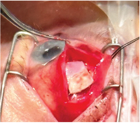
When placing tissue on the globe, the scleral portion is oriented posteriorly, and the corneal section is anteriorly approximated to the limbus (demonstrated in Figure 1, picture of patch graft sitting on the globe). The corneal component of the patch graft is usually half the total size of the graft. Subsequently, there is an option to anchor the corneoscleral graft to underlying sclera, and then the overlying conjunctiva and tenon’s are closed with dissolvable sutures.

Figure 1: Surgical implantation of corneoscleral patch graft.
Corneoscleral patch graft has the benefits of high tensile strength (decreased tube erosions) cosmetic outcome due to transparency, permission of visibility of underlying tube, low cost, and relatively good availability. Lam et al described use of corneoscleral patch grafts for Ahmed glaucoma valve implantation in patients with refractory and complicated glaucoma (sixteen consecutive patients). At a mean follow up of 6 months, they found no complications related to the corneoscleral patch, with corneal portion of the graft remaining transparent with good visualization of the underlying silicone tube [3]. The benefits of purely corneal donor tissue (including partial thickness corneal patch grafts) have also been described in the literature [4].
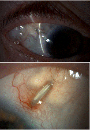
Tube erosion can be one of the most serious complications related to glaucoma valve implantation (Figures 2 & 3) illustrating tube erosions in a purely scleral graft). The intent of the donor material is to decrease the erosion. Observations of the donor material would show that donor cornea rarely thins and degrades with our eye bank preparation, unlike the sclera in which the tube can be seen migrating through the scleral material. Additionally, if the conjunctiva is not completely covering the donor material the healing can be reasonable over donor sclera but poor over donor cornea. The intent is to have the tougher corneal portion protect the tube towards the limbus, while the sclera is more posterior in the area of the limbus-based incision, 6-7mm back from the limbus, if that is the surgeon’s preferred technique. Further observation has been that the wound heals better over the scleral patch material. The hybrid offers decreased tube erosion at the limbus, but better healing posteriorly. Therefore, the optimal approach is believed to be a half-and-half donor patch, with the cornea positioned towards the limbus to reduce erosion, while the sclera placed more posteriorly to promote healing over a limbus-based flap. This conclusion is supported by clinical observations.

Figure 2 & 3: Tube erosions through scleral patch graft.
Furthermore, the transparency of the corneal grafts provides the benefits of cosmesis favourable to patients, in addition to direct visualization of the underlying tube as previously mentioned (Figure 4) comparing scleral graft versus corneoscleral graft in the same patient). Visualization could allow suturelysis in non-valved tubes.

Figure 4: Cosmetic difference of corneoscleral graft (right eye) and scleral graft (left eye) in same patient.
Sclera as a donor tissue by itself has the downside of variability in tissue thickness, which when approximated near the limbus can result in uneven tissue contour and the subsequent formation of dellen. Additionally, glycerin preserved donor sclera also results in a poor cosmetic appearance that is noticeable and bothersome to patients. When using corneoscleral patch grafts with a posteriorly located scleral component, the thickness of the corneal patch can prove beneficial for preventing erosion with the upper lid covering the scleral component of the patch graft.
In summary, we believe the corneoscleral patch graft provides several benefits to patients undergoing Ahmed glaucoma valve implantation. Its tensile strength decreases erosion, improved cosmesis and transparency, allowing visualization of the tube. Further research is required into assessing conjunctival erosion rates and long-term viability of these donor tissues.
Author Statements
Funding
This research did not receive any specific grant from funding agencies in the public, commercial, or not-for-profit sectors.
Conflict of Interest
No conflicting relationship exists for any authors.
References
- Riva I, Katsanos A, Floriani I, Quaranta L, Konstas AG, Airoldi G. Ahmed glaucoma valve implant: Surgical technique and complications. Clinical Ophthalmology. 2017; 11: 357.
- Wolf A, Mehta S, Khaimi MA, Molinari A, Mancini R, Sheng L. Use of autologous scleral graft in Ahmed glaucoma valve surgery. Journal of Glaucoma. 2016; 25: 365-370.
- Lam DS, Cheuk W, Lai JS. Short-term results of using lamellar corneo-scleral patch graft for the Ahmed glaucoma valve implant surgery. Yan Ke Xue Bao. 1997; 13: 109-112.
- Spierer O, Waisbourd M, Golan Y, Newman H, Rachmiel R. Partial thickness corneal tissue as a patch graft material for prevention of glaucoma drainage device exposure. BMC Ophthalmology. 2016; 16: 20.