
Case Report
Austin J Orthopade & Rheumatol. 2015;2(2): 1018.
Calcific Tendinitis of the Gluteus Maximums. Do we need to Investigate?
Chougule S* and Drabu KJ
Department of Orthopaedic Surgery, East Surrey Hospital, UK
*Corresponding author: Chougule S, Department of Orthopaedic Surgery, East Surrey Hospital, Redhill RH1 5RH, UK
Received: April 28, 2015; Accepted: June 30, 2015; Published: July 09, 2015
Abstract
Calcific tendinitis of gluteus maximus is not very common condition and there are only few cases reported till now. . In these patients radiological appearance does mimic malignant changes so we should only investigate these patients if they have significant past medical history i.e. Weight loss, history of malignancy or known primary elsewhere. Calcific tendinitis is quiet common in shoulder mainly distal supraspinatus; other sites have been reported but are rare in occurrence. It can present in two forms either with only amorphous calcification in tendon or calcification of tendon with cortical erosion which can be confusing with metastasis or primary malignant tumour.
Keywords: Calcific tendinitis; Gluteusmaximus; Malignancy; Tumour
Introduction
Calcific tendinitis of gluteus maximus is not very common condition and there are only few cases reported till now. We did have a patient who presented with this condition and we searched literature and there is hardly any material published on this. We think it is important to know detailed history of patient and if they have any history suggestive of malignancy then only we need to investigate these patients. We think this is important learning point in this article which will help reduce un-necessary investigations.
Case 1
A 55 years old white European female developed new onset of thigh and hip pain, she is normally fit and active. Patient described the pain as a dull ache without radiation; she tried over the counter pain medications with no real improvement, pain increased in intensity and radiated to her inner thighs and groin. She did not have fever or sweats and was systemically well. She was particularly anxious about her pain as her sister presented with back pain caused by metastasis.
Conclusion 1
Calcific tendinitis of gluteus maximus can have unusual presentation. We do not think all the patients with above radiological changes need further investigations but only those patients with significant cancer history do need further investigations.
Case 2
A 55 years old fit and well white European female developed pain in thigh and hip region. Patient described the pain as a dull ache without radiation; she tried over the counter pain medications with no real improvement, pain increased in intensity and radiated to her inner thighs and groin. She did not have fever or sweats and was systemically well. She did not have any malignancy in the past but was quiet anxious as her sister had back pain caused by metastatic tumour. As she was very anxious we carried out blood tests in the form of CRP/ESR White cell count, all were within normal limit, X-ray showed soft tissue calcification and lucent lesion in cortex of femur, CT scan showed cortical defect in the femur (lytic lesion), MRI revealed bone and soft tissue oedema, IV Gadolinium revealed increased signal within marrow and periosteum and bone scan showed focal area of increased uptake in lateral aspect of femur. (Bone scan was triple phase and there was only small area of increased uptake, myositis ossificans was ruled out).
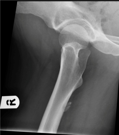
Figure 1: Lateral View Radiograph.
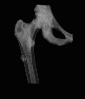
Figure 2: CT scan.
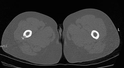
Figure 3: Axial view MRI Non contrast.
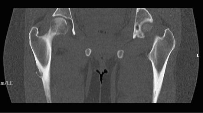
Figure 4: Coronal view MRI Non contrast.
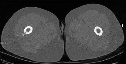
Figure 5: Axial view MRI Non contrast.
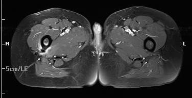
Figure 6: Axial view MRI With contrast.
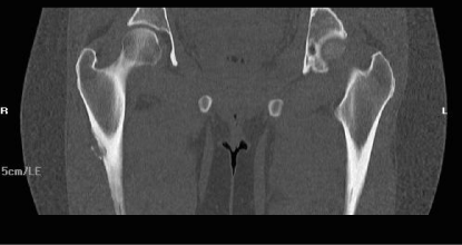
Figure 7: Coronal view MRI Non contrast.
We treated our patient with anti inflammatory medications and activity modification (she was advised to stop running as she was regular runner), this helped her and she recovered completely.
(Anti-inflammatory probably helped our patient in improving her pain in her initial episode, she denies any injury).
Conclusion 2
All the patients with calcific tendinitis doesn’t need investigations but only those with significant family history of malignancy or malignancy in the past or weight loss needs investigations to rule out primary or metastatic lesions. Most of the patients without this history and symptoms of only pain will resolve spontaneously. These patients need analgesics and modification of activity (stop running and jogging); time duration for resolution could be up to a year [1-3].
Discussion
Calcific tendinitis we see so commonly but mainly in shoulder and supraspinatus tendon in particular [6], but it can be found in other places as well [3], there are few cases reported on calcific tendinitis of gluteus maximus but it is important to know whether we need to investigate these patients further.
There are two parts of insertion of gluteus maximus tendon proximal is greater portion which is superficial and inserts on the iliotibial band of tensor fascia lata and smaller distal portion inserting on the gluteal tuberosity of linea aspera and is deeper [4].
This condition is primarily due to deposition of calcium hydroxy apatite crystals within the tendon in a relatively avascular area and no predisposing factors have been identified. There are theories as to why this occurs which include trauma and soft tissue necrosis due to hypovascularity and degeneration. It is important to appreciate bone changes reactive to calcification [5,6]. Calcification occurring intraosseous soft tissues could be myositis ossificans, periostealchonderal tumours and could be soft tissue or bony sarcoma. It is been shown that calcification in gluteus maximus has got unusual presentation with cortical bone erosion, in these circumstances it is important to know whether we need to investigate further.
Our patient had x-ray changes of soft tissue ossification/ periostitis and lucent lesion in cortex [7,8], CT scan had appearances of soft tissue calcification and loss of normal cortex ( lytic lesion ).
MRI scan revealed bone and soft tissue oedema at the site of lesion, thickened periosteum, IV Gadolinium revealed increased signal within marrow and periosteum. Bone scan showed focal area of increased uptake in lateral aspect of femur.
All the above radiological features can be seen in calcific tendinitis. Our patient was treated with anti inflammatory and responded well within three weeks, her pain improved and at three months radiographs did show reduction in calcification.
References
- Wepfer JF, Reed JG, Cullen GM, McDevitt WP. Calcific tendinitis of the gluteus maximus tendon (gluteus maximus tendinitis). Skeletal Radiol. 1983; 9: 198-200.
- Mizutani H, Ohba S, Mizutani M, Otake S, Otsuka T, Nakamura T. Calcific tendinitis of the gluteus maximus tendon with cortical bone erosion: CT findings. J Comput Assist Tomogr. 1994; 18: 310-312.
- Hayes CW, Rosenthal DI, Plata MJ, Hudson TM. Calcific tendinitis in unusual sites associated with cortical bone erosion. AJR Am J Roentgenol. 1987; 149: 967-970.
- Last RJ. Anatomy; Regional and Applied. 7th edn. 1984;145-146.
- Fritz P, Bardin T, Laredo JD, Ziza JM, D’Anglejan G, Lansaman J, et al. Paradiaphyseal calcific tendinitis with cortical bone erosion. Arthritis Rheum. 1994; 37: 718-723.
- Holt PD, Keats TE. Calcific tendinitis: a review of the usual and unusual. Skeletal Radiol. 1993; 22: 1-9.
- Wepfer JF, Reed JG, Cullen GM, McDevitt WP. Calcific tendinitis of the gluteus maximus tendon (gluteus maximus tendinitis). Skeletal Radiol. 1983; 9: 198-200.
- Hodge JC, Schneider R, Freiberger RH, Magid SK. Calcific tendinitis in the proximal thigh. Arthritis Rheum. 1993; 36: 1476-1482.