Case Presentation
Austin J Otolaryngol. 2018; 5(2): 1103.
Dysphagia by Giant Osteophyte: A Case Report
Lacerda NSO¹*, Fernandes FL¹, Aniteli MB¹ and Sakano E²
¹Otorhinolaryngologist, Otorhinolaryngology Discipline, Head and Neck, UNICAMP, Brazil
²Otorhinolaryngologist, Boss of the Rhinology Service, Otorhinolaryngology Discipline, Head and Neck, UNICAMP, Brazil
*Corresponding author: Nayara Lacerda, Department of Otorhinolaryngology, State University of Campinas, Brazil
Received: May 31, 2018; Accepted: June 18, 2018; Published: June 25, 2018
Abstract
Introduction: Dysphagia is a modification in the swallow that can occur in the oral, pharyngeal and esophageal phase. The investigation of the etiology depends on the location, being the oropharyngeal one mainly caused by structured and neuromotor anomalies, whereas the esophageal ones are caused by structural and inflammatory changes. Between elderly patients, the complaint about dysphagia is common, because of the associated comorbidities; in young population, it should be researching mainly for inflammatory and autoimmune causes. Disturbs in the cervical spine are caused by extrinsic pressure and are rare in the young population.
Case Report: 57-year-old female patient, with high dysphagia and associated weight loss. She was diagnosed with fusion and prominence of vertebral osteophytes, being subjected to surgery treatment with postoperative clinical improvement.
Discussion: Changes in the anterior cervical spine can cause dysphagia, trismus and dyspnea, due to external pressure of the neck structures. In literature, there are few similar cases and among the changes already described are: osteophytes by degenerative joint disease, ankylosing spondylitis, Diffuse Idiopathic Skeletal Hyperostosis and hereditary multiple exostoses.
Conclusion: In situations of investigation to diagnose dysphagia, we always recommend the effectuation of a detailed physical exam, accompanied by image examination, Nasofibrolaryngoscopy and Computed Tomography.
Keywords: Oropharyngeal Dysphagia; Osteophyte; Cervical Spine; Esophageal Compression; Cervical Spine
Introduction
Dysphagia is a modification in the swallow that can occur in the oral, pharyngeal and esophageal phase [1]. To elucidate the etiology, it is necessary to distinguish the location of the dysphagia, because the oropharyngeal is mainly caused by structural and neuromotor anomalies, such as cerebrovascular accident, Myasthenia gravis and multiple sclerosis. Among the esophageal ones are found mainly structural head and neck tumors, esophageal webs and esophageal rings) and inflammatory (esophagitis, achalasia and rheumatological diseases) causes [2,3].
Among elderly patients, the complaint about dysphagia is common, mainly due to the high incidence of associated comorbidities; in young population, it should be researching mainly for inflammatory and autoimmune causes. Disturbs in the cervical spine are caused by extrinsic pressure and are rare in the young population [1].
Abnormalities in the cervical spine can cause the direct pressure of the esophagus and trachea, resulting besides dysphagia, dysphonia and trismus. Despite it is a rare diagnosis, it is always important to consider about the cervical spine injuries as the possible causer of esophageal and oropharyngeal dysphagia [4]. About 20% to 30% of the populations present cervical osteophytes, with rare progression to dysphagia and obstruction of the upper respiratory tract [5]. In patients with hyperostosis of the cervical spine, 17% to 28% will present dysphagia [6]. Other cases of bone changes were already described in literature; it is mostly related to the bad formation on the anterior arch of the cervical spine. The examples are the degeneration of the spine (large osteophytes, clefts, intervertebral mergers and aplasia) ankylosing spondylitis and Diffuse Idiopathic Skeletal Hyperostosis (DISH) [5]. Abnormalities of the posterior arch are more difficult to be found and are usually due from surgical complications, but can also cause dysphagia [7].
In general, patients with this type of injury are asymptomatic and these modifications can be unnoticed lifelong, being many times found accidentally on exams made for another purpose. The diagnosis can be done by simple radiograph and Computed Tomography of the neck [4].
This article describes a case of high dysphagia, on a 57-year-old patient, who posteriorly was diagnosed with fusion and prominence of vertebral osteophytes, being subjected to surgery treatment for correction.
Case Presentation
A 57-year-old patient, female sex, treated on the clinic of otorhinolaryngology complaining about high progressive dysphagia initially with solids and then to liquids, with weight loss of 37.5 pounds in 5 months without antecedent of cervical traumas and previous surgeries without comorbidities. The physical exam did not present mass or palpable lymph nodes in the neck and oroscopy without changes.
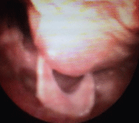
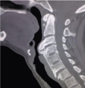
The patient then was subjected to a Flexible Nasofibrolaryngoscopy (FNF) with visualization of retropharyngeal vault at the level of epiglottis, without injuries or vegetation in the mucosa (Figure 1). The investigation was concluded with cervical Computed Tomography (CT) evidencing arthrodesis of vertebral bodies of C4 to C6, with prominent osteophytosis in the anterior side of the vertebral bodies of C3/C4 and C6/C7, with pressure of the posterior side of the cervical esophagus (Figure 2).

Figure 1: Preoperative FNF.

Figure 2: Preoperative CT.
The patient was guided to the neurosurgery and programmed surgical approach. The surgery was performed by cervical access with an oblique incision of the anterior border of the sternocleidomastoid muscle on the left, blunt dissection of lateral carotid to cervical esophagus and drilled of osteophytes of C3, C4 and C5.
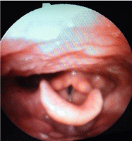
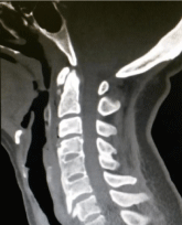
After the surgery, the patient progressed with improvement of the dysphagia, presenting weight recovery and good feed acceptance to solids and liquids. After performing the postoperative control tomography with bone windowing and Nasofibrolaryngoscopy (Figures 3 and 4), which demonstrates the success of the reached decompression after resection of the bone protuberance of C3, C4 and C5.

Figure 3: Postoperative FNF.

Figure 4: Postoperative CT.
Discussion
Changes in the cervical spine can cause dysphagia, trismus and dyspnea due to direct pressure of the neck structures. In literature, there are few similar cases reported and among the changes already described are: Osteophytes by degenerative joint disease, ankylosing spondylitis, Diffuse Idiopathic Skeletal Hyperostosis (Forestier’s disease) and hereditary multiple exostoses [4,8].
These pathologies are more common in elderly individuals, usually after the seventh decade of life. In children, this event is even rarer than in adults. In these cases, the pathologies usually are presented as congenital malformations or by a variation of the Atlas, as a bone prominence on the anterior tubercle. Recently Ílbay et al showed a case of abnormal protuberance of the Atlas, causing dysphagia on an 11-year-old child [4]. Besides these variations of the bone normality, other possible causes of injury in this topography would be metastases or bone tumors [9]. The metastases of spine are generally due to breast adenocarcinoma, pulmonary neoplasias, of prostate, of the kidneys and of the haematopoietic tumors, mainly lymphomas and multiple myeloma. Pulmonary and breast neoplasias usually occur with cervical or thoracic injuries, while in prostate, colon and pelvic tumor have more predilection for the lumbosacral vertebrae [10].
In patients who were subjected to surgeries in the cervical spine, there is an important frequency of dysphagia, due to the edema and change on the angulation between vertebrae, especially when it involves from C2 to C7, both on the anterior cervical spine and posterior spine surgeries [7].
Bone changes of anterior cervical vertebral spine shows a large variety of symptoms that are directly related with the different levels of pressure on the adjacent structures. Dysphagia is the most prevalent among those. To help in the therapeutic decision of the patient with dysphagia it is necessary primarily to classify the symptom in mild, moderate or severe.
In cases of mild or moderate gravity of dysphagia, the clinical treatment can be the first choice; it embraces the modification of the diet, nonsteroidal anti-inflammatory drugs, muscle relaxants and proton pump-inhibitors. In cases of severe dysphagia or moderate that were unresponsive to clinical treatment, the surgical intervention is the best choice [11]. The patient of this report presented a severe dysphagia, with absolute recommendation to surgical treatment. The procedure was successfully performed.
Conclusion
In situations of investigation diagnosis to dysphagia, we always recommend the effectuation of a detailed physical examination, accompanied by image examination, Nasofibrolaryngoscopy and Computed Tomography. Besides the dysphagia by abnormality in the anterior cervical spine is a rare event, it should be remembered as a differential diagnosis, mainly by the possibility of bone metastases, due to growing cancer incidence in the world population.
References
- Papadopoulou S, Exarchakos G, Beris A, Ploumis A. Dysphagia associated with cervical spine and postural disorders. Dysphagia. 2013; 28: 469-80.
- Abraham Khan, Richard Carmona, Morris Traube. Dysphagia in the Elderly. Clin Geriatr Med. 2014; 30: 43-53.
- Carucci LR, Turner MA. Dysphagia revisited: Common and unusual causes. Radiographics. 2015; 35: 105-122.
- K Ílbay, C Evliyaoglu, V Etus, H Ozkarakas and S Ceylan. Abnormal bony protuberance of anterior atlas causing dysphagia. A rare congenital anomaly. Spinal Cord. 2004; 42: 129-131.
- Granville L, Musson N, Altman R, et al. Anterior cervical osteophytes as a cause of pharyngeal stage dysphagia. J Am Geriatr Soc. 1998; 46: 1003-1007.
- Demuynck K, Van Calenbergh F, Goffin J, et al. Upper airway obstruction caused by a cervical osteophyte. Chest. 1995; 108: 283-284.
- Haruo Misawa, Masato Tanaka, Yoshihisa Sugimoto, Kouichiro Koshimune, and Toshifumi Ozaki. Development of Dysphagia and Trismus Developed after C1-2 Posterior Fusion in Extended Position. Acta Med. Okayama. 2013; 67: 185-190.
- Bethge KC. Jungehülsing KW. Atlantoaxial malformation as a rare cause of dysphagia and snoring. HNO. 2011; 59: 280-282.
- Barros Filho TE, Oliveira RP, Taricco MA, Gonzales CH. Hereditary multiple exostoses and cervical ventral protuberance causing dysphagia. A case report. Spine. 1995; 20: 1640-1642.
- Joaquim AF, Paula Maturana FA, Anderle DV, Lessa Zambelli HJ, Calfat Maldaun MV. Metástases na coluna vertebral. Rev Neurocienc. 2007; 15: 240-245.
- Ilknur Albayrak, Sinan Bagcaci, Ali Salli, Sami Kucuksen, and Hatice Ugurlu. A rare cause of dysphagia: Compression of the esophagus by an anterior cervical osteophyte due to ankylosing spondylitis. Korean J Intern Med. 2013; 28: 614-618.