Case Report
Phys Med Rehabil Int. 2015;2(4): 1045.
Dandy-Walker Syndrome: A Case of Conservative Management
Tam E1* and Mohammad I2
1New York Medical College, MD Candidate 2016, USA
2Department of Physical Medicine and Rehabilitation Metropolitan Hospital Center, USA
*Corresponding author: Tam E, New York Medical College, USA
Received: April 01, 2015; Accepted: April 27, 2015; Published: April 28, 2015
Abstract
Case: Diagnosis: Dandy-Walker Variant vs. Mega Cisterna Magna.
A 3 year 8 month old female with waddling gait, hypotonia, lower extremity clonus and global developmental delays was referred to PM&R. Physical examination showed generalized mild hypotonia and weakness throughout. MRI revealed hypoplasia of inferior vermis with prominent communication of 4th ventricle, leading to a preliminary diagnosis of Dandy-Walker Variant vs Mega Cisterna Magna. Treatment options for Dandy-Walker syndrome were discussed with the patient’s parents who refused surgery and opted for conservative management.
Discussion: Dandy-Walker syndrome (DWS) is the most common congenital malformation of the cerebellum, occurring in 1 of 30,000 live births. The disease causes dilation of the 4th ventricle, and patients present with waddling gait, hypotonia and developmental delays. The etiology of the disease is speculated to be the loss of Zic1/4 genes.
Conclusion: This is a case of DWS managed conservatively, with surgery being the usual treatment of choice. DWS should be suspected in patients with waddling gait, hypotonia and developmental delays. Further investigation is required to determine if breast malformations, amblyopia and family history of cerebral palsy are associated with a diagnosis of DWS.
Keywords: Dandy-Walker Syndrome; Waddling gait; Dandy-Walker variant; Mega cisterna magna
Abbreviations
DWS: Dandy-Walker Syndrome; ETV: Endoscopic Third Ventriculostomy; PM&R: Physical Medicine and Rehabilitation
Case Presentation
Diagnosis: Dandy-Walker Variant vs. Mega Cisterna Magna. A 3 year 8 month old female with waddling gait and history of poor appetite, hypotonia, lower extremity clonus and global developmental delays was referred to PM&R by Pediatrics. She was delivered full term by normal spontaneous vaginal delivery to a 27 year old G1P0 mother. Her mother a history of Gower’s sign when the patient was learning to stand up. The patient first walked at 2.5 years old with a waddling gait and, frequent falls and unsteadiness. Her family history was significant for a father with cerebral palsy.
On physical examination the patient had generalized mild hypotonia throughout, weakness in upper (3+/5) and lower extremities (4/5) bilaterally. Her feet were pronated and display bilateral pes planus and a valgus deformity. On ambulation she had out-toeing of the feet with a waddling gait. She had full range of motion in all extremities and was around 25th percentile for height and 5th percentile for weight.
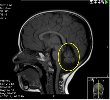
The presenting symptoms were indications for MRI which revealed hypoplasia of inferior vermis with prominent inferior retrocerebellar CSF space with prominent communication of 4th ventricle (Figure 1). These findings lead to a preliminary diagnosis of Dandy-Walker Variant vs Mega Cisterna Magna.

    Figure 1:  MRI without intravenous contrast showing a coronal view of the
head. A classic Dandy-Walker cyst is visible surrounding the cerebellum, and
communicating with the fourth ventricle. Note the dilation of the area around
cerebellum (circled in yellow), and compare with normal anatomy.
The patient’s family was informed of treatment options of DWS, who refused surgery and opted for conservative management. The patient receives physical therapy and occupational therapy and is followed by speech-language pathologists. PM&R prescribed bilateral foot orthosis and a pediatric brace for safe ambulation. The patient lives with her parents and goes to a special school, although a workup for intellectual disability has not been started at the hospital. In outpatient follow-up, her parents report she is compliant with the brace and foot orthoses, she is falling less often, and her gait abnormalities have been steadily improving.
She is currently being followed by many other teams in the hospital, such as orthopedics for a right hip click, but bilateral hip x-ray showed no abnormal findings. She also follows up with ophthalmology that performed surgery for amblyopia at 8 months old. The patient had mammogram for left breast hypertrophy and a palpable mass. The mammography showed an oval area in the left breast at 12 o’clock anterior depth of mixed echogenicity that correlates with the palpable mass. These findings represent gynecomastia, which requires further workup including correlation of estrogen levels.
Discussion
DWS is a congenital anomaly characterized by hypoplasia or complete absence of the cerebellar vermis and dilation of the 4th ventricle that occurs sporadically in 1 of 30,000 live births [1]. With treatment, DWS patients develop normally and have lifespans similar to unaffected adults [2]. DWS is separated into four categories.
- Dandy-Walker Malformation is cerebellar dysfunction due to the complete absence of the cerebellar vermis, cystic dilation of the 4th ventricle and enlargement of the posterior fossa. Absence of the cerebellar vermis can result in hypotonia and poor coordination. Cystic dilation of the 4th ventricle can cause an increase in intracranial pressure, manifesting as irritability, vomiting and seizures. These symptoms can develop insidiously or spontaneously [1,2,3].
- Dandy-Walker Variant presents in a similar fashion, but is characterized by hypoplasia of the cerebellar vermis rather than its complete absence. The posterior fossa is normal in size, but retains the cystic lesion that communicates with the 4th ventricle.
- Mega Cisterna Magna is a variation of normal anatomy which is classified by enlarged retrocerebellar cisterns in the posterior fossa with normal cerebellar morphology. Cerebellar hemispheres and vermis are normal, but the cisterna magna is enlarged, measuring greater than 10 mm on oblique transverse plane. Isolated mega cisterna magna has favorable prognosis, but one third of children with CNS and non-CNS anomalies with mega cisterna magna have cognitive, motor and language delay as well as cerebellar ataxia [4].
- Blake’s Pouch Cyst is a posterior ballooning of the superior medullary velum into the cisterna magna [5]. Patients present with delayed neurological development, with normal outcomes at 1-5 years old.
Diagnosis
DWS can be diagnosed prenatally with fetal ultrasound at the 18th week of gestation when the inferior vermis should normally form [6]. CT scan or MRI can also make a diagnosis of DWS after birth with the presence of dilation of the 4th ventricle and the characteristic Dandy- Walker cyst in the posterior fossa cyst extending from the cisterna magna to the fourth ventricle. Other visible features on CT and MRI include an elevated tentorium and dilation of the third and lateral ventricles. Abnormalities associated with DWS include various neurological anomalies such as the absence of the corpus callosum, as well as heart, and limb malformations [7].
Pathology
The proposed mechanism of DWS is the deletion of Zic1/4 genes on chromosome 3q24, which may have roles in cerebellar size and patterning in controlling cerebellar growth mediated by the Sonic Hedgehog pathway. Complete loss of Zic1 in mice was proven to cause cerebellar hypoplasia as well as ataxia, growth retardation and decreased viability. Loss of Zic4 possibly has a contributory role in the presentation of symptoms in DWS [8].
Differential diagnosis
The differential diagnosis of DWS should include arachnoid cyst, which is a congenital disorder causing a collection of cerebral spinal fluid in the arachnoid membrane [9]. Unlike in DWS, this cyst will not communicate with the fourth ventricle. Most cases are asymptomatic, but can present with similar symptoms as DWS, including headache, weakness, seizure, hydrocephalus, scoliosis, cognitive decline and visual loss [10].
Management and treatment
Surgical management of DWS is considered first line, which consists of cystoperitoneal shunting and endoscopic third ventriculostomy (ETV) [11]. Cystsoperitoneal shunting is a procedure in which a catheter is inserted into the dilated cyst while the other end of the tube allows the cerebrospinal fluid to empty into the chest or abdomen. ETV, on the other hand, consists of creating an opening in the floor of the third ventricle, allowing cerebrospinal fluid to bypass any obstructions to the interpeduncular cistern. ETV is considered less invasive, has a lower chance of infection, and the ETV shunt does not migrate and cannot be displaced, which can be issues with cystoperitoneal shunting [11]. Therefore cystoperitoneal shunting is only when ETV fails to resolve the hydrocephalus [12]. If the preoperative MRI shows obstruction of the cerebral aqueduct, a stent is recommended between the third ventricle and the posterior fossa cyst in addition to the ETV procedure [13].
Conservative management of patients with DWS includes occupational therapy, physiotherapy, speech therapy, specialized education, back brace and foot orthosis, with the goal of reducing falls and correcting gait abnormalities.
Conclusion
The patient presented shows a classic presentation of DWS, including cerebellar ataxia, hypotonia and developmental delays. DWS can present with developmental delays and is associated with congenital abnormalities such as neurological anomalies, and heart and limb malformations. Nevertheless, isolated DWS is associated with a good developmental outcome and patients have similar lifespans and quality of life as the normal population if diagnosed and treated properly [7]. Diagnosis can be made using fetal ultrasound or CT scan, and treatment includes both medical and conservative management. The first line treatment of DWS is surgery, with ETV preferred over cystoperitoneal shunting. Conservative management includes foot orthoses and correction of gait abnormalities. Examples of breast anomalies and amblyopia comorbid with DWS are sparse in the literature, and further investigation is required to determine if they are associated with DWS. Finally, there are many examples in the literature of patients with cerebral palsy concurrent with DWS. In light of the patient’s family history of her father having cerebral palsy, further investigation is required to determine if there is a relationship between the two diseases.
References
- Menkes JH, Sarnat HB. Neuroembryology, genetic programming, and malformations. Menkes JH, Sarnat HB, editors. In: Child Neurology. 6th ed. Philadelphia, PA: Lippincott Williams and Wilkins; 2000; 354-377.
- Parisi MA, Dobyns WB. Human malformations of the midbrain and hindbrain: review and proposed classification scheme. Mol Genet Metab. 2003; 80: 36-53.
- Warf BC, Dewan M, Mugamba J. Management of Dandy-Walker complex-associated infant hydrocephalus by combined endoscopic third ventriculostomy and choroid plexus cauterization. J Neurosurg Pediatr. 2011; 8: 377-383.
- Long A, Moran P, Robson S. Outcome of fetal cerebral posterior fossa anomalies. Prenat Diagn. 2006; 26: 707-710.
- Calabrò F, Arcuri T, Jinkins JR. Blake's pouch cyst: an entity within the Dandy-Walker continuum. Neuroradiology. 2000; 42: 290-295.
- Russ PD, Pretorius DH, Johnson MJ. Dandy-Walker syndrome: a review of fifteen cases evaluated by prenatal sonography. Am J Obstet Gynecol. 1989; 161: 401-406.
- Sasaki-Adams D, Elbabaa SK, Jewells V, Carter L, Campbell JW, Ritter AM. The Dandy-Walker variant: a case series of 24 pediatric patients and evaluation of associated anomalies, incidence of hydrocephalus, and developmental outcomes. J Neurosurg Pediatr. 2008; 2: 194-199.
- Blank MC, Grinberg I, Aryee E, Laliberte C, Chizhikov VV, Henkelman RM, et al. Multiple developmental programs are altered by loss of Zic1 and Zic4 to cause Dandy-Walker malformation cerebellar pathogenesis. Development. 2011; 138: 1207-1216.
- Hayashi Y, Kita D, Watanabe T, Yoshikawa A, Hamada J. Symptomatic foramen of Magendie arachnoid cyst in an elderly patient. Surg Neurol Int. 2015; 6: 7.
- Pradilla G, Jallo G. Arachnoid cysts: case series and review of the literature. Neurosurg Focus. 2007; 22: E7.
- Etus V, Ceylan S. Success of endoscopic third ventriculostomy in children less than 2 years of age. Neurosurg Rev. 2005; 28: 284-288.
- Hu CF, Fan HC, Chang CF, Wang CC, Chen SJ. Successful treatment of Dandy-Walker syndrome by endoscopic third ventriculostomy in a 6-month-old girl with progressive hydrocephalus: a case report and literature review. PediatrNeonatol. 2011; 52: 42-45.
- Hankinson TC, Vanaman M, Kan P, Laifer-Narin S, DeLaPaz, R, Feldstein, N, et al. Correlation between ventriculomegaly on prenatal magnetic resonance imaging and the need for postnatal ventricular shunt placement. Journal of Neurosurgery: Pediatrics. 2009; 3: 365-370.