Research Article
Phys Med Rehabil Int. 2015; 2(7): 1059.
Evaluation of Hip Muscles using Torque and Power- Velocity Relationships in Chronic Low Back Pain Subjects
Lemaire A1,2*, Lisembart A¹, Ritz M² and Rahmani A¹
¹LUNAM Université, Université du Maine, Laboratoire "Motricité, Interactions, Performance" EA4334, France
²Centre de l’Arche, Pôle Régional Spécialisé en Médecine Physique et Réadaptation, France
*Corresponding author: Lemaire A, Département STAPS, University of Maine, Avenue Olivier Messiaen 72085 Le Mans, France
Received: August 17, 2015; Accepted: September 22, 2015; Published: September 25, 2015
Abstract
The aim of the present study was to evaluate hip muscles in chronic low back pain (CLBP) patients, thanks to the torque- and power-velocity relationships and to determine whether a possible imbalance can be related to the trunk muscles. Twelve CLBP patients and fifteen healthy subjects (Control Group, CG) participated to the study. CLBP and CG performed isokinetic trunk and hip flexions and extensions at several velocities. Mechanical parameters, such as theoretical maximal isometric torque (T0) and maximal power (Pmax) were extrapolated from torque- and power-velocity relationships. T0 and Pmax, were significantly higher in CG than CLBP (p<0.05), whatever the considered movement, showing a muscle weakness in CLBP. A significant difference was obtained between the two lower limbs for CLBP when considering Pmax during hip extension. Although no relationship was clearly observed between hip weakness and trunk weakness for CLBP, results obtained for CLBP could be attributed to a deconditioning syndrome and to muscle inhibition typically observed in these subjects. This study shows the importance to take hip muscle rehabilitation into account in CLBP support.
Keywords: Hip; Trunk; Low back pain; Isokinetic; Torque- and powervelocity relationships
Abbreviations
CG: Control Group; CLBP: Chronic Low Back Pain; e: Extension; f: Flexion; Pmax: Maximal Power; Ppeak: Instantaneous Peak Power; T0: Theoretical Maximal Isometric Torque; Tpeak: Peak Torque
Introduction
Chronic low back pain (CLBP) is characterized by a strength imbalance between trunk flexor and extensor muscles [1-3]. This imbalance is responsible for significant functional impairment [4-7] which should be treated with a complex rehabilitation program [8]. Trunk rehabilitation is essential for CLBP subjects [1,3]. Nevertheless, other muscles should be considered. In a previous study [9], observed relationships between lower limb and pelvis movement in CLBP. Indeed, people with low back pain who play rotation-related sports are able to move their lumbopelvic region with a greater extent and earlier during lower limb movements than people without low back pain. Recently, focusing our attention on the leg extensor muscles, we observed higher torque (19.2%) and power (19.8%) productions in healthy subjects compared with CLBP patients [10]. Even if no relationship was revealed between the weakness of the leg extensor muscles and the trunk imbalance in CLBP patients, it can’t be denied that these two statements are probably linked, and can be explained by a CLBP inactivity, a fear relative to daily activity movements, and finally to the CLBP deconditioning syndrome.
These previous studies suggest that lower limb and pelvis muscles must be considered in the management of CLBP rehabilitation. Some previous studies focused on lower limbs in CLBP [9-11]. However, to the authors’ knowledge, no study focused on hip muscles assessments, which are anatomically directly linked to the trunk muscles. Indeed several hip flexor and extensor muscles have their proximal insertions on the trunk [12]. Deconditioning syndrome that occurs on trunk muscles and its consequences on muscle properties [13] would also have an impact on hip muscles. This impact could probably also be linked to low back pain, since these muscles have an important postural and dynamic role especially at the lumbar spine [14].
The aim of the present study was then i) to evaluate hip flexor and extensor muscles torque production capacity using torque- and power-velocity relationships in both CLBP and healthy subjects, and ii) to determine whether a possible imbalance between the two sides in CLBP hip muscle strength can be related to the trunk muscles.
Methods
Subjects
Twenty-seven subjects signed an informed consent to participate in this study. A control group (CG) was composed of fifteen healthy males with no prior history of low back pain. Healthy subjects were recruited in the rehabilitation centre or from a call for volunteers, and should respect the anthropometric characteristics (40.5 ± 5.0 years, 1.8 ± 0.6 m, 72.4 ± 8.7 kg) of the low back pain patients to allow group’s comparison. To also avoid any bias in the study, subjects of control group were not involved in any physical training.
Chronic Low Back Pain (CLBP) group was composed of twelve subjects (42.4 ± 7.4 years, 1.7 ± 4.5 m, 91.8 ± 29.6 kg) involved in a five weeks multidisciplinary rehabilitation program. This program was proposed by The Centre de l’Arche (Le Mans, France), following the Lombaction’s protocol from Angers (France) hospital. All the CLBP patients were recruited by the head clinician in charge of CLBP program of the rehabilitation center following the low back pain definition proposed by the French Society of Rheumatology (i.e., back pain for at least 3 months) [15]. For the present study, inclusion criteria for the CLBG patients were presence of chronic pain defined as a daily or almost daily pain for at least six months and a lumbar or lumbosacral pain before the start of the treatment. Selected patients had reported lower back pain for at least five years and had not been free of pain for a year. None of the subjects had any hip or lower limb injury or surgery, and no other associated pathology (e.g., multiple sclerosis, Guillain-Barre, muscular dystrophy, pelvic inflammatory disease). They were able to exert a maximal effort on isokinetic device. The rehabilitation program proposed at the Centre de l’Arche does not take into account current daily pain as an exclusion criterion. For the present study, exclusion criterion was a history of spine surgery. Testing was carried out in accordance with the ethical standards laid down in the 1964 Helsinki Declaration. Since the two groups presented a significant difference in weight values, results were normalized relatively to the subjects’ body weight in order to allow comparison between groups.
Protocol
The protocol of the present study was carried out during three different sessions on three following different days. Both groups performed the same protocol. Trunk flexions and extensions were tested on day 1 and 2 respectively, as recommended by Ripamonti et al. [16]. Assessment of hip flexions and extensions was performed on the third day. Each measurement was done within a week following the admission of the CLBP patients in the CLBP program. To limit errors due to faulty equipment and inherent wrong measurement, all the measurements were realized by the same highly qualified investigator.
Trunk flexion and extension measurements: Trunk flexions and extensions were both conducted on a Biodex® isokinetic dynamometer (Model 900-240, Biodex Corporation, Shirley, NY, USA). Subjects were seated on a chair, with the dynamometer aligned bilaterally with the anterior superior iliac crests at the mid-axillary line of the trunk. The upper body was then strapped to the back of the chair equipped with three fixable pads at the head, the dorsal region and the bottom of the back. Legs were also strapped and positioned on the footrest with a maximal knee angle of 15 degrees to minimize leg involvement in trunk movements [17]. Subjects were asked to hold the trunk strap with their hands, without contracting the upper limbs. Upper limb muscles activity was not controlled by any device but one experimenter made sure that subjects did not use their upper limbs as an extra help to produce force. If any arm movement was observed, the trial was not recorded and was repeated after a rest period of at least 4 minutes. Once the subject position was properly established, mechanical stops were positioned to allow a range of motion of 60 degrees (from 90 to 30 degrees relatively to the horizontal axis). This amplitude was selected to prevent subjects working in non-conventional zones. This amplitude was recorded by the computerized monitor after the Biodex lever arm had been placed in front of the 90 and 30 degrees graduations around the rotation axis.
Trunk extensors and flexors were assessed on two different days. On the first day, flexor muscles were evaluated, whereas the extensor muscles were assessed at the same time on the following day. After a period of standardized warm-up (10 minutes warm-up on a cycle ergometer (50 watts at 50 to 60 rpm) each subject performed sub maximal trunk flexions and extensions before the testing session. During the test, subjects performed five contractions [18] at 120, 105 and 90 deg.s-1 and three at 75, 60 and 45 deg.s-1. Only the concentric part of the movement was considered. At a given preset velocity, subjects were asked to perform the concentric contraction as rapidly and forcefully as possible, and to return to their initial position without any effort during the eccentric phase (isokinetic velocity was then fixed at 300 deg.s-1). A one-second break was set between two consecutive contractions to avoid any possible influence of the eccentric part of the movement. A 4 minutes rest period was observed between two preset-velocity trials. Verbal encouragements were given to the participants during all trials.
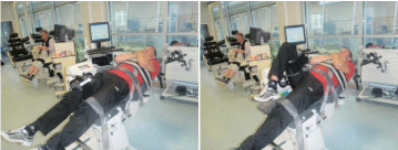
Hip flexion and extension measurements: Hip flexions and extensions were conducted using a calibrated Biodex System 4 dynamometer (Biodex Medical Systems, Inc., Shirley, NY). The motor axis was aligned with the major trochanter of the femur. Subjects were lying on their back on the dynamometer with an angle of 15 degrees at the hip joints to prevent lordosis. Subjects were fastened with belts at the chest, pelvis and thigh to minimize the contribution of the body parts that were not supposed to be involved in the movement. Subjects had to keep their hands crossed on the chest. The thigh of the tested hip was attached to the level arm of the Biodex (Figure 1). The tested hip was attached to the lever arm of the dynamometer. If any compensation with the back was observed, the trial was not recorded and was repeated after a rest period of at least 4 minutes. The range of motion during hip flexions and extensions evaluation was fixed at 75 degrees, with a starting position corresponding to the leg extended at an angle of 15 degrees relatively to the hip joint (0 degree corresponding to a complete hip extension). The final position of the leg was defined for an angle of 90 degrees at the hip joint.

    Figure 1:  Initial subject’s positions (left picture) and final position (right
picture) during hip flexion contractions.
After a standardized warm up (i.e., the same as the one done before the trunk assessment), subjects were asked to perform three sub maximal trials at each preset velocity (i.e., 180, 150, 90, 60 and 45 deg.s-1) during a familiarization session preceding the criterion test. During the testing session, subjects performed four maximal contractions at the five preset velocities. Velocity order was randomized to avoid any fatigue or velocity effect. Hip side evaluation (i.e., left or right) was also randomized. At each velocity, subjects were asked to perform maximal concentric flexions and extensions as rapidly and forcefully as possible. A 1-s break was set between two consecutive contractions to avoid any possible influence of the stretch-shortening cycle.
Mechanical parameters determination
To avoid any underestimation of torque [19-21] and to ensure data reliability, inertia of the lower limb and of the isokinetic lever arm was measured ahead of testing. This calibration was conducted with the participant lying on the back with a hip angle of 70 degrees (90 degrees corresponding to a complete hip extension), straight leg for hip measurements. Inertia of the trunk was also calculated with the subject seated in the starting position for the trunk flexions. In both conditions, subjects were asked to be completely relaxed. Gravitational errors of the tested body segment and dynamometer input arm were estimated by measuring the dynamometer moment in these specific positions. Data were then recorded and automatically stored on a PC (sampling at 100 Hz) via an electronic interface card (Biodex Medical Systems Inc. X2151, Shirley, NY, USA).
Torque- and power-velocity relationships
For each preset velocity, and whatever the considered movement and limb, peak torque (Tpeak) was identified as the highest value attained during the period of constant velocity, which was determined from the raw data. Instantaneous peak power (Ppeak) was then calculated as the product of Tpeak and the corresponding preset velocity.
Linear torque-velocity relationships were then determined using Tpeak measured at each preset velocity, for both lower limbs and trunk evaluations. Therefore, Tpeak was expressed as Tpeak = aV + b, where V was the preset velocity, a and b the linear regression coefficients. From those relationships, the theoretical maximal isometric torque (T0) was extrapolated as the intercept with the torque axis (null velocity) and hence equal to the coefficient b. The maximal velocity was also extrapolated from this relationship and corresponds to the intercept with the velocity axis (null torque). This parameter is not detailed in this article as we consider more interesting to focus our attention on torque and power only. Power-velocity relationships were defined from second order polynomial relationships, and were determined using Ppeak values measured at each preset velocity. The maximal power (Pmax), corresponding to the apex of the power-velocity relationship, was extrapolated from the second order polynomial equation and were expressed as: Ppeak = cV² + dV + e, where c, d and e were the second order polynomial regression coefficients. Throughout this paper, the indices "f" and "e" are used to identify flexion and extension results, respectively.
Statistical analysis
Data are presented as their mean ± Standard Deviation). Skewness and Kurtosis analysis were used to verify the normality of the distribution and the homogeneity of variance in the data sets. Torque-velocity and power-velocity relationships were described by linear and polynomial regressions, respectively. Corresponding coefficients of correlation (r²) and levels of significance (p) were also calculated. A factorial analysis of variance (ANOVA) was used to analyze results obtained for right (R) and left (L) hip side values during flexions and extensions, hip flexor/extensor ratio obtained for both sides, and trunk flexion and extension parameters for each group. A factorial ANOVA was also used to compare results obtained between CG and CLBP for each mechanical parameter. Finally, simple and multiple regressions were also used to identify relationships between hip and trunk mechanical parameters for each group. The significant level was set at p < 0.05.
Results
Trunk measurements
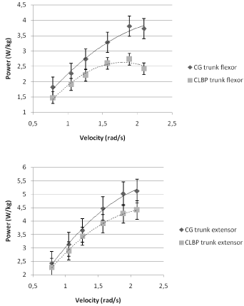
Considering all subjects, the torque- and power-velocity relationships exhibited a significant linear shape (r = 0.63-1; p < 0.05) and a second-order function (r = 0.86-1; p < 0.05) whatever the considered movement (i.e., flexion and extension), respectively. The torque and power-relationships obtained from mean values for both CG and CLBP groups and for each velocity are presented in Figure 2 and 3, respectively.

    Figure 2:  Torque-velocity relationships obtained for control group (CG) and
low back pain group (CLBP) for trunk flexion and extension movements.

    Figure 3:  Power-velocity relationships obtained for control group (CG) and
low back pain group (CLBP) for trunk flexion and extension movements.
Values obtained for T0 in CG during flexion (2.7 ± 0.5 Nm/kg) and extension (3.6 ± 0.6 Nm/kg) were significantly higher (p < 0.05) than those obtained in CLBP (2.0 ± 0.6 Nm/kg and 3.0 ± 1.1 Nm/kg for flexion and extension, respectively). In the same way, Pmax obtained in CG was significantly higher (p < 0.05) during both flexion (4.8 ± 1.5 W/kg) and extension (7.1 ± 2.5 W/kg) than Pmax obtained in CLBP (3.0 ± 1.1 W/kg during flexion and 4.7 ± 1.9 W/kg during extension). Thus, whatever the considered group, T0,e and Pmax,e were significantly higher than those measured during trunk flexion (p < 0.05).
Finally, Flexor/Extensor ratio showed no statistical difference between the two groups (0.77 ± 0.22 vs. 0.70 ± 0.16 for CG and CLBP, respectively), whatever the considered velocity.
Hip measurements
Values obtained for right and left sides for T0 and Pmax during hip flexions and extensions for both groups are presented in Table 1.
    Hip


    CG
    CLBP


    Flex
    Ext
    Flex R
    Ext R
    Flex L
    Ext L


    T0 (Nm.kg-1)
    1.6
    2.6
    0.9
    1.4
    0.9
    1.3


    (0.4)
    (0.6)
    (0.3)‡
    (0.5)‡
    (0.3)‡
    (0.4)‡


    Pmax (W.kg-1)
    4.5
    9.1
    2.0
    2.7
    1.6
    1.8*


    (1.4)
    (6.5)
    (1.8)‡
    (1.7)‡
    (1.1)‡
    (0.8)‡


    *p<0.05,    significantly different in a paired group.
      ‡:p<0.05, significant difference between the two    groups within the considered movement.
Table 1:  Mechanical parameters extrapolated from torque- and power-velocity
relationships: maximal torque (T0) and power (Pmax) for trunk and right (R) and
left (L) hip flexions (Flex) and extensions (Ext) in chronic low back pain patients
(CLBP) and control group (CG). Values are Mean (SD).
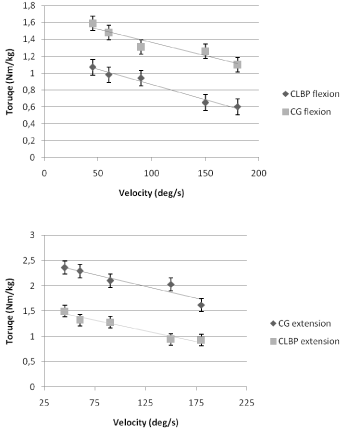
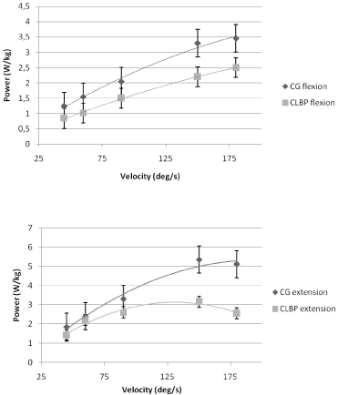
The torque- and power-velocity relationships obtained, for both CG and CLBP, are presented in Figure 4 and 5, respectively. Considering all subjects, the torque-velocity relationships exhibited a significant linear shape whatever the considered movement (flexion: r = 0.8 - 1; p <0.05; extension: r = 0.78 - 1; p<0.05). The power-velocity relationships were then significantly described by a second-order function during both trunk flexion and extension (r = 0.95 - 1; p <0.05 and r = 0.94 - 1; p <0.05, respectively).

    Figure 4:  Torque-velocity relationships obtained for control group (CG) and
low back pain group (CLBP) during hip flexion and extension movements for
the right side.

    Figure 5:  Power-velocity relationships obtained for control group (CG) and
low back pain group (CLBP) during hip flexion and extension movement for
right side. Results obtained for left side are identical.
Tpeak obtained for each preset velocity (i.e., 45, 60, 90, 150 and 180 deg.s-1) for CG was significantly higher (p <0.001) than that measured for CLBP for both flexion and extension movements. Consequently, T0 obtained for CG was significantly higher (p<0.05) than the one measured for CLBP (difference obtained between groups during flexion: 24% for right side and 29% for left side; difference obtained between groups during extension: 42% for right side and 45% for left side). This significant difference was also obtained for Pmax with higher values obtained for CG compared to CLBP (difference obtained between groups during flexion: 43% for right side and 62% for left side; difference obtained between groups during extension: 82% for dominant side and 74% for left side). Considering all subjects, results obtained for both hip flexion and extension showed no difference between right and left sides except for Pmax in CLBP during extension.
Hip muscle ratios showed statistical difference between CG and CLBP for right (0.6 ± 0.2 vs. 0.8 ± 0.2, respectively; p<0.05) and left sides (0.6 ± 0.2 vs 0.7 ± 0.1, respectively; p<0.05).
Finally, no relationships between lower limb and trunk strength or power were observed whatever the considered group.
Discussion
The purpose of the present study was to analyze hip muscle strength of CLBP patients, assuming that hip strength and/or power of CLBP should be lower than those measured in a healthy control group, presenting the same anthropometrical characteristics. It was also hypothesized that this muscle weakness would be linked to trunk strength muscle deficit in CLBP. Comparison between groups (i.e., CG and CLBP) was based on mechanical parameters extrapolated from torque-and power-velocity relationships established during isokinetic hip and trunk flexions and extensions. Results showed that relationships obtained from trunk and hip muscles were in accordance with those previously obtained in several studies focusing on trunk (Ripamonti et al. (16)) or lower limbs [22-24]. Our study showed that low back pain has no influence on torque- and powervelocity relationships for hip muscles.
Regarding trunk movements, results obtained in the present study were in accordance with those reported in previous studies comparing healthy subjects and CLBP. Indeed, strength and power produced by healthy subjects were significantly higher than those obtained from CLBP patients [11,25-27]. These significant differences were also observed from torque- and power-velocity relationships analysis. Results showed that i) extrapolated T0e was significantly higher than T0f, whatever the considered group, and that ii) T0 and Pmax obtained for CG were significantly higher than those determined for the CLBP (p<0.05) during both trunk flexion and extension movements with a greater impairment for trunk extensors relatively to trunk flexors. These expected results are in accordance with previous studies [1,28,29], and are explained by the CLBP deconditioning, defined by a loss of function of the trunk muscles [3] with specific deficits of lumbo-pelvic extensors and decreased resistance to fatigue [6]. Lumbar syndrome probably caused the further loss of lumbar muscle strength and may explain the imbalance of trunk musculature and the inability for CLBP to develop power. Finally, these expected results raise the question of the impact of the low back pain on the entire body, and particularly, on muscles of the lower limbs also connected to the trunk.
In the present study, torque and power measurements of CLBP showed significant lower values than those obtained in CG. This is probably a consequence of the deconditioning syndrome conventionally referred in CLBP [3,8,13]. Since trunk muscles are obviously not the only ones affected by this deconditioning [30], decline of daily activity in CLBP increased the wasting syndrome that can be found in trunk and in lower limbs [10], and probably cause the decrease in strength and power at these levels [1,10,13]. In the same way, hip muscle weakness found in this study for CLBP can also be explained by muscle atrophy, which is supported by the lower values obtained for T0 in CLBP. Thus, physical changes in the back muscle composition [2,31] such as fatty involutions or muscle fiber atrophy [32], or the alteration of muscle contractile properties found in CLBP for trunk muscles [3,33] could be extended to hip muscle and explain results obtained for T0 and Pmax during hip evaluation.
Since, to the authors’ knowledge, hip muscles have never been studied in CLBP patients, it should be noted that results obtained for healthy subjects in both hip flexion and extension, were in accordance with those obtained in previous studies [34-37], supporting the protocol used in the present study, and its possible application to CLBP patients. Moreover, results obtained for CG showed no difference between right and left sides, whatever the considered mechanical parameter, in agreement with a previous study [34], whereas a statistical difference between right and left sides was detected for Pmax during hip extension measurement in CLBP. The higher values obtained for the right extensor hip muscles probably induce muscular compensation and/or imbalance during daily activities such as walking, and generate abnormal movement and pain. Muscular compensation can also be due to calcaneal spurs and tarsal retinaculum syndrome or gonarthrosis. This can also be deducted from the hip muscle ratios. Indeed, ratios obtained for CLBP are significantly higher (around 0.8) than those obtained for CG (around 0.6, in agreement with previous study [37]. This ratio value indicates, as for trunk muscles, a larger deficit of extensor muscles, sustaining the fact that CLBP are not able to mobilize their entire extensor muscular chain in the same way as healthy subjects. Scholtes et al. [9] showed that healthy subjects were able to demonstrate a greater maximal lumbopelvic motion modification after instructions than CLBP. This could also be linked to kinesiophobia [33] which may be responsible for inhibitions and for a decrease in strength and power that can contribute to immerse patients in the spiral of deconditioning [2]. It seems important to offer a rehabilitation program including strength and power for hip muscles because strength and power muscle weakness highlight in the present study remain constant after the rehabilitation. Strength training proposed during the multidisciplinary program could, for example, include some hip strength session, to bring some changes in the force reinforcement. This can be done using isokinetic dynamometer, to take care on the subject abilities and security. Another way can be by including bath therapy, since working on water allows relaxation and easier work.
Finally, no relationship was observed between trunk and hip muscles imbalance. This can be due to the subject’s position imposed by the isokinetic device during the hip flexion and extension assessments. Indeed, subjects were asked to perform these movements lied on the back. This was probably one limitation of the present study, since these positions are far from these muscles physiological functioning conditions. Moreover, it would be more interesting to investigate the possible link between trunk and hip impairment during everyday life activities, such as walking or lifting tasks. This can be done by studying muscular activity and/or using video analysis to determine whether a segmental and/or muscular strategy can help to differentiate CLBP subjects from healthy ones. This should also allow investigating whether there is an effective physiological modification at hip muscle level, as it is clearly observed in trunk muscles. Nevertheless, results of the present study support the point that a specific attention should be brought on hip muscles during low back pain rehabilitation, as it was previously observed that rehabilitation program based on the complete body muscular program and training improved the well-being of CLBP patients [8,13]. Another potential limitation of the current study is that we do not take into account low back pain history. It should be interesting to determine if there is a relationship between the length of back pain and reduction in strength. Further studies are necessary to study the impact of pain history on reduction in strength.
Conclusion
The present study revealed a hip flexor and extensor muscles weakness for CLBP in addition to the classical trunk imbalance. Despite no relationship was observed between trunk and hip muscular imbalance, the present study enlightened new perspectives in the CLBP support. It should be interesting to evaluate hip muscles systematically at the beginning of the rehabilitation so as to propose the most suitable program for patients. It should also be interesting to introduce specific torque and power exercises for these muscles so as to limit trunk muscular compensations induced by muscle weakness at hip level that could occur in low back pain.
References
- Ripamonti M, Colin D, and Rahmani A. Maximal power of trunk flexor and extensor muscles as a quantitative factor of low back pain. Isokinetic and Exercise Sci. 2011; 19: 83-89.
- Poiraudeau S, Rannou F, Revel M. Low back pain: disability and evaluation methods, socio-economic impact. EMC-Rhumatologie Orthopédie. 2004; 1: 320-327
- Mayer TG, Smith SS, Keeley J, Mooney V. Quantification of lumbar function. Part 2: Sagittal plane trunk strength in chronic low-back pain patients. Spine (Phila Pa 1976). 1985; 10: 765-772.
- Poiraudeau S, Rannou F, Revel M. Functional restoration programs for low back pain: a systematic review. Ann Readapt Med Phys. 2007; 50: 425-429, 419-24.
- Genty M, Schidt D. Utilisation de l’isocinétisme dans les programmes de rééducation du rachis, modalités pratiques, protocoles proposés. Isocinétisme et rachis. Ed Masson. 2001.
- Vanvelcenaher J, Raevel D, O’Niel G, Voisin P, Struck P, Weissland T, et al. Programme de restauration fonctionnelle du rachis dans les lombalgies chroniques. Encyclopédie Médico-Chirurgicale, kinésithérapie Médecine Physique et Réadaptation. 1992; 26-294-8-10; 13.
- Nachemson A, Lindh M. Measurement of abdominal and back muscle strength with and without low back pain. Scand J Rehabil Med. 1969; 1: 60-63.
- Poiraudeau S, Lefevre MM, Fayad F, Rannou F, Revel M. Low back pain. EMC-Rhumatologie Orthopédie 1. 2004; 295-319.
- Scholtes SA, Gombatto SP, Van Dillen LR. Differences in lumbopelvic motion between people with and people without low back pain during two lower limb movement tests. Clin Biomech (Bristol, Avon). 2009; 24: 7-12.
- Lemaire A, Ripamonti M, Ritz M, Rahmani A. Influence of lower limbs strength on trunk flexion and extension in chronic low back pain patients. Comput Methods Biomech Biomed Engin. 2012; 15 Suppl 1: 206-207.
- Yahia A, Jribi S, Ghroubi S, Elleuch M, Baklouti M, Elleuch MH. Évaluation posturale et des forces musculaires du tronc et des membres inférieurs chez le lombalgique chronique. Revue du rhumatisme. 2011; 78: 166-172.
- Vitte E, Chevallier JM, Barnaud A. Nouvelle anatomie humaine. Atlas médical pratique. Nomenclature international, française classique et anglo-saxonne. Ed Vuibert - Pippa. 2007.
- Poiraudeau S, Nys A, Revel M. Évaluation analytique des moyens thérapeutiques dans la lombalgie: prise en charge physique et fonctionnelle. Rev Rhum [Ed Fr] 2001; 68: 154-159.
- Marieb EN. Anatomie et physiologie humaine. De Boeck Université. Version française de Human Anatomy and Physiology 4ème ed. 1998; 10: 342-354.
- Bernard JC, Dusquenoy. Classification of low back pain. Rev Rhum. 2001; 68: 145-149.
- Ripamonti M, Colin D, Rahmani A. Torque-velocity and power-velocity relationships during isokinetic trunk flexion and extension. Clinical Biomechanics. 2008; 23: 520-526.
- Smith SS, Mayer TG, Gatchel RJ, Becker TJ. Quantification of lumbar function. Part 1: Isometric and multispeed isokinetic trunk strength measures in sagittal and axial planes in normal subjects. Spine (Phila Pa 1976). 1985; 10: 757-764.
- Baltzopoulos V, Brodie DA. Isokinetic dynamometry. Applications and limitations. Sports Med. 1989; 8: 101-116.
- Parker MG, Ruhling RO, Bolen TA, Edge R, Edwards SW. Aerobic training and the force-velocity relationship of the human quadriceps femoris muscle. J Sports Med Phys Fitness. 1983; 23: 136-147.
- Kellis E, Baltzopoulos V. Resistive eccentric exercise: effects of visual feedback on maximum moment of knee extensors and flexors. J Orthop Sports Phys Ther. 1996; 23: 120-124.
- Delitto A. Isokinetic dynamometry. Muscle Nerve. 1990; 13 Suppl: S53-57.
- Yamauchi J, Mishima C, Nakayama S, Ishii N. Force-velocity, force-power relationships of bilateral and unilateral leg multi-joint movements in young and elderly women. J Biomech. 2009; 42: 2151-2157.
- Thorstensson A, Arvidson A. Trunk muscle strength and low back pain. Scand J Rehabil Med. 1982; 14: 69-75.
- Perrine JJ, Edgerton VR. Muscle force-velocity and power-velocity relationships under isokinetic loading. Med Sci Sports. 1978; 10: 159-166.
- Yahia A, Ghroubi S, Kharrat O, Jribi S, Elleuch M, Elleuch MH. A study of isokinetic trunk and knee muscle strength in patients with chronic sciatica. Ann Phys Rehabil Med. 2010; 53: 239-244, 244-9.
- Roques F, Felez A, Gleizes S, Van Des Bossche T, Boissezon X, Chatain M. Isokinetic assessement of the muscles of the trunk in chronic low back pain patients. Isokinetic exerc Sci. 2005; 13: 51.
- Thorstensson A, Arvidson A. Trunk muscle strength and low back pain. Scand J Rehabil Med. 1982; 14: 69-75.
- Crossman K, Mahon M, Watson P, Oldham J, Cooper R. Chronic Low Back Pain-Associated Paraspinal Muscle Dysfunction is not the Result of a Constitutionally Determined "Adverse" Fiber-type Composition Spine. 2004; 29; 628-634.
- Elfving B, Dedering A, Németh G. Lumbar muscle fatigue and recovery in patients with long-term low-back trouble--electromyography and health-related factors. Clin Biomech (Bristol, Avon). 2003; 18: 619-630.
- Netter FH. Atlas d'Anatomie Humaine. Ed Maloine.2002.
- Revel M. La rééducation dans la lombalgie commune: mise au point. Revue du rhumatisme. 1995; 62: 37-47.
- Bibré P, Voisin P and Vanvelcenaher J. Ischio-jambiers et lombalgies chroniques. Annales de Kinésithérapie.1997; 24: 328-334.
- Linton SJ, Melin L, Götestam KG. Behavioral analysis of chronic pain and its management. Prog Behav Modif. 1984; 18: 1-42.
- Costa RA, Oliveira LM, Watanabe SH, Jones A, Natour J. Isokinetic assessment of the hip muscles in patients with osteoarthritis of the knee. Clinics (Sao Paulo). 2010; 65: 1253-1259.
- Julia M, Dupeyron A, Laffont I, Parisaux JM, Lemoine F, Bousquet PJ, et al. Reproducibility of isokinetic peak torque assessments of the hip flexor and extensor muscles. Ann Phys Rehabil Med. 2010; 53: 293-305.
- Dugailly PM. Isokinetic assessment of hip muscle concentric strength in normal subjects: a reproducibility study. Isokinetics and Exercise Science. 2005; 5; 13: 129-137.
- Arokoski MH, Arokoski JP, Haara M, Kankaanpää M, Vesterinen M, Niemitukia LH, et al. Hip muscle strength and muscle cross sectional area in men with and without hip osteoarthritis. J Rheumatol. 2002; 29: 2185-2195.