Special Article – Physical Rehabilitation
Phys Med Rehabil Int. 2016; 3(2): 1081.
Physical Therapy in Caudal Regression Syndrome: Case Report
Mendelevich A* and Peralta FG
Department of Rehabilitation, Instituto de Rehabilitación Psicofísica, Buenos Aires, Argentina
*Corresponding author: Mendelevich A, Department of Rehabilitation, Instituto de Rehabilitación Psicofísica, Sanchez de Bustamante 2229 (1425), Ciudad Autónoma de Buenos Aires, Argentina
Received: April 03, 2016; Accepted: May 09, 2016; Published: May 12, 2016
Abstract
Case Presentation: F.V. 5 years of age diagnosed with caudal regression syndrome, confirmed at 2 days of life through a computed axial tomography scan of the spine. At 3 months of age she was admitted to Instituto de Rehabilitación Psicofísica and she began and interdisciplinary treatment. At 5 months of age she began physiotherapy focused on the acquisition of maturational patterns in a timely manner. At 4 years of age, after orthopaedic surgeries, she achieved assisted gait with reciprocal orthoses and walker. Currently, the goal is focused on achieving a limited community gait with equipment.
Discussion: The physiotherapy is still based on empirical principles due to the limited evidence available. Literature consulted is case reports, which most of them do not describe the evolution of those who survive. Functional achievements were due to the early beginning and continuity of the treatment, prompt surgical intervention and strict compliance with post-surgical protocol, within a continent social environment.
Keywords: Physical therapy; Caudal regression syndrome; Rehabilitation; Gait
Case Presentation
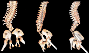
Our female patient was born at 37 weeks gestational age by elective caesarean delivery on November, 2009. During pregnancy, the mother reported not having complications. The girl was born with an Apgar 9/10 and weighed 1890g. In the newborn period she required phototherapy for 48 hours and was diagnosed with a displaced fracture of the left femur of unknown aetiology, which was reduced with plaster. 2 days after birth, the diagnosis of caudal regression syndrome was established, after a CT scan of the spine was performed. The CT scan revealed the absence of the sacral vertebrae and the last two lumbar vertebrae, and partially showed the structure of the three first lumbar vertebrae with alteration and dimorphism (Figure 1). It was also evident at the level of T11 a mayor morphological alteration of the vertebral body with displacement of T11 and T12 and with almost total obliteration of the medullary canal. On the same day, a karyotype was performed, showing normal result and, on subsequent days, neurological consultations were performed which showed no distinctive features, and cardio logical consultations showed patent foramen ovale and mild valvular pulmonary stenosis. It wasn’t reported any family history.

Figure 1: 3D-CT Reconstruction.
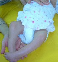
In February 2010, when the baby was 3 months old, inter consultation were made with the Instituto de Rehabilitación Psicofísica (I.Re.P). Until then, an early stimulation treatment was being conducted at another institution. Hip flexion at 45°, limitation of knee extension of more than 70° and equinovarus-supinated feet were observed during the evaluation (Figure 2). She was referred for inter consultation with physiotherapy, occupational therapy, speech therapy and psychology.

Figure 2: Baseline evaluation.
She began physiotherapy at the age of 5 months. During the initial evaluation, major retractions detailed by the physiatry department, together with increased anteroposterior and anterolateral diameters of the trunk. A motor and cognitive developmental impairment was also observed. She showed hyper responsiveness to external, tactile and auditory stimuli. It was decided to start treatment addressing soft tissue retraction, which limited the acquisition in a timely manner of maturational patterns and her relationship with the environment. Myofascial release techniques for hip, knees and feet flexors were performed. To reinforce this, progressive back plaster splints were applied, indicating night use. However, its use had to be suspended because they began to cause injury and discomfort. A soft derotation strap was also indicated to align the lower limbs that had excessive external rotation. General skin desensitization techniques were also applied. Rolling, tolerance for ventral decubitus position, and incorporation into sitting were stimulated in a timely manner achieving them all with great difficulty, and, in some cases, assistance was necessary due to lower limbs retraction.
She started to sitting with back support at 15 months old, during 2011. In this stage the use of a corset was indicated to achieve greater thoracolumbar stability and give the child the possibility to release her upper limbs. At the end of this year, improvement in cognitive resources was evident, noticing her more communicative and interested in toys.
During 2012, we continued with all previously detailed work, making the acquisition of quadruped static position. At this moment, it was notified that the girl had reached a therapeutic plateau, so the possibility of surgery of lower limb alignment and another of spine stabilization was raised.
Another important milestone in the same year was the prescription of a wheelchair, which gave her some movement independence, as she was able to propel her wheelchair.
On September 2, 2013, at 3 years and 10 months of age, deflection surgery of both knees and feet alignment surgery were performed at Hospital de Pediatría José Pedro Garrahan.
In the immediate post-operative preparation of a reciprocating orthosis, short AFO splints, knee stabilizers and a walker were indicated. During the 3 months after surgery the girl wore hip spica casts including feet, to maintain the alignment successfully achieved in surgery and to allow early standing. These casts were only removed to prepare the equipment, and applied again until the patient received such equipment. During this period the patient attended the Servicio de Kinesiologia of I.Re.P, where bipedalism was held in supine stander and in parallel bars to encourage the use of the upper limbs for support.
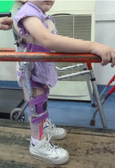
Currently, she attends kindergarten and continues with a weekly physiotherapy, working on stability in standing and sitting postures and training assisted walk with reciprocating orthosis and walker, with the aim of moving from a current home gait to a community gait (Figure 3).

Figure 3: Standing.
From having achieved bipedalism, improvement in cognitive resources and her language was observed, which in turn allowed a breakthrough in motor function. She doesn’t achieve yet bladder or bowel control.
As next goal, surgery is being planned for spine stabilization.
Discussion
Caudal Regression Syndrome was first described in 1852 by Geoffroy Saint-Hilaire and Hohl. In 1910, Joshy and Yadav described it as the total absence of the lumbosacral spine. However, only in 1964 Duhamel introduced the term making reference to malformations of the lower end of the spine, characterized by the absence of caudal vertebrae. Most of these malformations are found at sacral level, consequently also known as sacral agenesis. At the same time, there may be severe neurological, orthopaedic, gastrointestinal and neurological disorders leading to death. In literature, sacral agenesis or caudal regression syndrome, are often used as synonyms for a set of disorders with a wide range of variants ranging from asymptomatic coccygeal agenesis to sirenomelia [1,2].
The physical therapy approach for this pathology is still based on empirical principles due to the limited evidence available.
Works consulted are mainly case reports and contains information of a medical nature, where most of these descriptions date from the first moments of life, without describing in details the evolution of those few who survive.
Russell et al. [3] described bilateral lower limb amputation, with corresponding equipment to achieve the standing position as a treatment for this syndrome. We must consider that this type of intervention was performed in children aged 8 to 10 years of age, who had not received timely rehabilitation treatment.
In our view, functional achievements attained by this girl were due to several factors. An important one to consider was the early beginning and continuity of the treatment. She started at 5 months of age and attended 2 times per week without interruption until today. Probably, the fact of having started so early, avoided such a bloody intervention, as described by Russell et al3. Another relevant factor was the timely indication for surgery and strict compliance with the interdisciplinary post-surgical protocol for maintaining the benefits obtained with surgery.
It is worth stressing that the girl belongs to a continent social environment, with a mother committed to the treatment, who follows all the instructions given. It is also important to note that the girl has social security, which made it easier to get the equipment and to perform necessary examinations promptly. This shows the importance of starting treatment at an early stage, raising goals step by step, with an interdisciplinary team in constant communication and developing within an adequate family and social context.
Finally, it would be convenient conducting further studies on the rehabilitation of these kind of patients, to subsequently make therapeutic decisions on more scientific basis.
References
- Ramirez Rodriguez N, Pabon Uego C, Mendoza Vargas A. Síndrome de Regresión Caudal: Presentación de un Caso. CIMEL. 2005; 10: 69-72.
- Redondo CS, Arévalo A, Garavito P, Cortés A, Ordoñez J, Álvarez A, et al. Secuencia de displasia caudal: estudio clínico y radiológico de un paciente. Iatreia. 2010; 23: S-53.
- Russell HE, Aitken GT. Congenital absence of the sacrum and lumbar vertebrae with prosthetic management. The Journal of Bone & Joint Surgery. 1963; 45: 501-508.