Mini Review
Phys Med Rehabil Int. 2016; 3(4): 1092.
Stroke and Alzheimer’s Disease
Ikramuddin F*
Department of Physical Medicine and Rehabilitation, University of Minnesota, 420 Delaware St, Minneapolis MN 55455, USA
*Corresponding author: Farha Ikramuddin, Department of Physical Medicine and Rehabilitation, University of Minnesota, 420 Delaware St, Minneapolis MN 55455, USA
Received: June 20, 2016; Accepted: July 18, 2016; Published: July 20, 2016
Abstract
Stroke primarily affects the population over the age of 65 years, each year 795,000 people will experience new or recurrent stroke. It the most common cause of severe long-term disability (AHA 2010) in the US. Alzheimer’s disease (AD) is the most common form of dementia in the elderly. The prevalence of Alzheimer’s disease is known to increase as the population ages. Stroke and AD can occur together and result in deficits that appear to multiply. They share common vascular risk factors such as Hypertension, abdominal obesity and physical inactivity. It is difficult to differentiate the cognitive deficits that result from stroke and from Alzheimer’s disease individually and prognosticate function. The dysfunction and mismatch between the needs of the neural tissue to the mechanisms that regulate cerebral blood flow (CBF) have been postulated to result in cognitive deficits in both these disease processes. The neurovascular pathways are believed to be common to both the diseases and elucidate the potentiating effects that these two entities have on each other. This paper attempts to provide an understanding of the neurological pathways that may be common to the disease processes and pathology. Understanding of the mechanism of AD and cognitive deficits following stroke will help establish guideline and strategies in the prevention of these two diseases with similar and common neurological pathways.
Keywords: Stroke; Alzheimer’s disease; Hypertension; Cognitive deficits
Introduction
Stroke is the leading cause of severe long-term disability in United States and fifth leading cause of death according to the data from National Vital Statistics System, 2015. Although there has been a decline in the death rate from stroke in the last 50 years, it is not clear if these changes result from a decrease in the incidence or reduced case fatality rates [1-3].
While the age standardized rates of stroke mortality have declined in the world during the last two decades, the prevalence of stroke, overall global burden of stroke has increased with most remarkable increase in the population aged 75 and above. Additionally, with the aging population in US, based on the 2010 age distribution, the forecasted stroke incidence is expected to rise in the next 40 years by 2.25 times [4]. This increase in the incidence of stroke events is expected to be specifically in the white population as well as Hispanic population.
The epidemiology of AD is notable for AD being the 6th leading cause of death, and fifth leading cause in those over the age of 65 years (Alzheimer Association, 2015). AD affects 5.4 million Americans. The incidence of Alzheimer’s dementia dramatically increases after the age of 65 from 53 new cases per 1000 people aged 65-74 to 170 per 1000 people aged 75 to 84; 231 per 1000 people older than 85. It is the only one of the top ten diseases in US that does not have prevention, cure or clear guidelines to slow its progression. Based on an economic model created by an independent research firm, a report from the Alzheimer’s Association projects that Medicare spending on people with Alzheimer’s disease will more than quadruple in just over a generation to $589 billion annually in 2050.
It is clear that both stroke and AD affect similar patient populations within an age range. However, the effect of AD on a patient with premorbid stroke is not clears. Conversely, the studies that evaluate the effect of stroke in a patient with pre-existing AD are conflicting. Nevertheless, cognitive deficits are an important prognostic indicator to the ability of patients to be discharged home safely, particularly in this age range. The ability of a clinician to prognosticate the recovery of stroke while considering the cognition at baseline and eventual support needed to prevent institutionalization cannot be over stressed in this patient population.
Many studies have hypothesized the cause of AD, which remains unclear. The pathological characteristic of AD consisting of neurofibrillary tangles; tau protein deposits as well as acetylcholine diminution along with calcium dysfunction associated with reactive astrocytosis leading to inflammation may be enhanced in the presence of atherosclerotic disease. Roher et al have repeatedly found strong correlation between large vessel atherosclerotic disease and neurotic plagues [5].
In AD, mitochondrial dysfunction is an important factor in the formation of AD, through the Oxygen Reactive Species ROS. Increased and unregulated inflammatory free radicals accumulate in AD, which promote neuronal apoptosis at vulnerable regions such as the hippocampus and amygdala. In mild and moderate AD, there is diminution of choline acetyl transferase activity. There is in mild and moderate AD, diminution of choline acetyl transferase activity is also noted. Choline acetyl transferase is responsible for the synthesis of the neurotransmitter acetylcholine. This results in loss of acetylcholine, specifically in areas of the brain associated with memory and learning. However, the cholinergic dysfunction is not considered to be the cause of the illness, but rather a consequence. This mechanism of decreased cholinergic activity is the target of the currently approved treatments for AD.

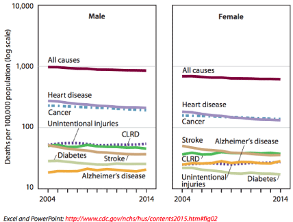
    Figure 1:  Age-adjusted death rates for selected causes of death for all ages,
by sex: United States, 2004-2014.
In addition to the dysfunction of the cholinergic system, increased loss of glutamatergic neurons is noted in AD. It is accompanied by disturbances in N-methyl-D-aspartate (NMDA) and α-amino-3- hydroxy-5-methyl-4-isoxazolepropionic acid receptor expression in the cerebral cortex and hippocampus. Increased depolarization of the postsynaptic membrane resulting from increased glutamate concentration and reduction in physiological NMDA receptor mediated signals is noted in AD [6].
Pathogenesis in AD has not been clear, the tau proteins and neurofibrillary tangles are considered the hallmarks of AD; however the occurrence of these pathognomic features in imaging does not correlate to the severity of AD. Similarly, the diminution of the acetylcholine and down regulation of the NMDA receptors is thought to be the consequential features and not the cause of the cognitive deficits. The biomarkers being studied in AD, are discordant to the cognitive deficits following stroke, are defined as those deficits that occur within three months of the stroke. These deficits are related to certain critical locations such as those involving the prefrontal and subcortical areas that mediate executive functioning. Other infarcts resulting in cognitive deficits are those involving the angular gyrus, medial frontal lobe, and infero medial portion of the temporal lobes. The third subtype of strategically located infarctions causing dementia after stroke involves bilateral hippocampal or thalamic infarctions [7]. However these locations do not account for the worsening cognition seen in the stroke patient involving other areas of infarction (L Pantoni 2001). Similarly, a study showed strong correlation between hippocampal volume and risk of developing AD [8].
In a systematic review and meta-analysis review of studies between the years 1975 to 2013, the relationship of AD and stroke was reviewed [9]. It was found that patients with AD were at increased risk for intracranial hemorrhage. However, the study showed that the risk of AD was higher in patients following stroke. It is well recognized that the risk factors for both disorders are similar, namely hypertension, diabetes, and heart disease and thus the results can be confounded. The amalgam of these co morbidities is part of the metabolic syndrome. Altered levels of glucose have been found to be predictive of worse cognition following stroke. There is an inverse relationship between the duration of diabetes and poor cognitive outcome following stroke in this study [10].
Brain derived neurotrophic hormone has been noted to be involved in the both the stroke and AD, and may point to the common pathways in these diseases. The BDNH has been seen to be high in patients with acute phase of stroke.
There appears to be a mismatch in both the AD and stroke individuals between the metabolic demands of the neural tissue and the ability of the neurovascular mechanisms that assure that the energy requirements of the neural tissue are commensurate with blood flow. This mismatch is seen in both the AD and patients with stroke [11].
AD incidence is as escalated with obesity associated with nonfatty liver disease, insulin resistance, and early cellular senescence. Environmental factors such as high cholesterol or high fat diets have been associated with high LDL and low HDL levels in AD populations, pointing to pathways common to both the entities.
The connection between AD and stroke is possibly related to low adiponectin and high density lipoprotein HDL cholesterol levels found in patients with hypertension, obesity, diabetics, and AD. It is postulated that there is down regulation of AD genes Sirtuin1, over expression of amyloid precursor protein APP, and mitochondrial apoptosis. Life style changes which include low fat diet, exercise and reduction of stress have been shown to increase adipose tissue mitochondrial biogenesis possibly by up regulation of the Sirtuin1 genes [12].
In a retrospective study, analysis of cerebrospinal CSF biomarkers, t-tau, p-tau and Aβ42 were assessed in patients with AD, stroke and vascular dementia. Similarities were reported in CSF profiles of those with AD and after stroke T tau levels were similar in stroke and AD, while phosphor tau levels were highest in AD, followed by MCI then stroke. Based on biomarkers alone, there was no difference noted between AD and stroke between the Aβ42 levels [13].
Interestingly, serum tau proteins have been detected in 48% of ischemic stroke patients, and those with detectable tau proteins were noted to develop more severe neurological deficits. Thus the presence of tau proteins points to a common pathway for the two diseases [14].
Conclusion
AD and stroke are important diseases that effect the US population beyond the ages 65. The seemingly disparate AD and stroke have a remarkable commonality in their underpinnings. Over- nutrition, and lack of exercise leading to ROS formation drive the pathologic finding of neurofibrillary tangles and tau proteins.
Metabolic syndrome has influence on the incidence of stroke and AD. There is increasing evidence that there are common pathways between the two diseases in the form of disequilibrium between the stroke and vascular damage and plague formation are strongly related to metabolic syndrome, an amalgam of Hypertension, Diabetes, and Hyperlipidemia, where the principle drive is adipose tissue dysfunction strongly linked to mitochondrial dysfunction, which in turn leads to ROS. Early identification and aggressive treatment of these risk factors may prevent stroke and delay AD.
References
- Roger VL, Go AS, Lloyd-Jones DM, Adams RJ, Berry JD, Brown TM. American Heart Association Statistics Committee and Stroke Statistics Subcommittee. Heart disease and stroke statistics--2011 update: a report from the American Heart Association. Circulation. 2011; 123: e18–e209.
- Hachinski V, Sposato LA, & Kapral M. Preventing both stroke and dementia. The Lancet. Neurology. 2016; 15: 659.
- Lee CDo, Folsom AR, & Blair SN. Physical activity and stroke risk: a meta-analysis. Stroke; a Journal of Cerebral Circulation. 2003; 34: 2475–2481.
- Howard G & Goff DC. Population shifts and the future of stroke: Forecasts of the future burden of stroke. Annals of the New York Academy of Sciences. 2012; 1268: 14–20.
- Roher AE, Esh C, Kokjohn T, Sue L, & Beach T. Atherosclerosis and AD: analysis of data from the US National Alzheimer’s Coordinating Center. Neurology. 2005; 65: 974.
- Wakabayashi K, Narisawa-Saito M, Iwakura Y, Arai T, Ikeda K, Takahashi H, et al. Phenotypic down-regulation of glutamate receptor subunit GluR1 in Alzheimer’s disease. Neurobiology of Aging. 1999; 20: 287–295.
- Kalaria RN, Akinyemi R, & Ihara M. Stroke injury, cognitive impairment and vascular dementia. Biochimica et Biophysica Acta. 2016; 1862: 915–925.
- Weinstein G, Beiser AS, Decarli C, Au R, Wolf PA, & Seshadri S. Brain imaging and cognitive predictors of stroke and Alzheimer disease in the Framingham Heart Study. Stroke; a Journal of Cerebral Circulation. 2013; 44: 2787–2794.
- Zhou J, Yu J-T, Wang H-F, Meng X-F, Tan C-C, Wang J, et al. Association between stroke and Alzheimer’s disease: systematic review and meta-analysis. Journal of Alzheimer’s Disease : JAD. 2015; 43: 479–489.
- Zietemann V, Wollenweber FA, Bayer-Karpinska A, Biessels GJ, & Dichgans M. Peripheral glucose levels and cognitive outcome after ischemic stroke--Results from the Munich Stroke Cohort. European Stroke Journal. 2016; 1: 51–60.
- Girouard H, & Iadecola C. Regulation of the Cerebral Circulation Neurovascular coupling in the normal brain and in hypertension, stroke, and Alzheimer disease. 2006; 10021: 328–335.
- Martins I. The Global Obesity Epidemic is Related to Stroke, Dementia and Alzheimer’s Disease. ECU Publications Post 2013. 2014.
- Kaerst L, Kuhlmann A, Wedekind D, Stoeck K, Lange P, & Zerr I. Cerebrospinal fluid biomarkers in Alzheimer ’ s disease , vascular dementia and ischemic stroke patients : a critical analysis. J Neurol. 2013; 260: 2722–2727.
- Bielewicz J, Kurzepa J, Czekajska-Chehab E, Stelmasiak Z, & Bartosik-Psujek H. Does serum Tau protein predict the outcome of patients with ischemic stroke? Journal of Molecular Neuroscience : MN. 2011; 43: 241–245.