Research Article
Phys Med Rehabil Int. 2018;5(2): 1142.
Effects of Different Low-Density Insoles on Foot Activation Time at Different Walking Speeds in Young Females
Güner S*, Alsancak S, Altinkaynak H, Güven E,özgün K and Aytekin G
1Department of Prosthetics and Orthotics, Vocational School of Health Services, Ankara University, Ankara, Turkey
*Corresponding author: Senem Güner, Prosthetics and Orthotics Department, Vocational School of Health, Ankara University, Fatih Street 197/A, 06290 Kecioren, Ankara, Turkey
Received: February 26, 2018; Accepted: March 22, 2018; Published: March 29, 2018
Abstract
Aim: Shoes for walking and other locomotor activities provide the only interaction between the body and the ground; therefore, shoes are typically constructed to provide stability and comfort to the user. However, studies investigating the optimal shoe insole material, particularly for flat shoes, based on different walking speeds are limited. Therefore, the purpose of this study was to evaluate the foot activation time using different low-density insole material and walking speed combinations.
Methods: Twelve healthy females participated in this study. The activation time measurements were obtained using 8 WalkinSense sensors in each trial and exported from the proprietary software (WalkinSense version 0.96, Tomorrow Options Microelectronics, S.A). Each subject underwent the same testing procedure on a treadmill (i.e., slow, normal and fast walking speeds) first without the insole and then in shoes with Insole I (6-mm flat noraLunatec EP insole), Insole II (6-mm flat nora Astro form 8 insole) and Insole III (6-mm flat nora Aero sorb M insole).
Results: The percentage of activation time was significantly higher under the fast walking condition than under the slow walking condition in the hallux with the shoe only, Insole I, Insole II and Insole III (p<0.05). The activation time of the lateral heel was significantly higher under the slow walking condition than under the fast walking condition with the shoe only, Insole I and Insole II. The activation time of the medial heel was significantly higher under the slow walking condition than under the fast walking condition with the shoe only, Insole I, Insole II and Insole III (p<0.05). The percentage of the activation time of the lateral midfoot was significantly lower under the shoe only condition than under the Insole II and Insole III conditions during slow walking.
Conclusion: Lower-density insoles may positively affect the loading activation time in the lateral midfoot. Custom-made insole surfaces produced with low-density material may be suitable for fast walking in people with flat shoes.
Keywords: Flat shoes; Insole material; Foot activation time
Introduction
Plantar loading evaluations are frequently used to assess the effectiveness of an insole in reducing the risk factors for soft-tissue mechanical trauma associated with weight-bearing activities of daily living. When the plantar pressure is reduced, the force is distributed over a larger area [1]. The materials used to fabricate insoles have been shown to improve the plantar pressure, reduce shock, and enhance comfort [2,3]. Pratt et al. quasi-quantitatively evaluated the shockabsorbing characteristics of Plastazote, Spenco, Sorbothane, Poron and Viscolas. Poron was rated the best insole material for shock absorption [4,5]. Leber and Evanski investigated 26 participants who complained of forefoot pain upon bearing weight, and all participants exhibited areas of increased pressure under one or more metatarsal heads when tested. While all materials tested reduced the overall plantar pressure compared to that under the barefoot condition, the authors ranked the materials as follows: PPT (an open cell, porous, firm foam material), Plastazote (foamed polyethylene material with a closed-cell structure) and Spenco (a neoprene sponge product with nitrogen-induced closed cells covered with a multi stretch nylon fabric on one side) were the most effective; Dynafoam (a polyvinyl chloride foam compound) was somewhat effective; and Ortho felt (a resilient fabric composed of a cotton-wool blend with a relatively low tensile strength) and latex foam (a cellular rubber material with an open-cell structure) were the least effective [6]. Gillespie and Dickey developed a filter bank procedure to determine the effectiveness of different foot orthotic materials. The materials were found to reduce the initial peak force, loading rate and frequency of transient impact during walking, and Plastazote was the most effective material for attenuating the high-frequency component of the initial ground reaction force during walking [7].
Ethylene-vinyl acetate (EVA) is a highly elastic copolymer with ethylene and vinyl acetate sintered to form a porous material similar to rubber but with excellent toughness. The porous elastomeric characteristic of EVA is much more flexible than that of low-density polyethylene, which is commonly used in shoe construction; due to its resistance, flexibility, and temperature toughness properties, EVA is among the most used copolymers in shoe midsole construction. The addition of EVA to elastomers could lead to the ideal softness and high resilience characteristics needed for a full-recovery capacity in the next foot step after a heel strike, while a less resilient (more viscous) material could attenuate more energy during the initial loading cycles, easily achieving compression flattening after some cycles [8,9].
Numerous studies have examined the plantar pressure during walking at different speeds, both barefoot and in shoes [10-15]. According to these studies, increasing the walking speed results in an increased plantar pressure in each of the foot regions examined [10,14]. The walking speed has been shown to linearly influence the loading patterns beneath the hallux and rear foot; however, the increases in the walking speed had a lower impact on the forefoot loading patterns [10,14-16]. Medial forefoot loading initially increases at slower walking speeds; however, the medial forefoot loading remains constant or decreases as individuals begin to walk faster, which is attributed to a decrease in the contact time as the walking speed increases [10]. While changes in the loading patterns have been observed in the hindfoot, hallux and forefoot, the forces beneath the medial and lateral midfoot were not significantly altered as the walking speed increased [14]. In addition to examining the influence of the walking speed, previous studies have examined the effect of different footwear on the plantar pressure patterns; the plantar pressure is significantly lower in participants walking in running shoes than in participants walking barefoot [14,17]. In contrast to studies examining plantar pressure distribution patterns during walking, very few studies have examined plantar pressure distribution patterns during running. Burnfield et al. examined the differences in plantar loading at different walking speeds [14] and found that as the walking speed increased, the plantar loading also increased.
Thus, the purpose of our study was to determine the effects of different insole material and walking speed combinations on the foot activation time under the hindfoot, midfoot, and forefoot plantar surface areas in individuals with normal feet. We hypothesized that a decrease in the insole density could increase the activation time distribution on the foot during fast walking.
Methods
Subjects
In total, 12 healthy female participants with an average age, weight, and height of 20.16, 54.7 kg and 159.7cm, respectively, without any known history of diseases or foot pathologies were recruited for this study. All subjects were undergraduate students at a local university. Ethical approval was obtained from University High School Committee. Written informed consent was obtained from the volunteers before the measurements were performed. The inclusion criteria included volunteers with no known diseases or foot deformities, who had the ability to walk on a treadmill independently without assistance or any walking aids. Participants with the following foot deformities were excluded: pes planus, pes cavus, plantar heel pain, metatarsalgia, hallux valgus/varus/limitus/rigidus and lesser toe deformities, such as hammer toes, clawed toes and mallet toes. The demographic data, including the ages, heights and weights of the subjects, were recorded. This study used flat shoes because these shoes are more popular and widely used than high heel shoes among university-aged women.
Activation time measurements
The activation time was calculated as a percentage of the walking cycle. The activation time measurements were performed using 8 WalkinSense sensors in each trial and exported from the proprietary software (WalkinSense version 0.96, Tomorrow Options Microelectronics, S.A.) [18]. The system consisted of a data acquisition and processing unit and eight individual sensors attached to the participants’ socks to measure the plantar pressure and activation time (Figure 1). The participants wore standard shoes and socks, which were fitted with the WalkinSense sensors and provided to the participants. The order of the trials without an insole and the three insoles (I, II and III) was counterbalanced in this study (Figure 2).

Figure 1: WalkinSense-system.

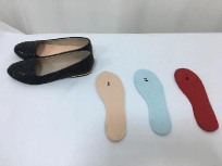
Figure 2: Flat- shoes and Insole I, Insole II, Insole III.
Insole material
This study consisted of the following 4 testing conditions involving participants walking on a treadmillShoes only: Shoes with a heel height of 0.8 mm and flat, thin, high-density polyurethane 0.4- mm outsoles.
- Shoes with Insole I: Shoes with a 6-mm flat noraLunatec EP insole made with an EVA material (Germany Medical Product, Approx. 22 Shore A, density approx. 0.20g/cm3). The material has a closed-cell structure and a high-restoration capability due to its particularly light weight. This material is highly resilient; has an excellent restoration capability, a low volume loss, and a smooth surface; is a closed-cell structure; and is durable, hygienic, washable, and thermoformable.
- Shoes with Insole II: Shoes with a 6-mm flat nora Astro form 8 insole made with a light cellular rubber exhibiting a truly unique combination of properties (Germany Medical Product, density 0.17g/cm3). This material is extremely soft; has an excellent recovery capability after compression, low compression capacity, and optimum shock absorbance; and is hygienic.
- Shoes with Insole III: Shoes with a 6-mm flat nora Aero sorb M insole made with a light cellular rubber exhibiting a unique combination of properties (Germany Medical Product, density approx. 0.16g/cm3). This material is extremely soft; exhibits both bedding and absorbent properties and a delayed recovery capability; and is shock-absorbing and hygienic.
- Roozbeh N, Kimberley L, Aoife H, Nachiappan C. The influence of slow recovery insole on plantar pressure and contact area during walking. Journal of Mechanics in Medicine and Biology. 2015; 15: 1540005.
- Mills K, Blanch P and Vicenzino B. Identifying clinically meaningful tools for measuring comfort perception of footwear. Med Sci Sports Exerc. 2010; 42: 1966-1971.
- Brodsky JW, Pollo FE, Cheleuitte D, et al. Physical properties, durability, and energy-dissipation function of dual density orthotic materials used in insole for diabetic patients. Foot Ankle Int. 2007; 28: 880-889.
- Pratt DJ. Long term comparison of some shock attentuating insoles. Prosthetics and Orthotics International. 1990; 14: 59–62.
- Pratt DJ, Rees PH, and Rodgers C. Technical note: assessment of some shock absorbing insoles. Prosthetics and Orthotics International. 1986; 10: 43–45.
- Leber C. and Evanski PM. A comparison of shoe insole materials in plantar pressure relief. Prosthetics and Orthotics International. 1986; 10: 135–138.
- Gillespie KA and Dickey JP. Determination of the effectiveness of materials in attenuating high frequency shock during gait using filterbank analysis. Clinical Biomechanics. 2003; 18: 50–59.
- Verdejo R, Mills NJ. Heel-shoe interactions and the durability of EVA foam running-shoe midsoles. Journal of Biomechanics. 2004; 37: 1379–1386.
- Wang L, Hong Y, Li JX. Durability of running shoes with ethylene vinylacetate or polyurethane midsoles. Journal of Sports Sciences. 2012; 30: 1787–1792.
- Segal A, Rohr E, Orendurff M, Shofer J, O’Brien M, Sangeorzan B. The effect of walking speed on peak plantar pressure. Foot Ankle Int. 2004; 25: 926–933.
- Grampp J, Wilson J, Kernozek T. The plantar loading variations to uphill and downhill gradients during treadmill walking. Foot Ankle Int. 2000; 21: 227– 231.
- Morag E, Cavanagh PR. Structural and functional predictors of regional peak pressures under the foot during walking. J Biomech. 1999; 32: 359–370.Morag E, Cavanagh PR. Structural and functional predictors of regional peak pressures under the foot during walking. J Biomech. 1999; 32: 359–370.
- Ledoux WR, Shofer JB, Ahroni JH. Biomechanical differences among pes cavus, neutrally aligned, and pes planus feet in subjects with diabetes. Foot Ankle Int. 2003; 24: 845–850.
- Burnfield JM, Few CD, Mohamed OS. The influence of walking speed and footwear on plantar pressures in older adults. Clin Biomech. 2004; 19: 78–84.
- Rosenbaum D, Hautmann S, Gold M, Claes L. Effects of walking speed on plantar pressure patterns and hindfoot angular motion. Gait Posture. 1994; 2: 191–197.
- Kernozek TW, LaMott EE, Dancisak MJ. Reliability of an in-shoe pressure monitoring system during Treadmill walking. Foot Ankle Int. 1996; 17: 204– 209.
- Perry J, Ulbrecht J, Derr J, Cavanaugh P. The use of running shoes to reduce plantar pressure in patients who have diabetes. JBJS. 1995; 77A: 1819–1828
- Healy A, Burgess-Walker P, Naemi R, Chockalingam N. Repeatability of WalkinSense in shoe pressure system: A preliminary study. The Foot. 2012; 22: 35-39.
- Cheung JT, Zhang M. Parametric design of pressure-relieving foot orthosis using statistics- based finite element method. Med. Eng. Phy. 2008; 30: 269- 277
- Healy AD, Dunning N, Chockalingam N. The effectiveness of footwear and other removable off-loading devices in the treatment of diabetic foot ulcers: a systematic review. Curr. Diabetes Rev. 2014; 10: 215-230
- Nicolopoulos CS, Black J, Anderson EG. Foot orthoses materials. Foot. 2000; 10: 1-3.
- Healy AD, Dunning N, Chockalingam N. Effect of insole material on lower limb kinematics and plantar pressures during treadmill walking. Prosthetics and Orthotics Int. 2011; 36: 53-62.
- McMillan A, Payne C. Effect of foot orthoses on lower extremity kinetics during running a systematics literature review. Journal of Foot and Ankle Research. 2008; 1: 13.
- TSung BYS, Zhang M, Mak AFT, Woung MWN. Effectiveness of insoles on plantar pressure redistribution. J Rehabil Res Dev. 2004; 41: 767-774.
- Gerych D, Tvrznik A, Prokesova E, Nemeckova Z, Jelen K. Analysis of peak pressure, maximal force and contact area changes during walking and running with conventional and shock-absorbing insoles in the combat boots of the czech army. Journal of Mechanics in Medicine and Biology. 2013; 13: 1350042.
- Curryer M, Leamire ED. Effectiveness of various materials in reducing plantar shear forces. A pilot study. J Am. Podiatr. Med. Assoc. 2000; 90: 346-353.
- Paton J, Jones RB, Stenhouse E, Bruce G. The physical characteristics of materials used in the manufacture of orthoses for patients with diabetes. Foot Ankle Int. 2007; 28: 1057-1063.
- Mills K, Blanch P and Vicenzino B. Influence of contouring and hardness of foot orthoses on ratings of perceived comfort. Med. Sci Sports Exerc. 2011; 43: 1507-1512.
- Lo Wai T, Wong D, Yick K, Ng S, et al. The biomechanical effects and perceived comfort of textile-fabricated insoles during straight line walking. ProsthetOrthot Int. 2017: 309364617696084.
- Lo WT, Yick KL, Ng SP,Yip J. New methods for evaluating physical and thermal comfort properties of orthotic materials used in insoles for patients with diabetes. JRRD. 2014; 51: 311-324.:
Testing procedure
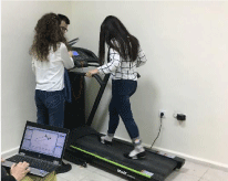
The subjects were guided to the treadmill (VOIT Company, Speed F1), and their resting heart rate (HR) and blood pressure (BP) were recorded. Throughout the testing procedure, the HR was monitored every 2min to gauge the subjects’ reaction to the testing protocol. The subjects were asked to straddle the treadmill belt and grasp the handrails as the treadmill started at a speed of. Each subject was asked to walk for five minutes at 0.6m/s on the treadmill. After the adaptation test, the subjects performed trials of slow walking (0.8m/s), normal walking (1m/s), and fast walking (1.8m/s) (Figure 3). Each speed was maintained for 45s, and the transition time between adjacent speeds was 15s. Each subject performed the same test procedure on the treadmill first without the insole and then with shoes containing insoles I, II and III.

Figure 3: Treadmill testing using WalkinSense-System.
Statistical analysis
All data are expressed as means (SD). The statistical analysis was performed using SPSS statistical analysis software (version 16). A one-factor repeated-measures analysis of variance (ANOVA) was performed to analyse each activation time measurement under the 4 conditions during three walking speeds. Bonferroni post hoc testing was performed for the multiple pair-wise comparisons following a significant difference in the ANOVA (p<0.05) among any of the 4 conditions.
Results
The mean age, weight, and height of the 12 healthy female participants were 20.16±1.8 years, 54.7±4.01kg and 159.7±4.6m, respectively (Table 1). The activation time percentages for sensors 1, 6, 7, and 8 are presented as the means ± standard deviations under the flat shoe only condition and the flat shoe with insoles I, II, and III at different walking speeds (Table 2).
    Participant characteristics (n = 12)
    Mean ± SD


    Age (years)
    20.16±1.8


    Weight (kg)
    54.7±4.01


    Height (cm)
    159.7±4.6
Table 1: Participant characteristics.
    Sensor number/
    1 (Hallux)
    6 (Midfoot laterally)
    7 (Heel medially)
    8 (Heel laterally)


    Walking type
    Mean ± SD
    Mean ± SD
    Mean ± SD
    Mean ± SD


    Slow walking


    Shoe only
    34,9± 18,9
    29,4± 22,6
    70,6 ±10,3
    66,8± 11


    Insole-I
    36± 18,7
    53,5± 18,4
    74, ±1 7,6
    72,6± 11


    Insole-II
    43,3± 26,7
    61,7± 14,8
    76,7 ±8,7
    75,3± 8,7


    Insole-III
    40,7± 24
    54,5 ±19,8
    74,4 ±9
    66± 15,6


    Normal walking


    Shoe only
    47± 17,5
    23,9± 20,7
    60,9± 9,9
    54,9 ±10,7


    Insole-I
    49,5± 12,3
    45± 15,9
    66,8 ±10,2
    63,4± 10,2


    Insole-II
    49,6± 15,6
    45,1± 19,5
    67,5± 8,2
    64,7± 6,9


    Insole-III
    47,8± 17,7
    41,5± 16,9
    66,6± 8,4
    60,6± 10,5


    Fast walking


    Shoe only
    64,2± 22,4
    31,3 ±22,3
    48,5± 8,4
    41,7± 11,5


    Insole-I
    65,4 ±22,4
    40,1± 12,9
    54,4± 9
    51,9± 5,6


    Insole-II
    74,6± 13,9
    40,8 ±10,7
    57,7± 7,9
    49,7 ±13,5


    Insole-III
    72± 8,5
    43,5 ±11,1
    58,4 ±7,1
    54,2± 10
Table 2: The mean and standard deviation of the activation percentege are 1 (hallux), 6 (mid foot laterally), 7 (heel medially) and 8 (heel laterally) according to walking
speed and insole type.
By comparing the three movement tasks (i.e., slow, normal and fast walking) under the 4 conditions, significant differences in most dependent variables were observed in various regions of the foot. The activation time percentage during fast walking was significantly higher than that during slow walking in the hallux (number 1) with the shoe only and insoles I, II and III (p<0.05) (Table 3). The lateral heel (number 8) activation time during slow walking was significantly higher than that during fast walking with the shoe only and insoles I and II. The medial heel (number 7) activation time during slow walking was significantly higher than that during fast walking with the shoe only and insoles I, II and III (p<0.05), as shown in Table 3. The lateral midfoot (number 6) activation time percentage under the shoe only condition was significantly lower than that under the insoles II and III conditions during slow walking. No significant differences were observed under the slow, normal and fast walking conditions among the shoe only, Insole I, Insole II and Insole III conditions in the metatarsal head 1–5 areas (numbers 2, 3, 4, and 5) (p>0.05).
Shoe only
Post hoc results
P value
Insole-I
(High density)
Post hoc results
P value
Insole-II
(Medium density)
Post hoc results
P value
Insole-III
(Low density)
Post hoc results
P value
Activation percentege (%) 1
SW SO< FW SO
0.01*
SW HD< FW HD
0.01*
SW MD< FW MD
.007*
SW LD<FW LD
.002*
Activation percentege (%) 8
SW SO > FW SO
.000**
SW HD > FW HD
.000**
S W MD> FW MD
.000**
No significant difference
NSD
Activation percentege (%) 7
S W SO > FW SO
.000**
SW HD > FW HD
.000*
SW MD> FW MD
.000**
S W LD > FW LD
.000**
SW SO< SW MD, SW SO<SW LD
.001*
No significant difference
NSD
SW MD> SW SO
0.01*
SW LD> SW SO
0.04*
S0: Shoe only; HD: Insole I; MD: Insole II; LD: Insole III; SW: Slow Walking; FW: Fast Walking; NSD: No Significant Difference.
*Significant difference (p<0.05) between condition;**Significant difference (p<0.001) between condition.
Table 3: The activation time percentages were 1 (hallux), 6 (mid foot laterally), 7 (heel medially), 8 (heel laterally) post hoc and p value values according to walking
speed and insole difference.
Discussion
The purpose of this study was to investigate the effects of different density insoles on the foot activation times at slow, normal and fast walking speeds. All 3 insole types used at different walking speed affected the activation time and loading current time on the foot. According to the results of this study, the activation time percentage in the lateral and medial heel decreases at a fast walking speed; however, fast walking increases the activation time percentage on the hallux. As the insole density decreased, the activation time percentage increased in the lateral midfoot.
Increasing the walking speed has been shown to increase the plantar pressure in each of the examined foot regions. The walking speed has been shown to linearly influence the loading patterns beneath the hallux and hindfoot. Segal et al. [10] showed that the medial forefoot loading increased at slower walking speeds and decreased at faster walking speeds, which was attributed to a decrease in the contact time as the walking speed increased. Bunfield et al. [14] showed that while changes were observed in the loading patterns in the hindfoot, hallux and forefoot, the forces beneath the medial and lateral midfoot were not significantly altered with increasing walking speeds. In this study, compared with the slow walking speeds, at fast walking speeds, the activation times were decreased, particularly in the hindfoot, while the activation times were increased in the hallux region of the foot. Using insoles, the activation time in the hindfoot, hallux, and midfoot regions increased at both fast and slow walking speeds. These findings may be explained by the increased support of the foot and the redistribution of pressure and activation time provided by the shock-absorbing insoles. Therapeutic footwear and/or foot orthoses are strongly dependent on the mechanical characteristics of the materials used to offer cushioning, and stiffness is among the most important characteristics [19-21]. Healy et al. showed that mediumdensity polyurethane could impact the loading characteristics of the material, indicating that medium-density polyurethane may be a suitable material for patients with a compromised ability to handle pressure [22]. According to this study, low-density insoles increase the activation time percentage in the lateral midfoot; thus, the foot loading increases in the lateral foot. Clinicians may consider this information when choosing a material that must be shock absorbent and have good pressure-reducing characteristics for patients who walk fast in their daily lives. McMillan et al. suggested that a reduction in the hindfoot inversion moment is the most consistent kinetic effect of foot orthotics during running [23].
Non-shock-absorbing insoles and shock-absorbing insoles are equally effective in preventing lower limb overuse injuries. Shockabsorbing insoles may be effective in preventing stress fractures and shin splints [24]. Gerych et al. applied two different insoles at a walking speed of 5km/h and running speeds of 8 and 12 km/h using the Pedar-X tensometric system and found that the shock-absorbing insoles contributed to a better plantar pressure distribution during walking and running and effectively prevented lower leg injuries [25]. In our study, the activation time in the midfoot lateral region was significantly increased with the low-density insole material. Soft insoles are believed to accommodate bony prominences and distribute plantar pressures over wider surface areas, thereby reducing the peak loading of high pressure [3,26].
The physical and mechanical properties of durability, resilience, compressive stiffness, and the coefficient of friction are generally considered key requirements in the evaluation and selection of suitable materials for the fabrication of orthotic insoles [3,27]. Factors, such as previous experience, the presence of pain or injury, neurophysiological and psychological issues, and the design, contour, and hardness of the insole fabrication, may contribute to the overall perception of the comfort of foot orthotics [28]. Although the use of orthotic insoles is well accepted in clinical practice, limited information is available regarding the selection of materials for the fabrication of orthotic insoles. Cellular polymer materials, such as EVA, are commonly applied as insole fabrication materials. Moreover, Lo et al. found that the plantar pressure and subjective measures of comfort provide insight into the advantages of using spacer-fabric insoles over insoles produced with traditional materials, such as EVA [29].
Lo et al. confirmed that the insole-shock interface has a lower coefficient of friction and shearing stress than the insole-skin interface. Furthermore, the material brand and corresponding density and cell volume are associated with the moisture absorption performance [30]. These authors emphasized that materials with low-density structures and large foam cell sizes are suitable for direct contact with feet because these materials absorb moisture, provide better comfort and enhance resistance to static build-up. In this study, lower-density insoles were shown to be effective during the loading activation time in the lateral midfoot. Thus, flat-type shoes containing custom-made insole surfaces covered with a low-density material are more effective for fast walking.
There were some limitations to this study. The sample size was relatively small and therefore might have limited the generalizability of the results. Only the immediate effect of the insoles was tested. Further studies are required to evaluate the long-term effects of the materials on the activation time and investigate the effects of prolonged wear. The current study investigated the normal foot, and future studies should focus on the effects of foot type, athletic activities, and the male gender on the activation time of the foot at different walking speeds.
Conclusion
This study looked at the mechanical properties of selected some commonly used insole materials in clinical practice. The results of the study have indicated that lower-density insoles may positively affect the loading activation time in the lateral midfoot. Custom-made insole surfaces produced with low-density material may be suitable for fast walking in people with flat shoes.
Acknowledgement
The authors would like to thank the people participating in this study.
Consent for Publication
All authors have approved this paper for submission. This work is original and has not been published elsewhere, nor is it currently under consideration for publication elsewhere.
References