Research Article
Austin J Public Health Epidemiol. 2016; 3(2): 1034.
Cutaneous Leishmaniasis Due to Leishmania Tropica in the Area of Ouezzane (North-Western, Morocco)
Jellouli A1,2*, Belghyti D¹, Mirat A²and Guamri YE1,3
¹Department of Environment and Renewable Energy, Ibn Tofail University, Morocco
²Health Center in the Rural District of Teroual. Provincial Delegation of Health in Ouezzane, Morocco
³Department of Life Sciences and Earth, CRMEF of Marrakech, Morocco
*Corresponding author: Jellouli A, Department of Environment and Renewable Energy, Ibn Tofail University, Morocco
Received: January 25, 2016; Accepted: February 02, 2016; Published: February 04, 2016
Abstract
The purpose of this work is to bring some clinical, diagnostic, therapeutic and evolutive and identify risk factors for cutaneous Leishmaniasis, observed Teroual (district of Ouezzane). We report an outbreak of 55 cases of indigenous cutaneous Leishmaniasis of Leishmania tropica occurred in 2005. The epidemiological situation has taken a more breadth and become epidemic in rural areas with 75% were women and 25% of men, 62% were between 0 and 15 years. The evolutionary stage of the lesions were classified into early lesions (26 cases including 12 with nodules and papules with 14) and advanced lesions (29 cases). The type of diagnostic certainty based on parasitological examination (73% positive) and clinical criteria.
The increase in the incidence of leishmaniasis in this region is due to several reasons including the influx of unimmunized population arriving in homes natural transmission, changes in the ecology of vectors, host reservoir, improved diagnosis and reporting of positive cases.
Keywords: Cutaneous leishmaniasis; Leishmania tropica; Ouezzane; Morocco
Introduction
Cutaneous Leishmaniasis (CL) is parasitic diseases caused by protozoa of the genus Leishmania flagellates. They present two different evolutionary stages: promastigote in the sandfly gut and intracellular amastigotes in the vertebrate host. In Morocco, a LC rampant endemic and epidemic fashion and are a public health problem. No fewer than 1011 cases of LC to L. tropica and 2140 cases of LC in L. major were recorded during of the year 2003 [1].
In the distinct of Ouezzane, the number of reported human LC significantly increased from three cases in 1992 to 75 in 2003. In the rural area of Teroual, the most common species is L. tropica (Ministry of Health, Morocco, 2003), head of a cutaneous ulcer with a main and beginners or advanced lesions. The only reservoir of Leishmania tropica is currently known human (Ministry of Health, Morocco, 1997). This is a strictly human parasitosis or anthroponose. The vector is Phlebotomus sergenti present in the Mediterranean basin [2], which more readily bites man indoors and outdoors. This work reports some clinical, diagnostic, therapeutic and evolutionary LC anthroponotic observed Teroual (ditrict Ouezzane).
Materials and Methods
Place of study
The cases of LC were observed in the rural commune of Teroual, north-east of the district of Ouezzane. Teroual is a rural town of 295 km² (5°N latitude 34°16 40 W longitude) with a hilly and mountainous limestone-sandstone hills (Lainson, 1981) belonging to the Tangier- Tetouan region Hoceima. The population of 12621 inhabitants is composed of 52.8% men and is very young, especially in rural areas [3]. The climate is sub-humid temperate winter type with an average of 546.6 mm of precipitation and an annual average temperature of the warmest month of 30.3°C and the coldest month of 9.5°C.
Patients
The prospective study included patients with inclusion criteria such as a skin lesion and isolation of Leishmania. The epidemiological, clinical and biological were collected in a standardized way. The local lesions were cleaned with antiseptic based Betadine and treated by intralesional route glucantime by means of a syringe fitted with a fine needle (insulin syringe type) of 1 to 3 ml of product per session, 1 to 2 times per week depending on the lesion. The injection is practiced in healthy skin, 1 cm from the edge of the lesion, to infiltrate the periphery where mostly sit Leishmania [4].
Techniques used
For each patient, a sample of the dermal juice was made by scraping the edge of the ugly lesion with a sterile lancet. A puncture - aspiration of dermal juice enables a smear, and this for all the 55 cases reported. The detection of parasites was performed by staining with May- Grunwald-Giemsa (MGG). All cases are confirmed by parasitological examination with search amastigote forms of Leishmania. Leishmania are sought optical microscope (target 100) with oil immersion. They come in amastigote form or micromastigote that are strictly immobile intracellular elements. During the preparation of the smears, host macrophage cells can burst and leishmania are thereby scattered on the smear. These elements are round or ovoid diameter of from 2 to 6μ. The cytoplasm is blue with 2 red dots: one is big eccentric, purplish red corresponding to the kernel; the other is red vermilion bacilliform corresponding to blepharoplast [5].
Results
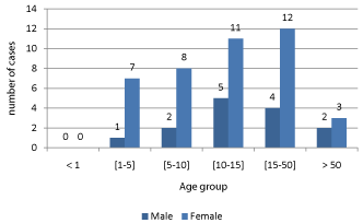
55 cases of LC have been fixed for 2005: 73% parasitologically positive, and 27% only clinics. There were significantly more women (75%) than men. The child population under 15 years old represents 61% of cases, confirming an active indigenous transmission in this outbreak. Ages [10-15] and [15-45] are equal (Figure 1). Women and children consult more frequently than men.

    Figure 1:  Distribution of 55 cases of cutaneous leishmaniasis by sex and age
group in 2005 during an outbreak in Teroual in northern Morocco.
Les aspects cliniques mettent en evidence les donnees suivantes:
- Nombre de lesions: lesion unique (37 CAS) ou lesions multiples ET multifocales.
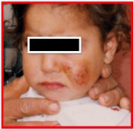
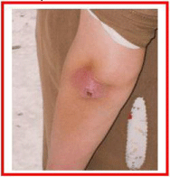
- Topographie des lesions: atteintes du membre superieur (25 CAS), du cou (20 CAS) ET de la region cephalique (5 CAS) prédominantes dans 45 CAS (Figure 2 and 3).

Figure 2: Ulcerative crusty ulcerated lesion (at face level).

Figure 3: Typical skin lesion in the left forearm.
- Stade evolutif: les lesions ont ete classees en lesions debutantes (26 cas don’t 12 avec nodules et 14 avec papules) et lesions evoluees (29 cas).
Discussion
Direct examination of dermal juice is the diagnostic reference and first line of cutaneous leishmaniasis due to its sensitivity and earliness. The polymorphic skin lesions justifies parasitological examination of the smear of the lesion and histological often.
The positivity of the direct examination of dermal juice is observed in 73% of cases, rates between those in the literature, ranging from 50 to 90.4% [6].
The non-immunized population influx arriving in natural foci of transmission, changes in the ecology of vectors and host reservoir and improved diagnosis and reporting of positive cases increase the incidence of LC. Men rarely or only in case of complications consult by occupation or negligence.
The validity of the clinical diagnosis is difficult to establish. This pathology appears well recognized in the region or physicians have a great diagnostic usual.
The treatment uses not devoid of side effects drugs requiring support by a specialized team.
The geographic distribution corresponds to the known affinities of Leishmania and sand flies for some ecosystems [7,8]. The distribution of leishmaniasis in Morocco with climate change was studied [9] since the sandfly-bioclimatic correlations [10] are established with sufficient precision in the ancient world.
CL due to L. tropica is a parasitic disease that plagues anthroponotic arid regions from southeastern Europe to Central Asia as well as in more limited foci in Morocco, Kenya, Namibia and south of the Arabian Peninsula. Skin lesions are readily erythematous scaly, more or less widespread (dry forms). They can simulate a lupus when they sit on the face. Their evolution is slower and can persist for years.
Our results, although limited to one year, suggest a periodicity of this endemic disease. The upsurge in the number of cases of LC anthroponotic and the possible spread of the disease in certain provinces bordering [11,12]. Conventional outbreaks require increased monitoring of its development and implementation of appropriate measures.
Furthermore, studies conducted in central Morocco [13] hypothesized the existence of L. tropica tank other than man. Indeed, Riyadh al., Suspect the dog as a potential reservoir of this species Leishmania considered until now as strictly anthropophilic.
A monitoring network geographically finest administrative level, locality, would determine a transmission rate by locality and a risk of exposure [14] and better understand the emergence of leishmaniasis in rural areas and fluctuations.
Conclusion
It appears from the analysis of our results that the rural commune Teroual (circle of Ouezzane, Morocco) is an LC home to L. tropica. However, retrospective and prospective epidemiological study (between 2005 and 2015), ongoing; in the laboratory environment and Parasitology of the Faculty of Science of Kenitra, will certainly verify the North of Morocco (district of Ouezzane) and to better elucidate this new epidemiology, as several species may LC be responsible in the rural town of Teroual. The LC routine typing will verify the results of this preliminary study and so offer epidemiologists an update of epidemiological clinical case for LC suspicion.
References
- Ministere de la Sante (Maroc) - Etat d'avancement des programmes de lutte contre les maladies parasitaires. Direction de l'epidemiologie et de lutte contre les maladies. Rapport annuel d'activite. Ministere de la Sante, Rabat, Maroc. 2003.
- DPA (Direction provinciale de l’Agriculture) de Sidi Kacem - Contribution de l’agriculture en zone bour favorable à l’emergence et au developement d’une agro-industrie a Sidi Kacem. - Terre Vie. 2003; 69: 5.
- Bulletin official (numero 6354, 23 avril 2015) - Recensement general de la population et de l’habitat (RGPH) Province d’Ouezzane. Maroc. 2014.
- Ministere de la Sante (Maroc) - Programme de lutte contre les leishmanioses cutanees et viscerales. - Plan d’action provincial, S.I.A.A.P, Delegation provinciale de Sidi Kacem, Maroc. 2015.
- Ministere de la Sante (Maroc) - Guide de leishmanioses et phlebotomes. Rabat: INH. 1997; 191p.
- Belhadj S, Helali JH, Kallel K, Kaouech E, Abaza H, Toumi ELN, et al. Place de la culture dans le diagnostic parasitologique des leishmanioses viscerales et cutanees. Rev Fr Lab. 2005; 369: 41-45.
- Lainson R. Ecological interactions in the transmission of the leishmaniases. Philos Trans R Soc Lond B Biol Sci. 1988; 321: 389-404.
- Lainson R. Epidemiologia ecologia das leishmaniose tegumentar Amazonas Hileia Medica. 1981; 3: 35-40.
- Rioux JA, Akalay O, Perieres J, Dereure J, Mahjour J, Le Houerou HN, et al. L’évolution éco-épidémiologique du ‘risque leishmanien’ au Sahara atlantique marocain. Interest heuristique de la relation ‘phlebotome-bioclimats’. Ecol Medit. 1997; 23: 73-92.
- Rioux JA, Rispail P, Lanotte G, Lepart J. Relations phlébotomes-bioclimats en écologie des leishmanioses. Corollaires épidémiologiques. L'exemple du Maroc Bull Soc Bot Fr. 1984; 131: 549-557.
- Jebbouri Y. Profile epidemio-clinique, therapeutique et evolutif de la leishmaniose cutanee (A propos de 52 cas) Expérience du service de Dermatologie de l’hôpital militaire Moulay Ismail-Meknès. These de Medicines University Sidi Mohammed bens Abdellah. Faculty de Medicines et de Pharmacie Fes. 2013.
- El Aasri A, Belghyti D, Hadji M, El Kharrim Kh. Etude de risque de la leishmaniose cutanée et viscérale dans la région de Sidi yahia du Gharb (province de Kenitra, Maroc. Science Lib Editions Messene. 2013; 5.
- Riyad M, Chiheb S, Bichichi M, et al. Evolution de la leishmaniose cutanée a Leishmania tropica au Maroc: L’exemple du foyer de Taza. J Prat. 2006; 15: 20-25.
- Broutet N, Ingrand P, Sousa Ade Q, Chabaud F, Lima JW. [Analysis of the monthly incidence of cutaneous leishmaniasis in Ceará (Brazil) between 1986 and 1990]. Sante. 1994; 4: 87-94.