
Case Report
Austin J Public Health Epidemiol. 2022; 9(1): 1120.
COVID-19 Pneumonia with a Focal Lung Lesion and a Periosteal Reaction Imitating a Neoplasm - Case Report
Magdalena Osowicka* and Agnieszka Gorzewska
Department of Palliative Medicine Medical University of Gdansk, Poland
*Corresponding author: Magdalena Osowicka, Department of Palliative Medicine Medical University of Gdansk, Poland
Received: January 03, 2022; Accepted: January 28, 2022; Published: February 04, 2022
Abstract
Introduction: Computer Tomography (CT) findings of COVID-19 are well described in the literature and include predominantly peripheral, bilateral Ground-Glass Opacities (GGOs), combination of GGOs with consolidations, and/or septal thickening creating a “crazy-paving” pattern [1]. COVID-19 pneumonia may mimic different infectious and non-infectious diseases [2]. However, it is rare for pulmonary changes to be accompanied by osteal changes suggesting malignant etiology.
It needs a special attention and vigilance in diagnostic process.
COVID changes may have misleading character and implicates the diagnosis and treatment.
Keywords: COVID-19; Osteal changes; Mimic; Cancer
Abbreviations
CT: Computer Tomography; GGOs: Ground-Glass Opacities; PCR: Polymerase Chain Reaction; mMRC: Modified Medical Research Council; VAS: Visual Analogue Scale
Introduction
Computer Tomography (CT) findings of COVID-19 are well described in the literature and include predominantly peripheral, bilateral Ground-Glass Opacities (GGOs), combination of GGOs with consolidations, and/or septal thickening creating a “crazypaving” pattern [1]. COVID-19 pneumonia may mimic different infectious and non-infectious diseases [2]. However, it is rare for pulmonary changes to be accompanied by osteal changes suggesting malignant etiology.
Case Presentation
43 years old patient without chronic diseases was diagnosed of SARS-CoV-2 infection confirmed by PCR test on 22th September 2020. During the infection patient felt severe dyspnea (mMRC 3) and pain on the right side of chest (VAS 6). After the 10-day isolation, he visited the pulmonary outpatient clinic due to the persistence of the above-mentioned symptoms. X-ray did not reveal any abnormalities and CT scan was performed. The CT image revealed the area of consolidation in segment 9 of the right lung (supra-diaphragm, subpleural) creating an image of lesion of size 17x12x18 mm, and in its vicinity subpleural small nodules up to 6 mm with adjacent area of atelectasis and fibrosis with accompanying periosteal changes of the X rib. There was an enlarged lymph node 14x11 mm in the right pulmonary hilum and segmentally weaker contrasting of the right segmental artery to segment 2 and sub segmental branches suggesting thromboembolic changes (Figure 1). Enoxaparin was applied at a dose of 2 x 0.1 g/ml.

Figure 1: Primary CT scan.
Two weeks later, angio-CT of the pulmonary arteries was performed, revealing the lesion of the size 14 x 10 x 17 mm and a slight periosteal reaction of the X rib at the level of the lesion. Additionally several non-specific small subpleural nodules in both lower lobes end lymph node (11x9 mm) in the right hilum were found, without no signs of pulmonary embolism. The CT image was ambiguous - both inflammatory and proliferative (Figure 2). Levofloksacine was used empirically at a dose of 500 mg per day for 10 days. In a follow-up CT examination after 4 weeks, there was a partial regression (to 6 mm) of the consolidation area in the ninth segment of the right lung (Figure 3).
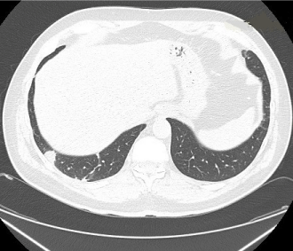
Figure 2: Angio CT scan.
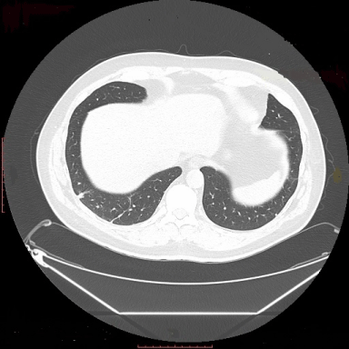
Figure 3: Final CT scan.
There was complete regression of the subpleural changes in the right lung and the disappearance of the periosteal reaction. On 4th January 2021, after the completed treatment, patient’s clinical condition was good. He reported reduced dyspnoea (1 mMRC) and pain in chest (2 VAS).
Patient has provided informed consent for publication of the case.
Discussion
The common chest CT findings of COVID-19 pneumonia are Ground Glass Opacity (GGO) with consolidation, bilateral distribution and multifocal lesions similar to presented case report [1,2]. Pulmonary embolism often accompanies SARSCoV- 2 infection. In presented situation, angio-CT did not confirm embolism-perhaps due to thrombotic treatment applied immediately and performing angio-CT two weeks later. COVID-19 causes varied radiological changes not only in the lung parenchyma but also in other internal organs, including the bone marrow [4]. Periosteal changes associated with COVID-19 pneumonia have not been reported so far. Described periosteal reaction at the level of the lung lesion suggested her proliferative character. Empirical treatment (levofloxacin) turned out to be effective - causing a reduction in pulmonary reactions and complete resolution of bone changes. COVID changes may have misleading character and implicates the diagnosis and treatment [3,5]. It is essential to discuss the features that may aid in the differential diagnosis.
Conclusion
CT lung findings in COVID-19 can be associated with periosteal changes and mimic cancer disease. Providing antibiotic therapy (levofloxacin) was necessary to exclude malignant etiology of lung and osteal changes.
Declaration
Patient concerns: 43 years old patient without chronic diseases diagnosed of SARS-CoV-2 infection.
Diagnosis: Subpleural parenchymal focal change associated by periosteal changes.
Outcomes: There was complete regression of the subpleural and osteal changes after the course of antibiotic therapy.
References
- Ye Z, Zhang Y, Wang Y, Huang Z, Song B. Chest CT manifestations of new coronavirus disease 2019 (COVID-19): a pictorial review Eur Radiol. 2020; 30: 4381-4389.
- Jiayi Liu, et al. Computer Tomography - Clinical and radiological changes of hospitalized patients with COVID-19 pneumonia from disease onset to acute exacerbation: A multicenter paired cohort study Eur Radiol. 2020; 30: 5702- 5708.
- Selin Ardali Duzgun, Gamze Durhan, et al. COVID-19 pneumonia: The great radiological mimicker. 2020; 11: 118.
- Postmortem Findings in Italian Patients with COVID-19: A Descriptive Full Autopsy Study of Cases with and Without Comorbidities Falasca, Laura. The Journal of Infectious Diseases. 2020; 222: 1807-1815.
- Al-Smadi AS, et al. Correlation of chest radiography findings with the severity and progression of COVID-19 pneumonia Clin Imaging. 2021; 71: 17-23.