
Case Report
Austin Surg Case Rep. 2024; 8(2): 1061.
Spontaneous Healing of a Pathological Proximal Humerus Fracture Secondary to a Renal Cell Carcinoma Metastasis with no Oncological Treatment
Murray E*; Gupta S
Department of Trauma and Orthopaedics, Glasgow Royal Infirmary, United Kingdom
*Corresponding author: Elspeth Murray Department of Trauma and Orthopaedics, Glasgow Royal Infirmary, United Kingdom. Email: elspeth.murray2@ggc.scot.nhs.uk
Received: February 13, 2024 Accepted: March 16, 2024 Published: March 22, 2024
Abstract
A 74-year-old male was referred to our Orthopaedic Oncology service with a pathological fracture of his proximal humerus. This was in the context of a previous open partial nephrectomy (18 months prior to presentation) for clear cell renal cell carcinoma. The humeral lesion was a solitary lesion, and a biopsy of this was consistent with metastasis. The patient was medically unwell during investigations and struggled to lie flat for his diagnostic biopsy due to a recent respiratory tract infection. Due to his overall poor health, he was not felt suitable for en bloc resection and the plan for management of his fracture was a joint sparing curettage and fixation with a plate and screws. His disease management until then had not included any oncological treatment (chemo- or radiotherapy). The planned surgical date for our patient’s humeral stabilisation was 4 months after his first presentation of pathological fracture. This was due to delays including: the time to achieve initial investigations by the referring hospital, the patient being unwell including during investigations in our tertiary centre, the need for a repeated biopsy attempt, and finally difficulty allotting theatre space on an urgent basis due to our own service experiencing a high volume of patients requiring urgent and emergent surgery. At the time of admission to our hospital for surgery, our patient questioned the need for his operation as his humerus was now moving as one, and his pain had greatly reduced. A radiograph confirmed that surprisingly his fracture had bridging callus present.
Keywords: Pathological fracture; Secondary bone tumour; Spontaneous healing
Case Presentation
A 74-year-old patient was referred to his local Emergency Department with a 6-month history of atraumatic right arm pain with associated subjective weakness (see Figure 1 for timeline of events). This pain had become significantly worse in the past week. His past medical history included Crohn’s disease (with panproctocolectomy and ileostomy), chronic kidney disease stage 3 with a non-functioning left kidney, ureteric calculi, osteoarthritis, previous pulmonary embolism, previous deep vein thrombosis and clear cell renal cell carcinoma diagnosed 2 years prior to this presentation which was managed with a partial nephrectomy 18 months prior to this presentation. He was reviewed by the Orthopaedic doctor on-call and found to have a deformity of his right arm, pain on all shoulder movements, reduced power in his shoulder girdle but was neurovascularly intact distal to the injury. His initial radiograph (Figure 2) showed a pathological lesion. An urgent outpatient MRI, a staging CT chest/abdomen/pelvis and a bone scan were organised over 3 weeks, and he was referred to our tertiary Orthopaedic Oncology Team thereafter. He was discussed at the Musculoskeletal (MSK) Oncology multi-disciplinary team meeting the next day where his imaging was reviewed by MSK Radiologists and 7 days later at the National Renal MDT. He underwent an ultrasound guided biopsy 5 days later which unfortunately yielded a pathologically inconclusive sample, and an open biopsy was therefore carried out. At the time of open biopsy, he was suffering an acute respiratory illness and therefore the open biopsy could only be carried out under regional anaesthetic. The pathology returned 10 days following, confirming the diagnosis of metastatic clear cell renal cell carcinoma. With a now confirmed solitary lesion, he was counselled in our outpatient clinic and a date for surgery planned. Due to the patient’s illness and pressures on our local state-funded service, which was experiencing a high volume of patients with need for urgent and emergent surgery, the patient could only be scheduled for surgery 1 month later. The patient was admitted the night before his planned surgery and at this time he questioned the necessity of the surgery. He felt that his humerus was moving as one, and that his pain had greatly reduced. After obtaining an up-to-date radiograph, we concurred with his impression that the fracture was unexpectedly healing. After discussions with his Renal Oncologist and Urologist it was deemed difficult to justify proceeding with surgery, in the context of the risks of his co-morbidities, where the evidence suggested that his fracture was healing.
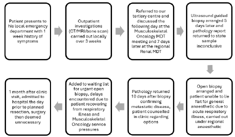
Figure 1: Timeline of events showing unavoidable delays that occurred.
Investigations
His initial radiographs showed a pathological lesion in the metaphyseal/diaphyseal region of his right proximal humerus with an associated transverse and comminuted fracture (Figure 2).
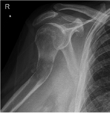
Figure 2: Radiograph from initial presentation to referring hospital.
He went on to have a staging CT which showed no local recurrence in his remaining kidney, nor any other suspicious lesion to suggest a separate primary pathology. This was followed by an MRI of his right upper arm, which was thought to be most likely in keeping with a renal cell metastasis. A bone scan was also carried out and showed no other involved areas. Serial radiographs were carried out of his right humerus. Figure 3 shows the fracture almost 12 weeks after initial presentation (whilst the patient was still symptomatic) and Figure 4 shows the lesion the day before his planned surgery when he was no longer symptomatic. This radiograph showed progressive callus formation and that his pathological fracture was on the way to uniting. Figure 5 shows the asymptomatic lesion at nearly 6 months after his initial presentation and Figure 6 shows the lesion at most recent follow up 14 months after his initial presentation when he was discharged from the Orthopaedic Oncology service.
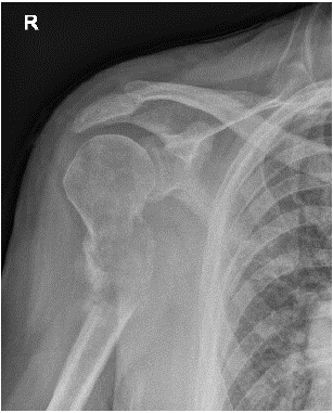
Figure 3: Fracture almost 12 weeks after initial presentation.
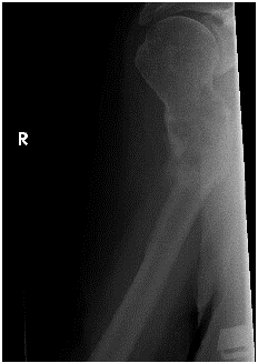
Figure 4: Bridging callus seen the day prior to surgery.
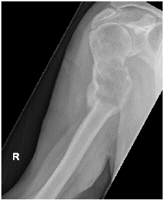
Figure 5: Follow-up at almost 6 months after initial presentation.
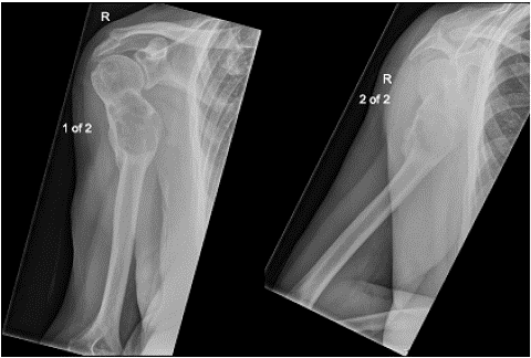
Figure 6: Overall survival, autologous stem cell transplant (ASCT) versus no ASCT (p=0.12).
Differential Diagnosis
In the context of this patient’s previous renal cell carcinoma and his age, a metastasis from this primary was at the top of the differential diagnosis list at first presentation. A skeletal metastasis from a renal cell primary is typically an osteolytic lesion which is hypodense on radiographs (illustrated well on the patient’s radiographs included in this report), with thinned out or absent trabeculae and ill-defined margins. Metastasis from a separate primary was another possible diagnosis. Other common primaries which may give a lytic lesion include non-small cell lung cancer, thyroid cancer and melanoma, and haematological primaries such as multiple myeloma and non-Hodgkin’s lymphoma. Lytic primary bone tumours are less likely, especially with this patient’s past medical history, but include benign lesions such as a unicameral bone cyst or aneurysmal bone cyst, and malignant lesions such as a chondrosarcoma – found especially in the axial skeleton and in limb-girdle regions.
Treatment
This patient was treated conservatively initially in a collar and cuff and then a humeral brace whilst awaiting surgical management. After discussion on his planned surgical date which resulted in his surgery being cancelled, he continued to wear a polysling at home. The patient was seen 8 weeks later in clinic when he reported that he had begun to wean himself from the sling and that this was only necessary when he was out for long periods of time.
Discussion
Renal cell carcinoma accounts for 2.2% of global cancer diagnoses and deaths, which can be contrasted against lung cancer, the most commonly diagnosed cancer which accounts for 11.6% of all cancer diagnoses globally [1]. Diagnosing patients early is challenging, and rather happens later in the disease course, where 25-30% of patients have metastatic disease already [2]. Metastases are most commonly found in the lung (45.2%), bone (29.5%), lymph nodes (21.8%), liver (20.3%), adrenal glands (8.9%) and brain (8.1%) [3]. The presence of metastasis in renal cell carcinoma confers a poorer outcome for the individual with a overall median survival of less than 1 year [2].
Metastatic renal cell carcinoma is a typically chemotherapy and radiotherapy resistant entity. Previous work with interleukin-2 and interferon alpha has shown low response rates, however progress in the ability to stabilise and shrink metastatic disease has been made with tyrosine kinase inhibitors [4]. In one published case report, metastatic disease has been histologically cured [5].
With increasing developments in the treatment of metastatic renal cell cancer, patients are benefiting from treatment of their pathological fractures for longer. Pathological fractures of metastatic deposits cause significant problems with pain, function, and mobility for this group of patients, who can be expected to suffer these symptoms for several months, and perhaps with ongoing developments in systemic therapy, even longer in the future. In patients who suffer a pathological fracture, and who are well enough, surgical fixation or endoprosthetic replacement are the recommended mainstays of treatment, to facilitate pain free function and mobility. Surgery, of course, comes with associated risks, which include but are not limited to; ongoing pain, infection, nerve injury, vessel injury, prosthesis dislocation and periprosthetic fracture, all of which can necessitate further surgeries for the patient.
The likelihood of spontaneous healing of a pathological fracture in this context is unknown but the chances are presumed to be low and so conservative treatment is not usually recommended. One cited study published in 1983, used a variety of management methods of pathological fractures (including operative and non-operative methods), reported on healing rates of these fractures due to metastases of different primary tumours, but the rate of healing in conservatively managed fractures alone is not reported here or elsewhere in the literature [6]. The chances of a fracture through a metastatic deposit healing are thought to be slightly greater should the patient be receiving systemic therapy, and thought not to be possible in patients who are not. In one case report, a patient receiving alpha-interferon treatment for a clear cell renal carcinoma, chose conservative management of his pathological fracture in his proximal humerus. Spontaneous healing was observed at three months post initial presentation [7]. In another case report, one patient underwent spontaneous healing of a missed femoral neck fracture through a metastatic lesion from a primary breast cancer. Her radiological evidence of healing was found incidentally, two years after her hip pain had begun. Treatment of her primary breast cancer included bilateral mastectomy, chemotherapy and radiotherapy [8]. A case report of a fracture through malignant transformation of an enchondroma to a chondrosarcoma (radiologically diagnosed without tissue diagnosis) showed radiological healing without surgical or oncological treatment after 8 weeks [9]. Notably, none of these case reports demonstrate healing of a fracture through a metastatic bone deposit with no surgical or oncological treatment.
Conclusion
In the field of Orthopaedic Oncology, the case numbers are small, reflecting the natural prevalence of disease, be this primary or secondary bone tumours. There are few published cases of pathological fractures secondary to metastases healing spontaneously with concurrent oncological treatment and none without concurrent oncological treatment. There are recently published British Orthopaedic Association clinical guidelines for the management of suspected Metastatic Bone Disease which focus on the investigation of suspected lesions, but do not provide guidelines on clinical treatment [10]. This fact reflects on the case-by-case nature in which decisions must be made for patients who have such diagnoses. These patients are often older, come with co-morbidities and are not all amenable to what would be first-choice management in a fitter and younger patient.
References
- Bray F, Ferlay J, Soerjomataram I, Siegel R, Torre LA, Jemal A. Global cancer statistics 2018: GLOBOCAN estimates of incidence and mortality worldwide for 36 cancers in 185 countries. CA Cancer J Clin. 2018; 68: 394-424.
- Gupta K, Miller JD, Li JZ, Russel MW, Charbonneau C. Epidemiologic and socioeconomic burden of metastatic renal cell carcinoma (mRCC): a literature review. Cancer Treat Rev. 2008; 34: 193-205.
- Bianchi M, Sun M, Jeldres C, Shariat SF, Trinh QD, Briganti A, et al. Distribution of metastatic sites in renal cell carcinoma: a population-based analysis. Ann Oncol. 2012; 23: 973-80.
- Kathuria-Prakash N, Drolen C, Hannigan CA, Drakaki A. Immunotherapy and Metastatic Renal Cell Carcinoma: A Review of New Treatment Approaches. Life (Basel). 2021; 12: 24.
- Asano Y, Yamamoto N, Hayashi K, Takeuchi A, Miwa S, Igarashi K, et al. Case report: Complete remission of bone metastasis from renal cell carcinoma in histopathological examination after treatment with immune checkpoint inhibitors. Front Immunol. 2022; 13: 980456.
- Gainor BJ, Buchert P. Fracture healing in metastatic bone disease. Clin Orthop Relat Res. 1983; 178: 297-302.
- Shareef S, Sinha S, Bramely D. Spontaneous healing of a pathologic fracture of the humerus. Current Orthopaedic Practice. 2012; 23: 66-67.
- Kong AC, Zarate SD, Belzarena AC. Missed pathological femoral neck fracture undergoes spontaneous healing. Radiol Case Rep. 2022; 17: 72-76.
- Herget GW, Haag M, Strohm PC, Uhl M, Knoeller S, Sudkamp N. Spontaneous healing of pathologic humerus fracture caused by a cartilaginous tumor. Acta Chir Orthop Traumatol Cech. 2006; 73: 400-2.
- British Orthopaedic Association, B. O. O. S. (2022, June 2022). “Management of Metastatic Bone Disease (MBD). 2023.