Case Report
Austin J Surg. 2024; 11(3): 1329.
Internal Supravesical Hernia Revealed by Small Bowel Obstruction: An Unusual Case Report in Surgery
Rebbani Mohammed1,2*; El Brahmi Yasser1; Rahali Anwar1; Njoumi Nourddine1; Abderrahman Elhjouji1; Aziz Zentar1; Ait Ali Abdelmouain1
¹Department of Visceral Surgery, Morocco
²HMIMV, Rabat, Morocco
*Corresponding author: Rebbani Mohammed, Department of Visceral Surgery, HMIMV, Rabat, Morocco. Email: mohammed.rebbani@gmail.com
Received: July 19, 2024 Accepted: August 26, 2024 Published: September 03, 2024
Abstract
Internal supravesical hernia is an unusual and exceptional cause of internal hernia that can lead to small bowel obstruction. Often diagnosed during surgery, it presents a clinical and radiological diagnostic challenge. We report a case of a 56-year-old patient with no notable medical history was admitted to the emergency department with an internal supravesical hernia revealed by obstructive syndrome.
Introduction
Internal supravesical hernia is an unusual and exceptional cause of internal hernia that can lead to small bowel obstruction. Often diagnosed during surgery, it presents a clinical and radiological diagnostic challenge. we discuss by this case report the clinical picture, diagnosis and treatment of this rare entity.
Case Report
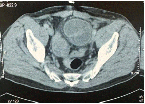
A 56-year-old patient with no notable medical history was admitted to the emergency department with an obstructive syndrome evolving over the past 2 days. At admission, the patient was stable hemodynamically, respiratory-wise, and neurologically. Blood pressure was 12/07, heart rate was 75 bpm, and oxygen saturation was 98% on ambient air. Abdominal examination revealed a distended abdomen with tympany upon percussion. A rectal examination showed an empty rectal ampoule and free hernial orifices. Laboratory tests showed white blood cells at 12,000, CRP at 50, with no other abnormalities. Abdominopelvic CT scan indicated small bowel distension measuring 42 mm with hydroaeric levels, with areas of disparity in the caliber of distended and flattened small bowel in contact with the bladder. The initial diagnosis was bowel obstruction due to adhesions.
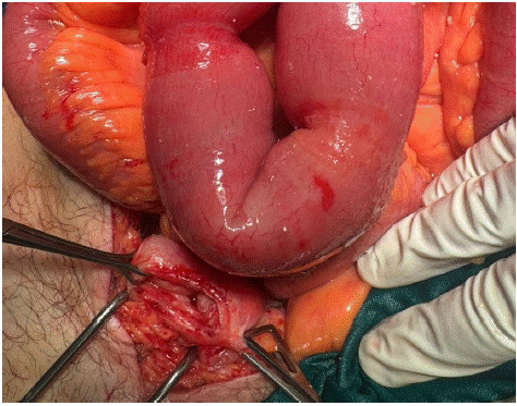
Initially, the decision was to manage the patient conservatively in hopes of spontaneous resolution of the obstruction, but due to its persistence, a decision was made for surgical exploration. The approach was a median subumbilical laparotomy. Exploration revealed small bowel distension upstream of an incarceration of the ileum at an internal supravesical hernia. The bowel loops were viable and non-strangulated. The bowel loops were released, and the hernial orifice was closed with separate stitches of slow-resorption sutures to prevent recurrence.

Figure 1: Abdominopelvic CT scan indicated small bowel distension measuring 42 mm with hydroaeric levels, with areas of disparity in the caliber of distended and flattened small bowel in contact with the bladder.

Figure 2: Small bowel distension upstream of an incarceration of the ileum at an internal supravesical hernia.
The postoperative course was uncomplicated. Feeding began on postoperative day 2, and discharge occurred on postoperative day 3.
Discussion
The supravesical internal hernia corresponds to the protrusion of the intestine through an abnormal intra-abdominal opening located in the supravesical compartment. This compartment forms between the remnants of the median umbilical ligament and the medial umbilical arteries, most often expanding into the Retzius space, but they can also extend laterally, forming external supravesical hernias or laterovesical hernias [1].
These hernias are rare, with an incidence of 4%, and they cause bowel obstruction in 0.5% to 1% of cases. They often occur in men around the age of 50 [2].
The most common presentation is small bowel obstruction due to incarceration, clinically manifesting as cessation of stool and gas passage, abdominal pain, and vomiting. Clinical examination confirms the obstruction by the presence of abdominal distension, tympanism, and an empty rectal ampulla on pelvic examination [3]. Thus, the diagnosis of obstruction is easily made, but attributing it to a supravesical internal hernia is almost impossible at this stage. In our patient, the obstruction was the presenting symptom that prompted an emergency consultation.
Radiology, particularly an injected abdominal CT scan, can alone confirm the diagnosis of an internal hernia preoperatively. CT remains the most sensitive and widely available examination in these situations. It confirms the obstruction and its etiology and helps identify radiological signs of severity, including ischemic bowel injury or even perforation [4]. However, the diagnosis may be challenging for young practitioners who have often never encountered this entity in their practice. In our case, the CT scan confirmed the diagnosis of obstruction but failed to identify the exact cause.
Surgical treatment, whether by laparotomy or laparoscopy, is the cornerstone of management [5]. It initially confirms the diagnosis, which is often missed until this point, reduces the hernia contents, resects the sac, and closes the hernial orifice, thereby preventing recurrence. In situations where the contents are necrotic, it allows for bowel resections with or without immediate anastomosis [6]. In our patient, the surgical exploration performed by laparotomy corrected the diagnosis by revealing a supravesical internal hernia with viable small bowel contents; the loops were reduced, and the orifice was closed with separate sutures using slow-absorption thread.
The prognosis is often favorable but can be influenced by the patient's condition and timeliness of management.
Conclusion
internal supravesical hernia is a rare and atypical condition, frequently discovered during surgery for acute intestinal obstruction. The standard surgical approach involves closing the hernia sac to avert the risk of recurrences.
Author Statements
Conflicts of Interest
The authors declare no conflicts of interest.
Author Contributions
All authors have read and approved the final version of the manuscript.
References
- H Kotobi, A Echaieb, D Gallot. Traitement chirurgical des hernies rares, EMC – Chirurgie. 2005; 2L 425-439.
- Bensardi F, Othmane E, Majd A, El Bakouri A, Mounir B, El Hattabi K, et al. Hernie sus-vésicale interne étranglée associée à une hernie inguinale gauche: un cas très rare d’obstruction intestinale aiguë. Annales de Médecine et de Chirurgie. 2021; 66: 102393.
- Sozen I, Nobel J. Inguinal mass due to an external supravesical hernia and acute abdomen due to an internal supravesical hernia: a case report and review of the literature. Hernia. 2004; 8: 389-92.
- Takeyama N, Gokan T, Ohgiya Y, Satoh S, Hashizume T, Hataya K, et al. CT of internal hernias. Radiographics. 2005; 25: 997-1015.
- Bouassida M, Sassi S, Touinsi H, Kallel H, Mighri MM, Chebbi F, et al. Internal supravesical hernia - a rare cause of intestinal obstruction: report of two cases. Pan Afr Med J. 2012; 11: 17.
- Cissé M, Konaté I, Ka O, Dieng M, Dia A, Touré CT. Internal supravesical hernia as a rare cauase of intestinal obstruction: a case report. J Med Case Rep. 2009; 3: 9333.