Case Report
Austin J Surg. 2024; 11(5): 1339.
A Case Report of Small Bowel Perforation Secondary to Ingested Fishbone Manifesting as Small Bowel Obstruction
Pamathy Gnanaselvam*; Pirahanthan Karunanithy; IPKB Thilakarathna; Saranga Eshanie Wickramaratne; AMPP Semasinghe
Department of Surgery, National Hospital of Sri Lanka, Colombo 10, Sri Lanka
*Corresponding author: Pamathy Gnanaselvam, Department of Surgery, National Hospital of Sri Lanka, Colombo 10, Sri Lanka. Email: pamathysha@yahoo.com
Received: October 23, 2024; Accepted: November 08, 2024 Published: November 15, 2024
Abstract
Small bowel obstruction secondary to ingested fish bone is an exceedingly rare occurrence, particularly among the elderly, accounting for less than 1% of cases necessitating surgical intervention. This article reports a case of an 84-year-old male who presented with acute abdominal pain and bilious vomiting for seven days after accidentally swallowing a fish bone. Computed Tomography (CT) of the abdomen and pelvis indicated sub-acute bowel obstruction, revealing a linear foreign body in the jejunum with a focal collection. An emergency exploratory laparotomy uncovered a pocket of pus containing the fish bone in the mesentery of the jejunum, with evidence of perforation at approximately 150 cm distal to the duodenojejunal flexure.
Introduction
Perforation of the gastrointestinal tract due to ingested foreign bodies is rare but may occur in patients with conditions such as hernia or Meckel’s diverticulum where the integrity of the wall tissue has been impaired. Small bowel perforation is more likely in individuals with intestinal diseases or at sites of acute angulation, such as the ileocecal and rectosigmoid junctions [3-5]. Ingested foreign body may not only cause perforation but also may cause obstruction, and fistula formation which may be fatal. We present a case of small bowel perforation due to a swallowed fish bone in a patient with no history of intestinal disease or prior abdominal surgery, along with the management approached for this uncommon condition.
Case Presentation
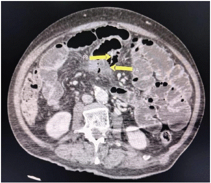
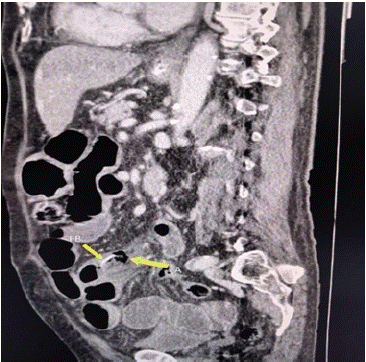
An 84-year-old male with diabetes and hypertension was admitted to the emergency room with progressive upper abdominal pain, bilious vomiting, and absent bowel opening for two days. Physical examination revealed tenderness in the left upper quadrant with rigidity, guarding and reduced bowel sounds. An erect abdominal X-ray indicated air-fluid levels in the proximal small bowel without evidence of pneumoperitoneum. CT scan of the abdomen demonstrated sub-acute bowel obstruction, transition in the distal jejunum, and a linear foreign body with a focal collection and diffuse inflammation of the jejunum and surrounding fatty tissues. This image was initially reported as foreign body causing perforation (Figure 1 & 2) An emergency exploratory laparotomy was performed, revealing small bowel perforation with a fish bone at the mesentery of the jejunum, 150 cm from the ligament of Treitz. Localized inflammation and abscess formation were noted, prompting a segmental resection of the affected bowel. The remainder of the intestine appeared grossly normal except for the perforation site, and end-to-end anastomosis was performed manually. The patient experienced no complications during the postoperative period and was discharged after eight days. A retrospective history taken postsurgery revealed that the patient had consumed fish seven days prior to admission.

Figure 1: Axial view of CECT abdomen shows fishbone [FB] and abscess [A].

Figure 2: Coronal CECT shows fishbone [FB] and abscess [A].
Discussion
Fish bones account for 84% of accidentally ingested foreign bodies, the majority of which are excreted without surgical complications [1]. Only 1.0% of foreign bodies lead to intestinal perforation, typically at the ileal level [16,2]. Fish bones are particularly prone to cause perforation due to their sharp edges and elongated shapes [6]. Factors such as advanced age, underlying inflammatory conditions causing increased fragility of the intestine, rushed eating habits, inadequate food preparation and the presence of dental prosthetics (cause loss of sensation of a foreign body) can increase the risk of accidental ingestion of fish bones[13].
Perforation can occur anywhere in the gastrointestinal tract, with areas of angulation – such as the ileum, ileocecal junction, and rectosigmoid–being most susceptible. In segments where a change in direction or transition from a mobile to an immobile segment occurs possibility of perforation increases. The most frequent sites of fish bone perforation are the ileum, the ileocecal junction and the rectosigmoid, however it can occur in all segments of the GI tract [8]. While perforation may occur at other sites, including hernia sacs, Meckel's diverticula, or the appendix [7] our case involved a jejunal perforation.
Clinical presentations of fish bone perforation vary widely, from unnoticed passage per rectum to severe peritonitis. Common symptoms include abdominal pain, vomiting, fever, and occasionally melena or bowel obstruction [4]. Depending on the location of the damage, different surgical conditions may occur including localized abdominal abscess, colorectal, Colovesical and entervesical fistulas, inflammatory mass or omental pseudo-tumor and bleeding. The diagnosis can be challenging, often mimicking appendicitis or diverticulitis, and cases can occasionally be asymptomatic[14]. The stomach, duodenum, and colon typically exhibit delayed presentations compared to small bowel perforations.[7]
Diagnosing fish bone perforation preoperatively is challenging and rare. Various imaging techniques can aid in detection, with each having its distinct advantages. While abdominal X-rays may reveal ingested metal foreign bodies or free air in the abdomen associated with perforation, or the obstruction, ultrasound can detect intraabdominal fluid and assist in excluding differential diagnoses. CT scans are particularly effective in visualizing perforations caused by foreign bodies, highlighting mucosal wall thickening, intestinal obstruction, and associated abscess formation.
Role of plain film radiography is an insignificant in the detection of fish bones in the aerodigestive tract is as low as 32%, false negatives are seen in up to 47% of cases [10]. The presence of pneumoperitoneum is not found in many cases, because the perforation is usually caused by the impaction and the progressive erosion of the FB through the intestinal wall, allowing it to be covered by fibrin, omentum and adjacent loops of bowel [6].
Because of their high reflectivity and various background shadows, foreign bodies that are not radio-opaque such as fish bone or toothpick can be detected by US [15]. Changes in tissues surrounding perforations and luminal contents of superficial intestines could be evaluated by US. However, deeper tissues may be hard to visualise. The morphological properties of the patient, localization of the perforation, and the experience of the observer may limit the functionality of the ultrasonography. Computed Tomography (CT) yields more useful information illustrating fish bones as demarcated linear, calcifications [18]. Furthermore, associated CT-scan findings at the site of perforation include: mucosal wall thickening of the bowel, intestinal obstruction, pericolic fat stranding, and at times there may be abscess formation [10]. Abdominal CT with contrast revealed a typical image of the perforation caused by ingested fish bone, as seen in our case, the linear lesion with hyper density surrounded by inflamed tissues. In some cases, imagery findings can be nonspecific; however, the finding of a foreign body with extra-luminal pockets of free air or an associated mass in patients with clinical signs of peritonitis, mechanical bowel obstruction or pneumo-peritoneum strongly suggests the diagnosis of foreign body perforation [4].
The management of ingested foreign bodies depends on the patient's symptoms and the type and location of the object [11]. Surgical intervention is the preferred approach for repairing perforations caused by foreign bodies. In cases of complications such as abscesses, fistulas, or ileus, treatment may include observation, medical management, or radiological interventions. Laparoscopy is often preferred [17], although patients who are hemodynamically unstable or in septic shock may require laparotomy for thorough peritoneal lavage and bowel resection and defunctioning colostomy if necessary. Depending on the size of the perforation, the degree of contamination, the underlying condition of the bowel and the judgement of the surgeon, an early intervention should be taken to prevent further morbidity and mortality [9]. Surgical treatment of small intestine perforations requires surgical repair or segmental resection [12].
In our patient, the ingested fish bone resulted in jejunal perforation and localized pus collection in the mesentery, early presentation and early advanced imaging led for early diagnosis and management causing better prognosis. Following advanced imaging, a laparotomy was performed, and the perforated segment was resected, with subsequent end-to-end anastomosis. The patient's recovery was uneventful.
Conclusion
Fish bones are common foreign bodies in the digestive tract, typically passing without incident. This case underscores the importance of considering intestinal perforations due to fish bone ingestion in the differential diagnosis of acute abdominal symptoms. Employing appropriate imaging techniques and conducting a thorough patient history can lead to accurate diagnosis. Early identification and appropriate management are crucial for improved outcomes.
Author Statements
Data Availability
All underlying data supporting the results of the study are included in the manuscript.
Consent
Informed written consent for publication was obtained from the patient prior to collecting data.
Conflicts of Interest
The authors declare that they have no conflicts of interests.
Authors' Contributions
Authors PG, PK, IPKBT, and SEW contributed to the collection of information and writing of the manuscript. Authors PG, PK and SEW contributed to the writing and final approval of the manuscript. AMPPS contributed equally to this work.
References
- SH Venkatesh, NK Venkatanarasimha Karaddi. CT findings of accidental fish bone ingestion and its complications. Diagn Interv Radiol. 2016; 22: 156-160.
- MA Pinero, JA Fernandez, PM Carrasco, RJ Riguelme, PP Parrilla. Intestinal perforation by foreign bodies. Eur J Surg. 2000; 166: 307-309.
- Schwartz GF, Polsky HS. Ingested foreign bodies of the gastrointestinal tract. Am Surg. 1976; 42: 236–238.
- Shariful Islam, Anthony Maughn, Patrick Harnarayn, Professor Vijay Naraynsingh. Fish Bone Perforation of Small Bowel Mimicking Acute Appendicitis. 2016; 4: 66-74.
- Maleki M, Evans WE. Foreign-body perforation of the intestinal tract. Report of 12 cases and review of the literature. Arch Surg. 1970; 101: 475–477.
- Shahid F, Abdalla SO, Elbakary T, Elfaki A, Ali SM. Fish bone causing perforation of the intestine and meckel’s diverticulum. Case Rep Surg. 2020; 2020: 8887603.
- Choi Y, Kim G, Shim C, Kim D, Kim D. Peritonitis with small bowel perforation caused by a fish bone in a healthy patient. World J Gastroenterol. 2014; 20: 1626–1629.
- Wada Yoshiki, Sasao Wataru, Oku Tadashi, Gastric perforation due to fish bone ingestion: a case report. J. General Family Med. 2016; 17: 315–318.
- Pulat H, Karakose O, Benzin MF, Benzin S, Cetin R. Small bowel perforation due to fish bone: a case report. Turk J Emerg Med. 2016; 15: 136–138.
- Sibanda T, Pakkiri P, Ndlovu A. Fish bone perforation mimicking colon cancer: a case report. SA J Radiol. 2020; 24: 1885.
- Kuwahara K, Mokuno Y, Matsubara H, Kaneko H, Shamoto M, Iyomasa S. Development of an abdominal wall abscess caused by fish bone ingestion: a case report. J Med Case Rep. 2019; 13: 369.
- KP Brian Goh, PKH Chow, Quah HM, Ong HS, Eu KW, Ooi LLPJ, et al. Perforation of the gastrointestinal tract secondary to ingestion of foreign bodies,World J Surg. 2006; 30: 372-377.
- B Coulier, MH Tancredi, A Ramboux. Spiral CT and multidetector-row CT diagnosis of perforation of the small intestine caused by ingested foreign bodies. Eur Radiol. 2004; 14: 1918-1925.
- M Hashmonai, T Kaufman, A Schramer. Silent perforations of the stomach and duodenum by needles. Arch Surg. 1978; 113: 1406-1409.
- B Coulier. Diagnostic ultrasonography of perforating foreign bodies of the digestive tract. J Belge Radiol. 1997; 80: 1-5.
- DE Mc Canse, A Kurchin, SR Hinshaw. Gastrointestinal foreing bodies. Am J Surg. 1981; 142: 335-337.
- Chin EH, Hazzan D, Herron DM, Salky B. Laparoscopic retrieval of intraabdominal foreign bodies. Surg Endosc. 2007; 21: 1457.
- Sierra-Solís A. Bowel perforations due to fish bones: rare and curious. SEMERGEN. 2012; 39: 117–118.