Clinical Image
Austin J Surg. 2025; 12(1): 1345.
Advancing Laparoscopic Cholecystectomy, A Few Messages
Yasser El Brahmi*; Hamada Abdelilah; Mehdi Ilahiane; Younes Laroussi; Yaka Mbarek; Abdelmounaim Ait Ali
Department of Visceral Surgery, Mohammed V Military Training Hospital, Rabat, Morocco
*Corresponding author: Yasser El Brahmi, Department of Visceral Surgery, Mohammed V Military Training Hospital, Rabat, Morocco. Email: yasserelbrahmi@gmail.com
Received: December 26, 2024; Accepted: January 07, 2024; Published: January 14, 2025
Clinical Image
Laparoscopic cholecystectomy is a safe and common procedure, but it can be associated with some complications. These complications may occur during the surgery or in the following days or weeks.
We will highlight some complications or risks of laparoscopic cholecystectomy through three clinical images while explaining the preventive measures [1,2].
Image 1

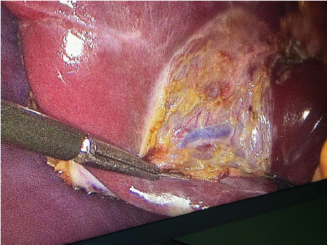
Figure 1: Identification of the segment VII portal branch during laparoscopic cholecystectomy.
The gallbladder is anatomically located in an area where critical structures pass through (main bile ducts, cystic artery, hepatic artery, portal vein). Dissection must be performed carefully when separating the gallbladder from the hepatic parenchyma by clearly identifying the anatomical structures before any cutting (the "Critical View of Safety" method). This approach helps prevent injuries to the main bile ducts, arteries, or veins [1,3].
Image 2

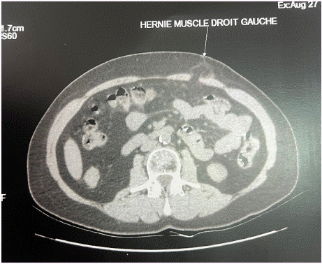
    Figure 2: Herniation on the left rectus abdominis muscle following a
laparoscopic cholecystectomy.
Trocar site herniation is preventable with rigorous techniques and appropriate preventive measures.
An excessively large trocar site is associated with an increased risk, especially when inserted in weak areas such as the midline. Care should be taken to avoid traumatic insertion that may damage surrounding tissues during trocar placement.
Systematic and proper closure of trocar sites, particularly larger ones, can effectively prevent this type of complication [3,5].
Image 3

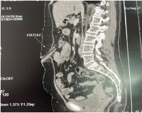
    Figure 3: Active fistulous tract in the left paraumbilical region extending
deeply into the preperitoneal space, measuring 7 mm in caliber and
approximately 5 cm in length, along the pathway of the laparoscopic trocar.
This is suggestive of a post-laparoscopy parietal fistula.
Using metal or absorbable clips is a common practice for ligating vascular and biliary structures (cystic duct, cystic artery) during laparoscopic cholecystectomy.
While clips are safe and widely used, improper application or technical issues can lead to serious complications, such as intraabdominal abscesses, especially if the clips become infected or irritate surrounding tissues.
The dislodgement of metal clips into the umbilical port site is a rare but possible complication during laparoscopic cholecystectomy. This situation may occur primarily during the extraction of the gallbladder or the manipulation of surgical instruments.
If the clip remains in the umbilical port site or surrounding tissues, it may cause local irritation or infection. This highlights the importance of systematically placing the gallbladder in a retrieval bag before extraction through the reduced umbilical trocar. This practice minimizes the risk of disseminating stones, bile, or clips into the umbilical site [1,4].
References
- François Dubois. Cholécystectomie et exploration de la voie biliaire principale par coelioscopie. 1993;
- L Barbier, N Tabchouri, E Salame. Technique de la cholécystectomie. 2020.
- Mascagni P, Longo F, Barberio M, et al. New intraoperative imaging technologies: Innovating the surgeon’s eye toward surgical precision. Journal of Surgical Oncology. 2018; 118: 265–282.
- Nijssen MAJ, Schreinemakers JMJ, Meyer Z, et al. Complications after laparoscopic cholecystectomy: a video evaluation study of whether the critical view of safety was reached. World Journal of Surgery. 2015; 39: 1798–1803.
- Pucher PH, Brunt LM, Davies N, et al. Outcome trends and safety measures after 30 years of laparoscopic cholecystectomy: a systematic review and pooled data analysis. Surgical Endoscopy. 2018; 32: 2175–2183.