Special Article - Surgery Case Report
Austin J Surg. 2018; 5(1): 1120.
An Unusual Case of Epidermoid Cyst of the Kidney: Case Report and Review of Literature
Venkata RMK¹*, Rajiv R¹, Pavani B2 and Srinivas G1
¹Department of Urology and Renal Transplantation, Kamineni Hospitals, India
²Department of Pathology, Kamineni Hospitals, India
*Corresponding author: Venkata RM Kusuma, Department of Urology and Renal Transplantation, Kamineni Hospitals, India
Received: December 04, 2017; Accepted: January 03, 2018; Published: January 10, 2018
Abstract
We report a rare case of epidermoid cyst of the left kidney in a 36-year-old female who presented with complaints of intermittent dull pain in the left loin for 6 months. Imaging revealed a complex cyst of the kidney. The cyst bearing kidney was removed uneventfully. Histopathology confirmed an epidermoid cyst which is an extremely unusual form of renal cystic mass with only few cases reported in the literature.
Introduction
Epidermoid cysts resemble the common epidermal inclusion cyst of the skin. They are characterized by a squamous cell lining and produce keratin debris filling the lumen [1]. The occurrence of these cysts in various internal organs has been reported [2-4]. Renal epidermoid cysts occur very rarely, only few cases have been reported [5-10]. We present a case of complex renal cyst which on histopathology was diagnostic of epidermoid cyst and also review the existing literature on this rare pathology.
Case Description
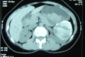
A 36-year-old female presented with history of dull aching pain in left loin of 6 months duration. She had no complaints of fever, loss of weight or appetite. She had no family history of cystic renal disease. An ultrasound scan of the abdomen showed an 84 mmx55 mm relatively well defined complex echo texture area arising from mid-pole of left kidney with multiple calcifications within, suggestive of complex cyst. CT scan of abdomen, revealed a large 7.2cmx6.2cm lesion, mostly hyper-dense with 110 HU. The cyst wall was thick and dense with calcification (Figure 1). With a provisional diagnosis of cystic renal cell carcinoma, laparoscopic radical nephrectomy was done. Post operatively she recovered well and doing well on follow up.

    Figure 1:  Computerized tomographic image showing the mixed density
lesion with calcification with adjacent compressed renal parenchyma.
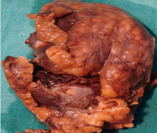
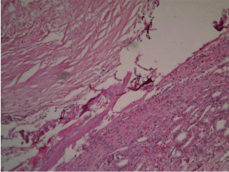
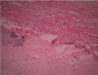
Histopathology revealed a gross specimen weighing over 100 gms, measuring 12x5x3 cms, with renal cortical thickness of 1.0 cm on cut section (Figure 2). The pelvicalyceal system was distorted by an ovoid mass measuring 10.5 cm x 7.3 cms, firm in consistency, gritty on cut section, filled with gelatinous material. Sections from cystic area showed compressed renal parenchyma with thyroidisation of tubules and the interstitium show lymphoid collections. The lining of cyst was atrophic and flattened. The lumen was filled with dense laminated strands of keratin material with areas of dystrophic material. (Figures 3 & 4) There was no malignancy or parasitic material. The final histopathologic impression was epidermoid cyst with chronic pyelonephritis.

    Figure 2:  Grossly excised specimen showing the cyst with adjacent renal
parenchyma.

    Figure 3:  The cyst lining showing keratinized squamous epithelium with
lamellated keratin. (H&Ex100 magnifications).

    Figure 4:  Cyst wall with adjacent renal parenchyma showing sclerosed
glomeruli and tubular thyroidization. (H&E x 100 magnifications).
Discussion
Epidermoid cysts of the kidney are extremely rare, unlike their preponderance in the rest of the body. Intrarenal epidermoid cysts have been encountered only in eight cases as reported in the English literature [5-11]. The origin of these cysts has been theorized to be due to either aberrant ectoderm implantation during embryogenesis or traumatic metaplasia [8]. According to some, the cyst originated from the embryonic remnant of Wolffian ducts and this hypothesis is considered the most acceptable [8].
The main presenting complaints are loin pain, haematuria and urinary frequency in the cases reported (Table 1). In our case, the patient present with dull aching left loin pain. Radiologically there are no characteristic features suggestive of epidermoid cyst. These cysts should be considered in the differential diagnosis of complex renal cysts, especially when they show thick wall and calcification.
    Author
    Age
    Sex
    Complaint
    Imaging
    Management


    Krogdah
    67
    M
    Recurrent renal colic
    Renal cyst
    Lower pole partial nephrectomy


    Duprat et al
    4
    M
    Frequency
    Calcified intrarenal mass
    Partial Nephrectomy


    Abdou And Assad
    67
    M
    Loin pain
    Multilocular cystic mass of the kidney
    Nephrectomy


    Dadali et al
    50
    M
    Loin pain, haematuria, dysuria
    Cystic mass
    Nephrectomy


    Lim and Kim
    51
    M
    Loin pain and haematuria
    Cystic mass lower pole of kidney
    Nephrectomy


    Emtage and Allen
    74
    F
    Loin pain
    Renal mass
    Nephrectomy


    Desai et al
    74
    M
    Haematuria
    Cystic renal mass
    Nephrectomy


    Rathod et al
    52
    M
    Dysuria, fever, vomiting
    Hydronephrotic kidney
    Nephrectomy
Table 1: Epidermoid cyst of the kidney reported in the literature.
As a result, the diagnosis is made most of the times on postoperative histopathology. The typical pathological features are that the cyst is lined by stratified squamous epithelium with a granular layer and filled with keratinous material that is arranged in lamina [8].
Because of the rarity of the cases, no definitive management guidelines can be made. Partial or radical nephrectomy seems to be the appropriate form of management. As these are benign cysts, the prognosis seems to be good.
Conclusion
Epidermoid cyst of the kidney is a very rare entity. High index of suspicion should be made while considering the differential diagnosis for complex renal cyst.
References
- Hagr A, Laberge JM, Nguyen LT, Emil S, Bernard C, Patenaude Y. Laparoscopic excision of sub diaphragmatic epidermoid cyst: A case report. J Pediatr Surg. 2001; 36: 8.
- Maskey P, Rupakheti S, Regmi R, Adhikary S, Agarwal CC. Splenic epidermoid cyst. Kathmandu Univ Med J. 2007; 18: 250-252.
- Tatgiba M, Iaconetta G, Samii M. Epidermoid cyst of the cavernous sinus: clinical features ,pathogenesis and treatment. Br J Neurosurg. 2000; 14: 571- 575.
- Loya AG, Said JW, Grant EG. Epidermoid cyst of the testis: radiologicpathologic correlation. Radiographics. 2004; 24: 243-246.
- Krogdahl AS. Epidermoid cyst in the kidney. Scand J urol Nephrol. 1979; 13: 131-132.
- Duprat G, Filiatrault D, Michaud J. Intrarenal epidermoid cyst. Pediatric Radiol. 1986; 16: 73-75.
- Emtage LA, Allen C. A renal epidermoid cyst. Br J Urol. 1994; 74: 125-126.
- LIM SC, Kim CS. Intrarenal epidermal cyst. Pathol Int. 2003; 53: 574-578.
- Abdou AG, Asaad NY: Intrarenal epidermoid cyst presented as an enlarged multicystic kidney. Saudi J kidney Dis Transpl. 2010; 21: 728-731.
- Rathod, Dharmendra, Rameshbhai, Pratyush Parmar. Epidermoid renal cyst: an unusual finding. Annals of Pathology and Laboratory Medicine. 2015; 2: 13-16.
- Mümtaz Dadali, Levent Emir, Melih Sunay, Elif Özer, Demokan Erol, Intrarenal Epidermal Cyst, In The Kaohsiung Journal of Medical Sciences, 2010; 10: 555-557.