Case Report
Annals Thyroid Res. 2025; 11(1): 1094.
A TIRADS V Thyroid Nodule Revealing Tuberculosis: A Rare Case of Thyroid Tuberculosis
Amine HM1*, Najout H2, Walid A3, Younes A4 and Seddiki R3
1ENT Departement, Hassan II Military Teaching Hospital, Laayoune, Morocco
2Anesthesiology and intensive care Unit, Mohamed V Military Teaching Hospital, Rabat, Morocco
3Anesthesiology and intensive care Unit, Hassan II Military Teaching Hospital, Laayoune , Morocco
4Pneumo-phthisiology Departement; Hassan II Military Teaching Hospital, Laayoune, Morocco
*Corresponding author: Hanine Mohamed Amine, ENT Departement, Hassan II Military Teaching Hospital, Laayoune, Morocco Email: hanmohami@gmail.com
Received: June 06, 2025 Accepted: June 19, 2025 Published: June 23, 2025
Abstract
Introduction: Thyroid tuberculosis is a rare form of extrapulmonary tuberculosis. Due to its nonspecific clinical and radiological presentation, it can easily mimic thyroid cancer.
Case Report: A 55-year-old female presented with a thyroid swelling. Neck ultrasound showed a 26 mm TIRADS V nodule. Total thyroidectomy was performed. Histopathological examination revealed isolated thyroid tuberculosis.
Discussion: This case highlights the rarity of thyroid tuberculosis and the diagnostic challenge it poses. It underlines the importance of considering this diagnosis in the differential, especially in endemic areas.
Conclusion: Even when clinical and imaging signs suggest malignancy, thyroid tuberculosis should be considered. Histology remains crucial for diagnosis and management.
Keywords: Tuberculosis; TIRADS; Thyroidectomy; Thyroid nodule
Introduction
Tuberculosis remains a global health concern, with a growing number of extrapulmonary manifestations in endemic areas. However, thyroid involvement is exceedingly rare, accounting for less than 1% of all extrapulmonary tuberculosis cases. This rarity, coupled with a nonspecific presentation, often leads to misdiagnosis, especially when nodular thyroid disease is suspected to be malignant. We present a case of isolated thyroid tuberculosis discovered postoperatively after total thyroidectomy surgery.
Case Presentation
A 55-year-old woman, with no particular medical history, presented with a progressively enlarging anterior neck mass. She reported no fever, weight loss, or night sweats. Clinical examination revealed a painless, enlarged thyroid gland with no cervical lymphadenopathy.
Neck ultrasound showed a 26 mm hypoechoic nodule with irregular margins and microcalcifications, categorized as TIRADS V. Given the high suspicion of malignancy, total thyroidectomy was indicated and performed without complication.
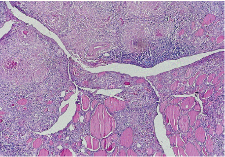
Histopathological analysis revealed granulomatous inflammation with caseating necrosis, consistent with thyroid tuberculosis (Figure 1). There were no malignant features.

    Figure 1: Caseating necrosis, consistent with thyroid tuberculosis further
evaluation including chest radiography and tuberculin skin test; which was
positive; showed no evidence of pulmonary or extrapulmonary tuberculosis.
Anti-tuberculosis therapy was initiated with and the patient remains with
good health.
Discussion
Thyroid tuberculosis is an exceptionally rare condition, even in countries where tuberculosis is widespread. Several theories have been proposed to explain this rarity including the thyroid’s high iodine content, the bactericidal properties of colloid, its rich blood supply, and the absence of native lymphoid tissue. Still, isolated cases are reported from time to time, often discovered by chance during surgery initially intended to investigate a suspected thyroid cancer [1].
In our case, the patient showed no obvious symptoms, and there was nothing in her background to suggest tuberculosis. It was a neck ultrasound that raised suspicion of a malignancy, as the nodule had features typical of a TIRADS V classification: hypoechoic appearance, irregular margins, and microcalcifications. Given these findings, and the nodule’s size (26 mm), surgery was the logical next step. As is often seen in the literature, the diagnosis of thyroid tuberculosis was only made after analyzing the removed tissue under the microscope.
While fine-needle aspiration is a useful tool for evaluating thyroid nodules, it’s not particularly helpful for detecting tuberculosis. It might suggest granulomatous inflammation, but actually identifying Mycobacterium tuberculosis this way is rare unless specific molecular tests (like PCR) or specialized cultures are performed [2].
Treatment involves starting a standard anti-tuberculosis drug regimen, as recommended by the WHO and national pulmonology societies. For our patient, we followed this schedule [3]:
¾¾ An initial two-month phase using four drugs – Rifampicin (R), Isoniazid (H), Pyrazinamide (Z), and Ethambutol (E).
¾¾ Followed by a four-month continuation phase with just Rifampicin and Isoniazid.
This 2RHZE/4RH regimen, lasting a total of six months, is the most commonly used for uncomplicated forms of extrapulmonary TB.
In localized cases like this one without widespread disease or significant lymph node involvement this treatment is usually sufficient and well tolerated. Our patient responded very well, and no further surgery or drainage was needed since the thyroidectomy had already removed the affected tissue.
Routine blood tests are necessary during treatment to monitor for possible liver toxicity, particularly from Isoniazid and Rifampicin. In our patient’s case, liver function remained normal throughout [4].
There are currently no specific guidelines for treating thyroid tuberculosis, but the evidence suggests that six months of therapy is generally enough, provided there are no risk factors for recurrence [5].
This case is a good reminder that when faced with an unusual thyroid nodule especially in regions where TB is common we need to keep tuberculosis in mind as a possible cause. While surgery often ends up being necessary when the diagnosis isn’t clear, better awareness of this rare condition might allow for earlier, less invasive diagnosis in the future and maybe even help avoid surgery in some well-documented cases
Conclusion
This case illustrates the importance of considering tuberculosis in the differential diagnosis of thyroid nodules, particularly in endemic regions. Surgery is often required for diagnosis, and antituberculosis treatment leads to excellent outcomes when initiated promptly.
Ethics
Ethical conditions are approved.
The Work was Reported in Accordance with the Scare Guidelines
During the preparation of this work the author used HANINE MOHAMED AMINE/ENT in order to VERIFY THE QUALITY OF WORK. After using this tool, the author reviewed and edited the content as needed and takes full responsibility for the content of the publication.
References
- Golden MP, Vikram HR. Extrapulmonary tuberculosis: an overview. Am Fam Physician. 2005; 72: 1761–1768.
- Russ G, Royer B, Bigorgne C, et al. Proposal of a TIRADS classification of thyroid nodules based on ultrasound features. Eur Radiol. 2013; 23: 2532– 2541.
- World Health Organization. Treatment of tuberculosis guidelines. 4th ed. WHO; 2010.
- Papi G, Corrado S, Carani C, et al. Thyroid tuberculosis: a diagnostic challenge. Clin Endocrinol (Oxf). 2004; 60: 552–556.
- Sharma SK, Mohan A. Extrapulmonary tuberculosis. Indian J Med Res. 2004; 120: 316–353.