
Research Article
Int J Nutr Sci. 2022; 7(1): 1064.
Palatal Implantation Using Platelet Rich Plasma and Radio Frequency Palatal Surgery for Management of Obstructive Sleep Apnea
Elsayad OA*, Bioumy OE and El Sayed MD
Department of Otolaryngology, Benha University, Egypt
*Corresponding author: Osama A Elsayad, Department of Otolaryngology, Benha University, Benha 13111, Egypt
Received: June 09, 2022; Accepted: July 07, 2022; Published: July 14, 2022
Abstract
Background: Obstructive Sleep Apnea (OSA) is a common health problem affecting large number of people all over the world with great psychological and physiological burdens. The aim of this study is to investigate the effect of radio frequency palatal Surgery and platelet rich plasma in the soft palate as a new technique in treatment of obstructive sleep apnea OSA.
Patients and Methods: This study included 30 patients with mild to moderate OSA Group A: n=15 (Tonsillectomy and palatal radio frequency surgery) and Group B n=15 (Tonsillectomy and Platelet Rich Plasma (PRP) palatal injection, this study investigate the effect of body mass index, retropalatal anteroposterior diameter, Epworth Steepness Scale Apnea Hypopnia Index in treatment of OSA in both groups.
This study found that both techniques improved day time sleepiness with reduction of Apnea-Hypopnea Index (AHI), also there was marked improvement in radiological parameters in the form of increase of the anteroposterior diameter and retro-palatal cross section area, there was reduction of Body Mass Index BMI in postoperative in both groups but there was superiority of group A over group B in improvement of this parameters, group A success rate was 86.6% and Group B was 80%.
Conclusion: Radio frequency palatal surgery and platelet rich plasma, both are effective as a treatment of obstructive sleep apnea due to retro-palatal collapse.
Keywords: Radiofrequency Ablation; Platelet Rich Plasma; Snoring; Obstructive Sleep Apnea; Body Mass Index; Apnea Hypopnea Index; Palatal; Retropharyngeal Diameter
Introduction
Obstructive Sleep Apnea (OSA) is a serious health problem affecting a large number of people all over the world. The prevalence ranged from 9% to 37%, the Apnea-Hypopnea Index (AHI) =5, being higher in men. It increases with age. OSA prevalence is directly proportional to the Body Mass Index (BMI), so the disease is more common among obese people [1].
Obstructive Sleep Apnea (OSA) is presented by pharyngeal collapse during sleep, Pharyngeal collapse can occur at the retropalatal level (soft palate), retroglossal level (tongue base), oropharyngeal lateral walls, and/or hypopharynx. [1].
Radiofrequency Ablation (RFA) is effective in treatment of OSA, The mechanism of action for RFA is due to a low temperature, high frequency current leads to local inflammation, fibrosis and stiffening of the tissue. RFA of the soft palate is minimally invasive, with few complications, and aims to stiffen the soft palate [2].
The treatment of patients with obstructive sleep apnea using palatal implantation in the of multi- level or stepwise surgery has been relatively little research on using palatal implants alone for the treatment of OSA [3].
Platelets are unique blood elements initiating homeostasis and healing processes. Platelet Rich Plasma (PRP) is plasma contains a high concentration of platelets. Data from human and animal studies provide both direct and indirect evidence that platelet rich plasma plays a considerable role in tissue regenerative processes [4].
The recent development of platelet concentrate for surgical use is an evolution of the fibrin glue technologies [5], these different technologies were tested in many clinical fields, particularly oral and maxillofacial surgery, Ear-Nose-Throat surgery, plastic surgery, orthopedic surgery, sports medicine, gynecologic and cardiovascular surgery and ophthalmology [6].
This study aimed to investigate the effect of palatal Radiofrequency technique and platelet rich plasma injection in the soft palate as a new technique in treatment of mild and moderate obstructive sleep apnea.
Patients and Methods
This study is a prospective randomized case series clinical study. The study was conducted on Otorhinolaryngology clinics of Benha University Hospitals, during the period from June 2019 June 2021. This study included 30 patients with mild to moderate OSA were divided into two groups
The study was approved by the local ethics committee of faculty of medicine, Benha University in accordance with the declaration of Helsinki. Oral and written consent were taken from all patients who participated in this study
Inclusion Criteria
• Both sexes
• Patients> 18 years old, and < 60 years.
• Body Mass Index (BMI) of patients less than 35 kg/m2.
• Patients diagnosed with OSAS with palatal flutter and collapse.
• Documented failure/refusal of attempts of conservative treatment measures (not limited to continuous positive airway pressure CPAP).
• Class 1 occlusion.
• Pharyngeal tonsillar size grade I–II- III
Exclusion Criteria
• Patient age<18 or >60 years old.
• Marked deviated septum or marked hypertrophy of inferior turbinate
• Modified Mallampati classification: class IV tongue position
• Retrognathia, craniofacial abnormalities, chronic rhino sinusitis, trismus, anesthetic allergies.
• Patient with central type apnea.
• Body mass index (BMI)>35Kg/m2.
• Failure to attend postoperative follow-up polysomnography
• Previous surgery to the palate or other surgical treatment of OSAHS.
• A history of malignancy of the head and neck region, laryngeal trauma, or other previous oropharyngeal/laryngeal surgery.
• Class 2 occlusion.
• Patients with severe medical illness.
Study Population
Thirty patients complaining of OSA (mild to moderate) degree, due to retropalatal collapse. After getting informed consent, patients were randomly allocated to two groups:
Group A: tonsillectomy with palatal radiofrequency technique
Group B: tonsillectomy with palatal PRP injection.
Preoperative Evaluation
• Full detailed history (personal, present and past history)
• Complete clinical and physical examination:
• Otorhinolaryngological examination.
• Endoscopic examination by Awake Fiberoptic
Nasopharyngoscopy with Müller’s maneuver:
• Epworth Sleepiness Scale
• Polysomnography (PSG):
• Radiological examination, by Volumetric CT for the upper airway to evaluate the retropalatal space:
• Other preoperative investigation
• Bleeding profile
• Liver functions
• Kidney functions
• Blood sugar
• Viral markers
• ECG
• Chest X-Ray
Operative Procedures
• Patients were operated upon through the period from June 2019 to June 2021
• All the operative procedures were done by same E.N.T. surgeon.
• Patients were randomly allocated to two groups each included fifteen patients.
• Group A: n=15 palatal radiofrequency technique
• Group B: n=15 Tonsillectomy and PRP injection
• All patients underwent transoral endotracheal intubation under general anesthesia.
Group A: As shown in (Figure 1,2)
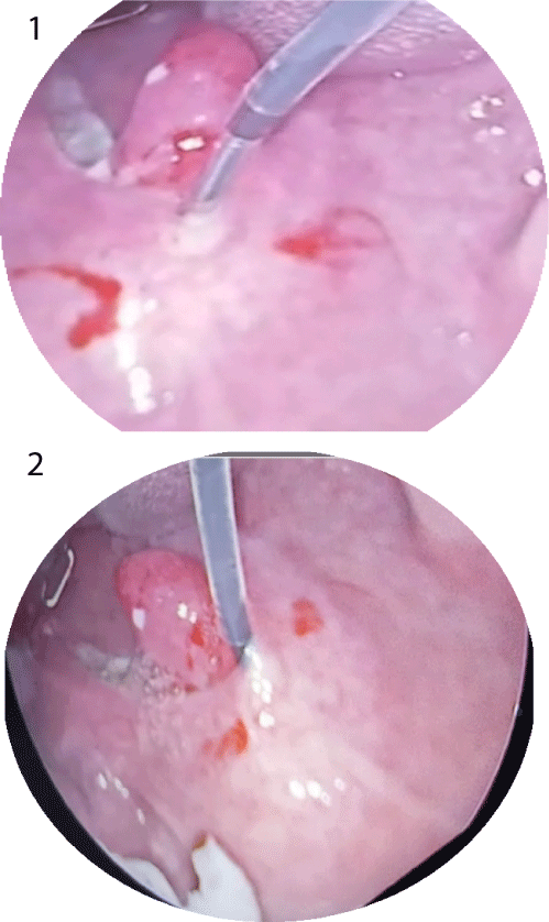
Group A: Figure 1 & 2: Palatal radio frequency technique.
• Tonsillectomy by cold dissection
• The procedure was performed in the operating room under general anesthesia. The Gyrus radiofrquency device was set to 81°C and 700 J. The probe was passed in the midline and 1 cm into the paramedian locations bilaterally (2100 J total). The patient was seen one week following surgery and was doing well without fever, pain, or complication [7].
Group B: As shown in (Figure 3,4)
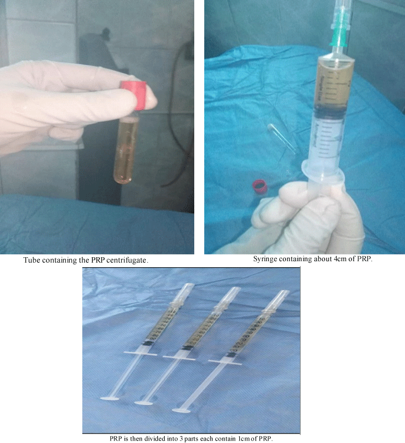
Figure 3: Group B.
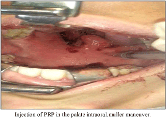
Figure 4: Group B.
• Tonsillectomy by cold dissection
• Prepared PRP 6cm is then divide into 3 insulin syringes each contain 1 cm of PRP.
• Injection of the PRP was done at three lines:
• 1st line is midline
• 2nd line rt paramedian
• 3rd line lt paramedian
• line starts 0.5 mm from the junction of soft and hard palate to the base of the uvula
• Aspiration then slowly injection of PRP in three lines as described.
Postoperative Evaluation
• Clinical examination: Follow-up visits were scheduled at 1, 2, 3 weeks, and 3 months postoperative.
• Epworth sleepiness scale (ESS): Daytime sleepiness Assessment Snoring loudness and ESS score were assessed at baseline, 3 months after surgery. In addition, the AHI and BMI between baseline and 3 months postoperative were also compared [8].
• Polysomnography (PSG): Polysomnography was done after 3 months, following the same protocol as preoperative one.
Surgical success was considered when there was 50% reduction of preoperative AHI index [9].
• Awake Fiberoptic Nasopharyngoscopy with Müller’s maneuver: Awake Endoscopic examinations with Müller’s maneuver to evaluate the site of collapse were done for all patients after 3 months [10].
• Radiologic investigations: volumetric CT was done 3 months postoperatively for all patients, following the same protocol as preoperative evaluation.
• Complications: We recorded; Postoperative infection, little bleeding, throat discomfort, foreign body sensation and velopharyngeal insufficiency were identified as an adverse event in this study.
Statistical Analysis
The clinical data were recorded on a report form. These data were tabulated and analyzed using the computer program SPSS (Statistical package for social science) version 25 to obtain descriptive data. Descriptive statistics were calculated for the data in the form of mean and standard deviation (SD±) for quantitative data, Frequency and distribution for qualitative data. In the statistical comparison between the different groups, the significance of difference was tested using student’s t-test, Chi square test and Correlation Study [11].
Results
The present study included 30 patients with obstructive sleep apnea ,17 male and 13 female with age range from 21-53 years, they included two groups: Group A=15 was subjected to tonsillectomy and palatal radiofrequency and group B =15 was subjected to tonsillectomy and palatal PRP injection , both groups were matched regarding age and sex distribution (Table 1).
      Gender

    Group A (n = 15)

    Group B (n = 15)


      Test of Sig.


      p


    No.

    %

    No.

    %


    Male

    9

    60.0

    8

    53.2

    χ2= 0.136


      0.713


    Female

    6

    40.0

    7

    46.7


    Age (years)


    Min. – Max.

    22.0 – 53.0

    21.0 – 42.0


      t= 1.124


      0.271


    Mean ± SD.

    35.93 ± 9.0

    32.80 ± 5.97


    Median (IQR)

    35.0 (31.0 – 40.50)

    33.0 (29.50 – 37.0)


    p: p value for comparing between the    studied groups, significant if >0.05.
Table 1: Comparison between the two studied groups according to demographic

data.
Comparison between the two studied groups according to decrease in Body mass index (BMI): The mean decrease in BMI of Group A was (0.47 ± 1.36 kg/m2). While the mean BMI of Group B was (0.67 ± 1.18kg/m2) which is not a significant difference (P-Value 0.967) (Table 2). Also, the comparison according to decrease in Epworth sleepiness scale (ESS); the mean decrease in ESS of group A was (6.27 ± 2.19). While it was (4.87 ± 2.56) in group B which is not a significant difference (P-Value 0.089) (Table 3).
BMI
A
B
U
P
Preop
Postop
P
Preop
Postop
P
Range
20.0 – 30.70
18.0 – 30.0
0.205
20.0 – 32.0
20.0 – 32.0
0.045*
111.0
0.967
Mean ± SD.
25.20 ± 3.60
24.73 ± 4.10
25.07 ± 4.02
24.40 ± 4.58
Median (IQR)
25.0 (22.50 –
28.0)24.0 (21.50 –
28.50)24.0 (22.50 –
28.50)23.0 (20.0 –
28.0)p: p value for comparing between Pre-op: pre-operative and Post-op: post-operative *: Statistically significant at p = 0.05
BMI: body mass index
Table 2: Comparison between the two studied groups according to change in BMI.
ESS
A
B
U
P
Preop
Postop
P
Preop
Postop
P
Range
8.0 – 17.0
3.0 – 13.0
0.001*
5.0 – 17.0
2.0 – 14.0
0.001*
71.50
0.089
Mean ± SD.
12.13 ± 2.77
5.87 )± 3.16
10.53 ± 3.93
5.67 ± 3.73
Median (IQR)
11.0 (10.0 –
14.0)5.0 (3.50–7.0)
9.0 (7.50 –
14.50)4.0 (3.0 –7.0)
p: p value for comparing between pre and post*: Statistically significant at p = 0.05
ESS: Epworth Sleepiness Scale
Table 3: Comparison between the two studied groups according to change in ESS.
The comparison between the two studied groups according to decrease in Apnea- Hypopnea Index (AHI); The mean decrease in AHI of group A was (5.91 ± 2.50). While it was (5.47 ±1.77) in group B which is not a significant difference (P-Value 0.713) (Table 4). Also according to Increase in Antro-Posterior diameter (A-P) (mm); The mean increase in A-P diameter (mm) of group A was (2.64 mm ± 1.57 mm). While it was (1.67 mm ± 1.0 mm) in group B which is not a significant difference (P-Value 0.106) (Table 5).
AHI
A
B
U
P
Preop
Postop
P
Preop
Postop
P
Range
7.0 – 22.0
3.0 – 16.0
0.001*
8.0 – 20.0
4.0 – 15.1
0.001*
103.0
0.713
Mean ± SD.
13.53 ± 5.32
7.62 ± 4.32
12.93 ± 3.97
7.47 ± 3.60
Median (IQR)
11.0 (9.0 –
17.50)6.0 (4.15 – 10.0)
12.0 (10.0
– 15.50)6.0 (5.0 – 8.50)
p: p value for comparing between pre and post*: Statistically significant at p = 0.05
AHI: Apnea-Hypopnea Index
Table 4: Comparison between the two studied groups according to change in AHI.
A-P
diameter (mm)A
B
U
P
Preop
Postop
P
Preop
Postop
P
Range
3.10 – 10.0
3.20 – 11.40
2.50 – 10.0
2.70 – 11.21
Mean ± SD.
6.59 ± 2.15
9.23 ± 2.54
0.001*
6.59 ± 2.25
8.26 ± 2.71
0.001*
73.50
0.106
Median (IQR)
6.20
(5.30 –
8.20)9.80
(9.0 –
11.0)
6.30
(5.50 –
8.45)8.50
(8.15 –
10.0)
p: p value for comparing between pre and post*: Statistically significant at p = 0.05
A-P diameter: Antro-posterior diameter of retropalatal (RP) area
Table 5: Comparison between the two studied groups according to change in A-P diameter (mm).
The Comparison between both groups according to degree and site of collapse in retropalatal area during Muller maneuver; Regarding) improvement in degree of collapse in retropalatal area: In group A the 1st degree became 86.7% while became 80.0 % in group B which was not a significant difference between both groups (P-Value0.592) (Figure 5), Also as Regards the site of collapse in retropalatal area during Muller maneuver: in group A; improvement with no detected site of collapse in 86.7% while it is 80.0% at group B, which is not a significant difference between both groups (P- Value0.592) (Figure 6).
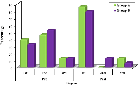
    Figure 5: Comparison between the two studied groups according to Degree

of collapse in retropalatal area during muller maneuver.
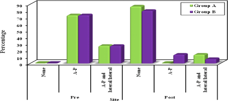
    Figure 6: Comparison between the two studied groups according to Site of

collapse in retropalatal area during muller maneuver.
The Outcome of both groups: Success rate = cured cases + success cases, In Group A: success rate was 86.6% while 80% in Group B: which is not a significant difference between both groups; P-Value:1.000 (Table 6).
      Outcome

    Group A (n = 15)

    Group B (n = 15)


      Χ2


      MCp


    No.

    %

    No.

    %


    Success

    8

    53.3

    8

    53.3


      0.435


      1.000


    Cure

    5

    33.3

    4

    26.7


    Fail

    2

    13.3

    3

    20.0


    2:Chi square test;MC:Monte    Carlo


      p: p    value for comparing between the studied groups *: Statistically    significant at p = 0.05


      Cure: AHI post < 5 and ESS post< 10 and reduction of both of them    > 50%


      Success: AHI post< 15 and ESS post< 10 and    reduction of both of them > 50%


      Failure: AHI post = 30 or ESS post = 10 or    reduction of both of them = 50%
Table 6: Comparison between the two studied groups according to outcome.
Discussion
As the field of sleep surgery expands and new procedures are being created to address the dynamic upper airway thorough understanding of the fundamental principles of this surgery, palatal surgeries focused on the lateral pharyngeal wall collapse, it varies from lateral pharyngoplasty, Barbed Reposition Pharyngoplasty and radiofrquency palatal surgery [12].
Our study was undertaken to show the effect of palatal stiffening using our new techniques (PRP) and palatal radiofrequency technique to determine the short-term success rate of the two procedures in the elimination of snoring and OSA, this study may address a clinical application of this biological product (PRP) in management of Obstructive sleep apnea due to retropalatal collapse .
The presence of palatal implants (PRP) does not affect or complicate subsequent palatal revision procedures [13], hence we adopt the non-respective manner for correction of the AHI with preservation of soft palate horizontal part and vertical part (uvula) and pillars.
The challenge of this procedure is preservation of soft tissue. Based on the above facts, the hypothesis of our study stands upon Evaluating a non-resective techniques of soft palate surgeries to prove which technique allows the best functional outcomes in OSA patients with single-level collapse (retropalatal collapse).
So the intrapharyngeal surgery (soft palate surgery) is a targetoriented procedure that needs to be performed precisely and combines with integrated treatment for OSA patients [14].
This study found that both techniques of palatal implantation using platelet rich plasma (PRP) and palatal radiofrequency technique improved daytime sleepness with reduction of AHI [15], also there was excellent improvement in radiological parameters in the form of enlarge the antro-posterior diameter and prove that BMI is an important factor in short term success of our techniques [16].
We found superiority of group A over group B in improvement of all parameters, that In Group A: success rate was 86.6% while in Group B was 80%. No major postoperative complications were recorded; as: severe bleeding which requires surgical intervention, severe edema which requires tracheotomy, or secondary infection. Some minor complications were recorded, mainly pain and dysphagia that improved in a few days.
Conclusion
palatal radiofrquency technique and platelet rich plasma injection technique, both are effective as a treatment of obstructive sleep apnea due to retropalatal collapse.
Sources of Funding
This research did not receive any specific grant from funding agencies in the public, commercial, or not-for-profit sectors.
Author Contribution
Authors contributed equally in the study.
Conflicts of Interest
No conflicts of interest.
References
- Rashwan MS, Montevecchi F, Cammaroto G, Deen MBE, Iskander N, Hennawi DE, et al. Evolution of soft palate surgery techniques for obstructive sleep apnea patients: A comparative study for single-level palatal surgeries. Clinical Otolaryngology. 2018; 43: 584-590.
- Lauren K. Reckley Camilo Fernandez-Salvador Edward T. Chang Macario Camacho, Soft palate fistula after radiofrequency ablation for primary snoring: a case report and literature, Brazilian Journal of Otorhinolaryngology. 2020; 86: 20-22.
- Lee Y, Lee L, Li H. The palatal septal cartilage implantation for snoring and obstructive sleep apnea. Auris, nasus, larynx. 2018; 45: 1199-1205.
- Everts PAM, Knape JTA, Weibrich G, Schönberger JPAM, Hoffmann J, Overdevest EP, et al. Platelet-rich plasma and platelet gel: a review. The journal of extra-corporeal technology. 2006; 38: 174-87.
- Simonpieri A, Corso MD, Vervelle A, Jimbo R, Inchingolo F, Sammartino G, et al. Current knowledge and perspectives for the use of platelet-rich plasma (PRP) and platelet-rich fibrin (PRF) in oral and maxillofacial surgery part 2: Bone graft, implant and reconstructive surgery. Current pharmaceutical biotechnology. 2012; 13: 1231-1256.
- Bielecki T, Ehrenfest DMD. Platelet-rich plasma (PRP) and Platelet-Rich Fibrin (PRF): surgical adjuvants, preparations for in situ regenerative medicine and tools for tissue engineering. Current pharmaceutical biotechnology. 2012; 13: 1121-1130.
- Vicini C, Hendawy E, Campanini A, Eesa M, Bahgat A, AlGhamdi S, et al. Barbed reposition pharyngoplasty (BRP) for OSAHS: a feasibility, safety, efficacy and teachability pilot study. “We are on the giant’s shoulders”. European Archives of Oto-Rhino-Laryngology. 2015; 272: 3065-3070.
- Yousuf A, Beigh Z, Khursheed RS, Jallu AS, Pampoori RA. Clinical Predictors for Successful Uvulopalatopharyngoplasty in the Management of Obstructive Sleep Apnea. International Journal of Otolaryngology. 2013; 2013: 1-6.
- Megwalu UC, Piccirillo JF. Methodological and statistical problems in uvulopalatopharyngoplasty research: a follow-up study. Archives of otolaryngology--head & neck surgery. 2008; 134: 805.
- Pang KP, Pang EB, Win MTM, Pang KA, Woodson BT. Expansion sphincter pharyngoplasty for the treatment of OSA: a systemic review and metaanalysis. European Archives of Oto-Rhino-Laryngology. 2015; 273: 2329- 2333.
- Cammaroto G, Montevecchi F, D’Agostino G, Zeccardo E, Bellini C, Meccariello G, et al. Palatal surgery in a transoral robotic setting (TORS): preliminary results of a retrospective comparison between uvulopalatopharyngoplasty (UPPP), expansion sphincter pharyngoplasty (ESP) and barbed repositioning pharyngoplasty (BRP). Acta Otorhinolaryngologica Italica. 2017; 37: 406-409.
- Yang L, Hu S, Yan M, Yang J, Tsou S, Lin Y. Antimicrobial activity of plateletrich plasma and other plasma preparations against periodontal pathogens. Journal of periodontology. 2015; 86: 310-318.
- Randerath WJ, Verbraecken J, Andreas S, Bettega G, Boudewyns A, Hamans E, et al. Non-CPAP therapies in obstructive sleep apnoea. European Respiratory Journal. 2011; 37: 1000-1028.
- Cho JH, Kim JK, Lee H, Yoon J. Surgical anatomy of human soft palate. The Laryngoscope. 2013; 123: 2900-2904.
- Sinkkonen ST, Ruohoalho J, Pietarinen P, Aaltonen L, Mäkitie AA, Bäck LJ. Limited long-term outcome of single-session soft palate interstitial radiofrequency surgery for habitual snoring in tertiary care academic referral centre: Our experience on 77 patients. Clinical Otolaryngology. 2017; 42: 470-473.
- Thorbjorn Eva Levring-Jäghagen, Karl A Franklin, Marie Lindkvist Effects of Radiofrequncy versus sham surgery of the soft palate on daytime sleepiness Laryngoscope. 2014; 124: 2422-6.