Case Report
Austin J Radiol. 2015;2(2): 1013.
A Rare Complication in the Treatment of Gastrointestinal Bleeding via Superselective Embolization: A Case Report
Cetinkaya OA1, Konca C1, Kocaay F1, Peker A2 and Akyol C1*
1Ankara University, Department of General Surgery, Turkey
2Ankara University, Department of Radiology, Turkey
*Corresponding author: Cihangir Akyol, Ankara University, Faculty of Medicine, Department of General Surgery, Division of Colorectal Surgery, 06100, Sihhiye, Ankara, Turkey
Received: December 10, 2014; Accepted: February 24, 2015; Published: February 27, 2015
Abstract
Acute lower Gastrointestinal (GI) bleeding is conventionally determined as a bleeding occured from a distal part of the ligament of Treitz. The bleeding stops spontaneously in most of the cases, and the mortality rate is reported as 2 to 4%. In the treatment of GI bleeding, local endovascular vasoconstrictive therapy, thermal cautery or bipolar injection delivered via an endoscope, and surgical resection are accepted as popular modalities. Superselective Mesenteric embolization (SME) has rapidly gaining acceptance as a treatment option for severe intestinal hemorrhage, especially for the patients with comorbidities. Surgical resection is still an option for the cases of severe bleeding but peroperative morbidity and mortality rates are higher in the patients with co-morbidities. Superiority of SME to vasopressin infusion is reported as the success of the durability of the bleeding. In superselective embolization series, clinical success ranged from 44% to 91% and major ischemic complications ranged from 0% to 6%. Enthusiasm for this technique continues to grow due to its inherent advantages as compared with vasopressin infusion. With the embolization, bleeding is stopped at the time of the procedure without a prolonged infusion or multi-angiograms.
We report here a case of a colonic bleeding treated by superselective embolization, but underwent surgery due to a complication of mesenteric ischemia.
Keywords: Gastrointestinal bleeding; Embolization; Acute ischemia
Case Report
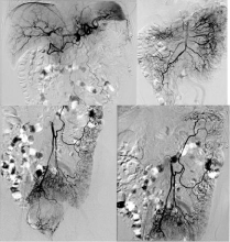
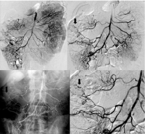
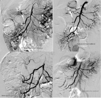
A 75-year-old male patient with type 2 diabetes mellitus and gallbladder neoplasia, admitted to our hospital with a complaint of spontaneous rectal bleeding consisting for 48 hours which occured only between 1 a.m. and 3 a.m.. Vital signs were found normal at the time of the administration. Hemoglobin level was 10,3 g/dL and hematocrit was %31,7. The patient hospitalized and observed for 2 days with a conservative management. In that period, he had recurrent hematochezia and needed two units (approximately 500 mL) of blood transfusion. Colonoscopy could not reveal the origin of the bleeding because of the colon was full of blood. There was no sign of hemorrhage in the upper GI endoscopy. A computerized tomographic angiogram was performed and the only pathologic finding was diverticulosis about the intestinal system without any proof of bleeding. In the need for ongoing blood transfusion and the unsuccessful endoscopic procuderes indicated for a digital substraction angiogram to diagnose and treat the problem. Bleeding could not be seen in the selective canulation of superior mesenteric and inferior mesenteric arteries (Figure 1). A negative colonoscopy was performed afterwards and the patient observed for that day. Massive bleeding attacks repeated at midnight and therefore conventional angiography was reperformed simultaneously. An extravasation originated from the distal branches of the right colic and the middle colic arteries was determined in our case. Embolization of the distal branches of the right colic artery was successfully done with the Polyvinyl Alcohol (PVA) molecules of 355-500 microns; however the middle colic artery could not be catheterized superselectively because of the angulation at the branching level and PVA was performed at this level (Figure 2). At the control angiogram, no signs of bleeding was determined (Figure 3).

Figure 1: First negative angiography images.

Figure 2: Second angiography images (Bleeding site marked by arrow).

Figure 3: Angiography images after PVA embolization.
In the following hours of the procedure, right lower quadrant pain and tenderness became clear. Clinical and laboratory findings indicated for an acute event, especially for a mesenteric ishchemia. A diagnostic laparoscopy was performed with a diagnosis of the ischemia in the right hemicolon and distal ileum without a perforated segment. Laparoscopic right hemicolectomy with a segment of 80-cm ileum resection was performed and side to side ileotransversostomy was established. Sessile serrated polyp located at the right colon specimen which could not be detected by the colonoscopy in the reason of colonic blood was examined in the histologic examination. Patient recovered without a bleeding or any other complication and discharged at the postoperative day 4.
Discussion
Traditionally, acute lower Gastrointestinal (GI) bleeding has referred to a blood loss of recent onset originating from a site distal to the ligament of Treitz. Bleeding is mostly ceased spontaneously by a conservative managment, approximately 85% of the cases. Superselective embolization has been preferred as a less invasive procedure comparing to a surgical approach, and found as succesful when compared to vasopressin infusion [1-3].
Superselective embolization is an alternative option in the cases of severe GI bleeding. Several reports in the literature citing indicates a technical success of over 90% without serious morbidity and mortality rates. Definition of the vascular area causes bleeding is the first and the most important step for the superselective embolization with the lower rates of complications. In our case, the definiton of the origin of the bleeding branches was correct; however the cannulation of the middle colic artery was problematic [4-8].
The incidence of ischemic events can be expected to be decreased with developing technology and experience, but the risk will be everpresent, and must be accepted considering the alternatives. Although the risk of ischemic complication is reported in lower rates, it carries a high risk of morbidity, with several reports highlighting a higher risk of anastomotic leak in the patients with primary anastomosis done initially for ischemic or infarcted bowel. In addition, the reported mortality among the patients who suffered postembolization ischemia was higher [5-7].
Conclusion
Minimal invasive tecniques have become important in the management of GI bleeding, especially patients with co-morbidities. Superselective embolization is a minimal invasive modality for the treatment of colonic bleeding. Although it has high rates of success, its ischemic complications are still remaining as a major problem.
References
- Zuccaro G. Management of the adult patient with acute lower gastrointestinal bleeding. American College of Gastroenterology. Practice Parameters Committee. Am J Gastroenterol. 1998; 93: 1202-1208.
- Farrell JJ, Friedman LS. Review article: the management of lower gastrointestinal bleeding. Aliment Pharmacol Ther. 2005; 21: 1281-1298.
- Funaki B. Superselective embolization of lower gastrointestinal hemorrhage: a new paradigm.Abdom Imaging. 2004; 29: 434-438.
- Bandi R, Shetty PC, Sharma RP, Burke TH, Burke MW, Kastan D. Superselective arterial embolization for the treatment of lower gastrointestinal hemorrhage. J Vasc Interv Radiol. 2001; 12: 1399-1405.
- Funaki B, Kostelic JK, Lorenz J, Ha TV, Yip DL, Rosenblum JD, et al. Superselective microcoil embolization of colonic hemorrhage. AJR Am J Roentgenol. 2001; 177: 829-836.
- Gordon RL, Ahl KL, Kerlan RK, Wilson MW, LaBerge JM, Sandhu JS, et al. Selective arterial embolization for the control of lower gastrointestinal bleeding. Am J Surg. 1997; 174: 24-28.
- Silver A, Bendick P, Wasvary H. Safety and efficacy of superselective angioembolization in control of lower gastrointestinal hemorrhage. Am J Surg. 2005; 189: 361-363.
- Tan KK, Wong D, Sim R. Superselective embolization for lower gastrointestinal hemorrhage: an institutional review over 7 years. World J Surg. 2008; 32: 2707-2715.