
Case Series
Austin J Radiol. 2015;2(2): 1015.
Computed Tomographic and Magnetic Resonance Imaging Findings in the Midline Nasopharyngeal Glial Heterotopia
Junfang Xian*
Department of Radiology, Capital Medical University, China
*Corresponding author: Junfang Xian, Department of Radiology, Beijing Tongren Hospital, Capital Medical University, 1 Dongjiaominxiang Street, District of Dongcheng, Beijing 100730, China
Received: January 06, 2015; Accepted: March 11, 2015; Published: March 17, 2015
Abstract
The glial Heterotopia is a rare developmental abnormality in the nasopharynx. We report two girls with this lesion. CT and MR imaging demonstrated an ovoid heterogeneously pedunculated mass attached to the left nasopharyngeal wall by a stalk of fibrous tissue, with no extension into the cranial vault. Scattered patchy fat tissues and pituitary tissues found in the glial heterotopia in one case were reported for the first time. Multiplanar images with multidetector CT, especially sagittal CT images never reported in the glial Heterotopia, might be very helpful in evaluating the adhesion between the mass and the structures in the nasopharynx, whether the bony defect of the skull base, and whether the communication between the mass and the intracranial structures.
Keywords: Glial heterotopia; Nasopharynx; Computed Tomography; Magnetic resonance imaging
Introduction
Glial heterotopia, used to refer to ectopic central nervous tissue arising as a developmental malformation, is a rare developmental abnormality seen in a wide age group but typically presenting at birth or in early childhood [1]. The most frequently affected site is the nasal area [2]. Less commonly, brain heterotopias have been reported in the tongue [3], palatopharyngeal complex [4-6], orbit [7,8], scalp [9], ear [10] and cervical spaces such as the infratemporal space [1,11] and the pterygopalatine fossa [12]. The glial Heterotopia in the nasopharynx has rarely been reported [4,6]. We herein present two additional cases of glial heterotopia arising in the nasopharynx and describe CT and MR imaging findings, in which fat tissues and pituitary tissues contained in the glial heterotopia were reported for the first time in this paper adding new information for the glial heterotopia. Moreover, Multiplanar images with multidetector CT, especially sagittal CT images never reported in the glial heterotopia, can be very helpful in evaluating the adhesion between the mass and the structures in the nasopharynx, whether the bony defect of the skull base, and whether the communication between the mass and the intracranial structures.
Case 1
A one year and three month old girl presented with nasal obstruction associated with mouth breathing and dyspnea during sleeping since birth. On physical examination, a 20x10 mm gray reddish mass above the soft palate in the nasopharynx obstructing the choana concurrently with cleft soft palate was seen. Multiplanar images including coronal, sagittal, and axial sections on 64-detector CT revealed a 22x21x16 mm heterogeneously isodense and hypodense ovoid mass attached to the left nasopharyngeal wall by a pedicle of fibrous tissue lying above the soft palate (Figure 1A, B). No communication between the mass and the intracranial structures or bony defect of the skull base was noted. On MR imaging, the mass showed mixed hyperintense and isointense signal intensity to the brain tissue on T1-weighted images (Figure 1C) and mixed hyper intense and isointense signal intensity on T2-weighted images with hyper intense regions on T1-weighted images becoming hypointense signal intensity on MR fat suppression images. No connection between the mass and the intracranial structures was demonstrated on MR imaging, either. No adhesion was found between the mass and the soft palate. Incomplete cleft soft palate was concurrently observed. The mass obstructed the nasopharyngeal cavity.
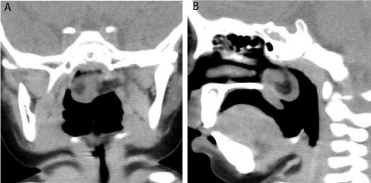
Figure 1A and 1B: Coronal (Figure 1A) and sagittal (Figure 1B) CT images demonstrated a mixed isodense and hypo dense ovoid mass attached to the left nasopharyngeal wall by a pedicle of fibrous tissue.
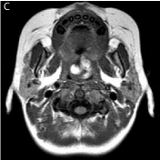
Figure 1C: Axial T1-weighted image showed a heterogeneously hyper intense and hypo intense signal intensity mass.
The initial diagnosis was Teratoma. The patient then received total excision of the mass. During surgery, a 22x21x16 mm mass with a pedicle adhered strongly to the left nasopharyngeal wall was observed. Also, incomplete cleft soft palate was found.
Grossly, the specimen appeared gray reddish and soft with a whitish gray solid lesion in the cut. On microscopy, serial sections showed neuroglial heterotopia in soft tissue composed of neuroglial tissues. Some pituitary tissues and choroid plexus-like elements showing cystic appearance with papillary formation inside were also noted (Figure 1D). Meningeal components were absent. Immunohistochemistry revealed intensive reactivity for glial fibrillary acidic protein (Figure 1E) and S100.
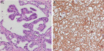
Figure 1E: Immunohistochemistry revealed intensive reactivity for glial fibrillary acidic protein (original magnification x200).
    Figure 1D and 1E:  Hematoxylin-eosin staining (original magnification x200) demonstrated "choroid plexus" like areas showing cystic appearance with papillary formation inside.
 Figure 1E: Immunohistochemistry revealed intensive reactivity for glial fibrillary acidic protein (original magnification x200).
Case 2
The case was an eight month old girl with cough during feeding and occasional dyspnea during sleeping since birth. Nasopharyngoscope showed a 15x15 mm whitish gray mass above the soft palate in the nasopharynx. Multiplanar CT images revealed a 17x15x12 mm heterogeneously isodense and hypodense ovoid pedunculated mass above the soft palate adhered to the posterior nasopharyngeal wall (Figure 2A&2B). No connection between the mass and the intracranial structures or bony defect of the skull base was seen. No stalk was demonstrated between the mass and the soft palate. The mass obstructed the nasopharyngeal cavity.
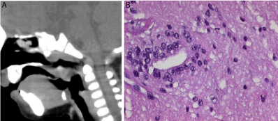
Figure 2B: Hematoxylin-eosin staining (original magnificationx200) showed cerebral tissues including glial cell and neural tube.
    Figure 2A and 2B:  Sagittal CT disclosed a heterogeneously isodense and hypodense ovoid pedunculated mass above the soft palate adhered to the posterior nasopharyngeal wall.
 Figure 2B: Hematoxylin-eosin staining (original magnificationx200) showed cerebral tissues including glial cell and neural tube.
The initial diagnosis was also Teratoma. The total excision of the mass was completed and a 17x15x12 mm mass with a pedicle adhered strongly to the left nasopharyngeal wall was observed during surgery.
Grossly, the mass was whittish gray and firm with a heterogeneous matrix in the cut. Microscopy identified neuroglial heterotopia in soft tissue composed of neuroglial tissues. Meningeal components were absent. The neuroglial tissue was intensely positive for Glial Fibrillary Acid Protein (GFAP) and S100.
Discussion
Glial heterotopias, representing developmental heterotopias of neuroglial tissue rather than true neoplasms, are rare conditions composed of differentiated derivates of the neuroectoderm outside the cranial vault or spinal canal and without communication with the brain, spinal cord, or meninges [1-12]. Unlike meningoencephaloceles, the key feature of the glial heterotopia is the lack of an extension into the cranial vault. A variety of terms such as nasal glioma, glial choristoma, cerebral heterotopy, ectopic brain, and heterotopic brain tissue have been used to describe this lesion [1-8]. We favor the term glial heterotopia, as it better reflects the lesion's pathologic nature. The most common location for the glial heterotopia is the nasal cavity, where it is traditionally, but erroneously, termed "nasal glioma" [1,2]. So-called "nasal gliomas" may indeed have communication with the intracranial structures, which some authors postulate is the reason why some of those lesions grow [1,2]. However, our two cases neither had bony defect in the skull base nor communication between the mass and the intracranial structures.
Grossly, the glial heterotopia is solid, relatively avascular and poorly encapsulated, and adherent to surrounding soft tissues, and may have cystic components containing cerebrospinal fluid-like clear fluid [1,3-6]. Histologically, the lesions were composed of mature glial tissue intermixed with fibrous or muscle tissues. Heterotopic pharyngeal neuroglial tissue might contain neurons and astrocytes as well as more complex central nervous system elements such as ependymal-lined structures, a functioning choroid plexus, and pigmented cells of retinal differentiation [3]. Mitoses, hemorrhages, and necroses were absent. Fat tissues and pituitary tissues were found in one of our cases, which have never been reported before.
The presence of glial heterotopia in this area could have disturbed the normal fusion process of palatal shelves causing cleft palate [3,6]. Therefore, the cleft palate might be the result rather than the cause of ectopic glial tissue.
Radiographic assessment of nasopharyngeal glial heterotopia is best performed using CT complemented by MRI [1,2,5,12]. Multiplanar images with multidetector CT scan delineate the location of the mass and its relationship to the skull base. Magnetic resonance characteristics of glial heterotopia resemble normal brain tissue in all pulse sequences. Cystic elements might be present and represent cerebrospinal fluid-like fluid-filled spaces.
Conclusion
The glial heterotopia is a rare developmental abnormality in the nasopharynx. However, it should be included in the differential diagnosis of the congenital mass in the nasopharynx including Teratoma, vascular or lymphatic malformations, and adenoidal hyperplasia [4,6,12]. CT and MR imaging, especially multiplanar CT images, can localize and characterize the lesion.
References
- Marina MB, Zurin AR, Muhaizan WM, Primuharsa Putra SH, Azizi AB, Kenali MS. Heterotopic neuroglial tissue presenting as oral cavity mass with intracranial extension. Int J Pediatr Otorhinolaryngol. 2005; 69: 1587-1590.
- Restrepo S, Martínez F, Herrera A, Palacios E. Nasal glioma. Ear Nose Throat J. 2004; 83: 88-89.
- Sun LS, Sun ZP, Ma XC, Li TJ. Glial choristoma in the oral and maxillofacial region: a clinicopathologic study of 6 cases. Arch Pathol Lab Med. 2008; 132: 984-988.
- Al-Ammar AY, Al Noumas HS, Alqahtani M. A midline nasopharyngeal heterotopic neuroglial tissue. J Laryngol Otol. 2006; 120: E25.
- Chen CY, Huang JH, Choi WM, Chen CL, Chan WP. Parapharyngeal neuroglial heterotopia presenting as a growing single locular cyst: MR imaging findings. AJNR Am J Neuroradiol. 2005; 26: 96-99.
- Uemura T, Yoshikawa A, Onizuka T, Hayashi T. Heterotopic nasopharyngeal brain tissue associated with cleft palate. Cleft Palate Craniofac J. 1999; 36: 248-251.
- Ghose S, Balasubramaniam ST, Mahindrakar A, Sharma V, Sen S, Sarkar C, et al. Orbital ectopic glial tissue in relation to medial rectus: a rare entity. Clin Experiment Ophthalmol. 2005; 33: 67-69.
- Bajaj MS, Kashyap S, Wagh VB, Pathak H, Shrey D. Glial heterotopia of the orbit and extranasal region: an unusual entity. Clin Experiment Ophthalmol. 2005; 33: 513-515.
- Rogers GF, Mulliken JB, Kozakewich HP. Heterotopic neural nodules of the scalp. Plast Reconstr Surg. 2005; 115: 376-382.
- Lee JI, Kim KK, Park YK, Eah KY, Kim JR. Glial choristoma in the middle ear and mastoid bone: a case report. J Korean Med Sci. 2004; 19: 155-158.
- Plontke SK, Preyer S, Pressler H, Mundinger PM, Plinkert PK. Glial lesion of the infratemporal fossa presenting as a soft tissue middle ear mass - rudimentary encephalocele or neural crest remnant? Int J Pediatr Otorhinolaryngol. 2000; 56: 141-147.
- Kallman JE, Loevner LA, Yousem DM, Chalian AA, Lanza DC, Jin L, et al. Heterotopic brain in the pterygopalatine fossa. AJNR Am J Neuroradiol. 1997; 18: 176-179.