Clinical Image
Austin J Radiol. 2015;2(5): 1028.
Lower Extremity Weakness Secondary to Isolated Spinal Involvement in Silk Road Disease: An Unusual Presentation of an Uncommon Disease
Manisha Patel, Mougnyan Cox* and Lisa Tartaglino
Radiology Resident, Thomas Jefferson University, USA
*Corresponding author: Mougnyan Cox, Radiology Resident, Thomas Jefferson University, PA, USA
Received: July 15, 2015; Accepted: July 31, 2015; Published: August 10, 2015
Clinical Image
A 35 year-old woman of Mediterranean descent with a past medical history notable for recurrent oral and vaginal ulcers was admitted to an outside hospital with fevers, headaches, and neck pain. She was diagnosed with aseptic meningitis and treated with intravenous steroids followed by a steroid taper upon discharge, with some improvement in her symptoms. Approximately 1 month after this admission, she presented to an outside hospital with new onset left lower extremity weakness. Her symptoms progressively worsened to involve weakness of both lower extremities and sensory hypersensitivity in both lower and upper extremities. She also complained of lower abdominal discomfort and urinary retention. She was readmitted to the outside hospital and a lumbar puncture was performed, which showed significant CSF pleocytosis. She was then transferred to our institution for further evaluation.
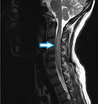
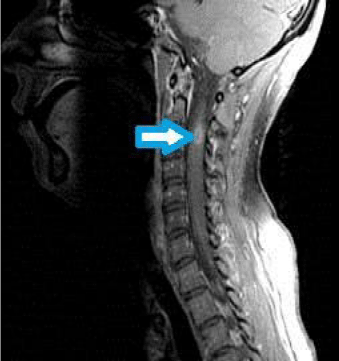
MRI of the cervical and thoracic spine performed at our institution revealed abnormal long-segment cord expansion with increased T2 hyperintense signal and areas of enhancement (Figure 1). Extensive infectious and autoimmune work-up was negative. She was presumptively diagnosed with Neuro-Behcet’s disease and was treated with intravenous immunoglobulin therapy and azathioprine, with subsequent improvement in motor function. Patient was transferred to a rehabilitation floor with continued management by neurology and rheumatology (Figure 2).

    Figure 1:  Figure 1 is a T2-weighted sagittal image of the cervical spine. The
arrow shows an expansilehyperintense lesion in the cervical cord. The lesion
extends from C2 through C7.

    Figure 2:  Figure 2 is a contrast-enhanced fat-supressed T1 weighted image
of the cervical spine. The arrow depicts a small focus of ring enhancement
at the level of C3.
Discussion
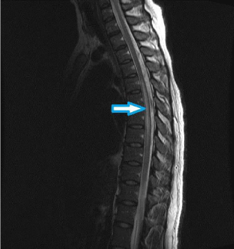
Behcet’s Disease (BD) is characterized by the clinical trial of recurrent oral ulcers, genital ulcers, and uveitis [1]. The disease is most common along the ancient Silk Road stretching from China to the Mediterranean. The underlying pathophysiology is an inflammatory perivasculitis that may involve virtually any organ. Neuro-Behcet’s Disease (NBD) is seen in up to 6% of women with BD, usually within the ages of 20 to 40 [2]. There is no definitive test for NBD, and diagnosis relies on the appropriate clinical history and exclusion of infection. The two major types of NBD are parenchymal (brain, spine) and nonparenchymal (cerebral venous thrombosis, aneurysm, dissection) [3]. The parenchymal form of NBD is more common, seen in approximately 80% of afflicted patients. However isolated spinal involvement in NBD is uncommon, reported in approximately 10% of cases of NBD [4]. Compared to patients with brainstem or supratentorial lesions, patients with spinal cord lesions appear to have a poorer prognosis. Early recourse to biological agents in addition to steroids is recommended by some providers (Figure 3).

    Figure 3:  Figure 3 is a T2-weighted sagittal image of the thoracic spine. The
arrow depicts a similar expansile lesion of the thoracic spine extending over
multiple thoracic segments.
Summary
In a patient with recurrent oral and genital ulcers, new expansile cord lesions should suggest the diagnosis of Neuro-Behcet’s disease.
References
- Al-araji A, Kidd D. Neuro-Behcet’s disease: epidemiology, clinical characteristics, and management. Lancet. 2009; 8: 192-204.
- Yesilot N, Mutlu M, Gungor O, Baykal B, Serdaroglu P, Akman-Demir G. Clinical characteristics and course of spinal cord involvement in Behcet's disease. Eur J Neurol. 2007; 14: 729–737.
- Borhani-Haghighi A, Samangooie S, Ashjzadeh N, Nikseresht A, Shariat A, Yousefipour G, et al. Neurological manifestations of Behcet’s disease. Saudi Med J. 2006; 27: 1542-1546.
- Mascalchi M, Cosottini M, Cellerini M, Paganini M, Arnetoli G. MRI of spinal cord involvement in Behcet’s disease: case report. Neuroradiology. 1998; 40: 255-257.