
Case Report
Austin J Radiol. 2015;2(5): 1029.
Ossicular Anomaly and Endolymphatic Hydrops as Risk Factors for Complications after Ossiculoplasty
Norihiko Inagaki¹, Tadao Yoshida¹*, Michihiko Sone¹, Satofumi Sugimoto¹, Hironao Otake¹, Masaaki Teranishi¹, Shinji Naganawa² and Tsutomu Nakashima¹
¹Department of Otorhinolaryngology, Nagoya University Graduate School of Medicine, Nagoya, Japan
²Department of Radiology, Nagoya University Graduate School of Medicine, Nagoya, Japan
*Corresponding author: Tadao Yoshida, Department of Otorhinolaryngology, Nagoya University Graduate School of Medicine 65, Tsurumai-cho, Showa-ku, Nagoya 466-8550, Japan
Received: July 06, 2015; Accepted: August 10, 2015; Published: August 12, 2015
Abstract
We report a case of endolymphatic hydrops with an ossicular anomaly, in which a hearing test showed fluctuating mixed hearing loss. A 42-year-old man with hearing impairment had experienced varying ear symptoms on his right side since elementary school. Evaluation by computed tomography showed an ossicular anomaly, and magnetic resonance imaging revealed endolymphatic hydrops in the symptomatic ear. Ossiculoplasty or stapes surgery is considered in patients with conductive hearing loss; however, the existence of endolymphatic hydrops is a risk factor for surgical complications. Preoperative magnetic resonance imaging examination may be beneficial when evaluating inner ear conditions such as ossicular anomalies, especially in cases accompanied by fluctuating hearing loss.
Keywords: Fluctuating mixed hearing loss; Endolymphatic hydrops; Ossicular discontinuity
Introduction
Endolymphatic Hydrops (EH) can be found by Magnetic Resonance Imaging (MRI) in patients with Meniere’s disease [1]. Mixed hearing loss with EH, as shown by histology, has been reported in patients with otosclerosis [2,3]. MRI provides useful preoperative information about EH in patients with cochlear and/or vestibular symptoms. We report a patient with an ossicular anomaly and EH for whom MRI analysis provided useful information about the existence of EH.
Case Presentation
A 42-year-old man visited our hospital because of fluctuating hearing loss on his right side. He had first become aware of this hearing impairment during elementary school. Thereafter, he occasionally had fluctuating hearing loss of his right side with vertigo. The tympanic membrane of his right ear appeared normal, and a puretone audiogram revealed the presence of mixed hearing loss of 60–80 dB with air–bone gaps at all frequencies (Figure 1A). Two months after the first visit, he complained of ear fullness on his right side and his hearing level had deteriorated (Figure 1B & C). High-resolution Computed Tomography (CT) showed an ossicular discontinuity, which appeared as a discontinuity between the long process of the incus and the stapes superstructure (Figure 2).
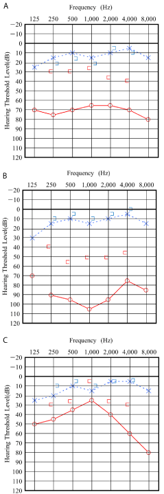
    Figure 1:  Pure-tone audiogram. (A) The first pure-tone audiogram showed
mixed hearing loss of 60–80 dB with air–bone gaps at all frequencies. (B)
Two months after the first visit, the bone conduction threshold had increased.
(C) Postoperative pure-tone audiogram showed improvement of air-bone gap
in all frequencies.
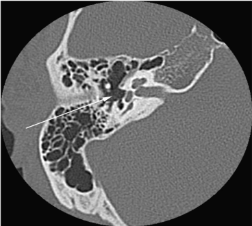
    Figure 2:  Computed Tomography (CT) of the affected ear. CT image shows
the deficit in the stapes of the right ear (arrow).
We planned an MRI examination with intratympanic and intravenous gadolinium injections. The MRI showed significant EH according to the grading scale of Nakashima et al. [4] in his right cochlea and vestibule (Figure 3); the contra lateral side appeared normal. These findings suggested that the ossicular anomaly had caused the air–bone gaps and that the EH caused sensorineural and fluctuating hearing loss detected in his audiogram. MRI was performed 24 h after the intratympanic injection and 4 h after the intravenous injection. Scans were performed on a 3-Tesla MRI scanner (MagnetomVerio; Siemens AG, Erlangen, Germany) using a receive-only, 32-channel, phased-array coil. Three-Dimensional (3D) Fluid-Attenuated Inversion Recovery (FLAIR), 3D real Inversion Recovery (real IR), and heavily T2-weighted 3D FLAIR were performed [5]. Because of good permeability of the gadolinium fluid from the middle to inner ear, intratympanic steroid injection was performed as therapy for the sensorineural hearing loss in his right ear, and the patient recovered hearing in this ear. After we provided the informed consent and signed about risk of surgery and no effect on remission of Meniere’s disease or EH, we performed ossiculoplasty by using tragal cartilage. Because the surgical findings revealed that a superstructure was disappeared and the foot plate mobility was present. He experienced several attacks in one year after surgery. The mean preoperative air conduction, bone conduction and air-bone gap were, respectively,66.7, 30.0 and 36.7 dB at three frequencies 500, 1,000, and 2,000 Hz. The mean postoperative air conduction, bone conduction and air-bone gap were, respectively, 33.3, 15.0 and 15.0 dB.
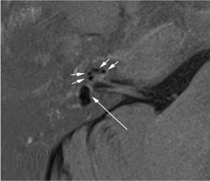
    Figure 3:  Axial Magnetic Resonance Imaging (MRI) of the affected ear. The
affected inner ear shown with 3D real inversion recovery (real IR) MRI. The
short arrows indicate significant Endolymphatic Hydrops (EH) in the cochlea,
and the long arrow indicates significant EH in the vestibule.
Ethics review
The Ethics Review Committee of Nagoya University School of Medicine approved the protocol of the study (approval no. 369-6). All patients gave their informed consent to their participation in the study. The written informed consent forms were attached to their electronic medical records.
Discussion
This is the first imaging report to find EH in a patient with an ossicular anomaly. Many studies have examined the relationship between otosclerosis and EH. Makarem and Linthicum [3] reported on a patient with otosclerosis and EH who experienced a sudden decrease in hearing acuity in his right ear and imbalance after a stapedectomy. Our patient had an ossicular anomaly in his right ear, and the characteristic finding on his audiograms was a fluctuating mixed hearing loss. MRI examination revealed significant EH in the cochlea and vestibule.
Our findings suggest that EH should be considered a risk factor for surgical complications after ossiculoplasty, especially in patients with cochlear and/or vestibular symptoms. Not considering such risk factors in these patients can produce unexpected results. The first possible risk involves damage to the endolymphatic space against the stapes footplate. Preoperative differentiation of the presence or absence of EH is essential to the hearing outcome. The results of histological examination showing otosclerosis in combination with Meniere’s disease suggest that a stapedectomy can cause severe inner ear hearing loss if saccule is in contact with the inner surface of the stapes footplate [6]. According to the report by Issa et al. [6], stapedectomy does not increase the risk of sensorineural hearing loss in patients with otosclerosis and Meniere’s disease who have bone conduction levels of 35 dB or better at 500 Hz and no high tone loss. Second, ossicular reconstruction can cause inadequate hearing recovery or unexpected sound-induced vertigo. When we perform the operation, local anesthesia is a better choice in terms of safety because it allows the surgeon to confirm the patient’s hearing and the presence of vertigo. In addition, use of cartilage instead of an artificial stapes may reduce the risk of inner ear damage. Imaging analysis provides useful information for the management of mixed hearing loss. Combined preoperative evaluation with CT and MRI can be beneficial by revealing the state of the middle and inner ear in cases of fluctuating mixed hearing loss.
Conclusion
Our case of ossicular anomaly demonstrates the need to study further the state of the middle and inner ear in patients with additional symptoms caused by EH. Careful history taking is necessary to identify the EH and to limit the risk of developing surgical complications.
References
- Nakashima T, Naganawa S, Sugiura M, Teranishi M, Sone M, Hayashi H, et al. Visualization of endolymphatic hydrops in patients with Meniere's disease. Laryngoscope. 2007; 117: 415-420.
- Pollak A. Otosclerosis associated with Ménière's disease: a histological study. Adv Otorhinolaryngol. 2007; 65: 50-52.
- Makarem A, Linthicum FH. Cochlear otosclerosis and endolymphatic hydrops. Otol Neurotol. 2008; 29: 571-572.
- Nakashima T, Naganawa S, Pyykko I, Gibson WP, Sone M, Nakata S, et al. Grading of endolymphatic hydrops using magnetic resonance imaging. Acta Otolaryngol Suppl. 2009; 5-8.
- Iida T, Teranishi M, Yoshida T, Otake H, Sone M, Kato M, et al. Magnetic resonance imaging of the inner ear after both intratympanic and intravenous gadolinium injections. Acta Otolaryngol. 2013; 133: 434-438.
- Issa TK, Bahgat MA, Linthicum FH, House HP. The effect of stapedectomy on hearing of patients with otosclerosis and Meniere's disease. Am J Otol. 1983; 4: 323-326.