Case Report
Austin J Radiol. 2015; 2(6): 1033.
Endolymphatic Hydropsin a Patient with a Small Vestibular Schwannoma Suggests a Peripheral Origin of Vertigo
Claudia Jerin1,3, Eike Krause1,3, Birgit Ertl- Wagner2 and Robert Gürkov1,3*
¹Department of Otorhinolaryngology Head and Neck Surgery, University of Munich Marchioninistr, Germany
²German Centre for Vertigo and Balance Disorders, University of Munich Marchioninistr, Germany
³Grosshadern Medical Centre, University of Munich Marchioninistr, Germany
*Corresponding author: Robert Gürkov, Department of Otorhinolaryngology Head and Neck Surgery, Grosshadern Medical Centre, University of Munich Marchioninistr. 15, 81377 Munich, Germany
Received: June 03, 2015; Accepted: August 27, 2015; Published: August 31, 2015
Abstract
Objective: To present audio vestibular and MR imaging findings in a case of vestibular schwannoma with endolymphtic hydrops.
Design: Case report with 9-years follow-up.
Study Sample: A 43 year-old male patient with a small stable intrameatal vestibular schwannoma who developed recurrent vertigo attacks.
Results: Inner ear MRI revealed vestibular endolymphatic hydrops. oVEMP responses showed an altered frequency tuning suggestive of endolymphatic hydrops. Audiometry revealed mild high frequency hearing loss and corresponding high-frequency loss of otoacoustic emissions, suggesting a cochlear lesion.
Conclusion: This case study supports a labyrinthine origin of audio vestibular function deficits and symptoms. Detection of endolymphatic hydrops in such patients may be clinically relevant for the management of vertigo symptoms possibly due to endolymphatic hydrops.
Keywords: Vestibular schwannoma; Vertigo; Hearing loss; Endolymphatic hydrops; MRI
Abbreviations
LEIM: Locally Enhanced Inner ear MRI; MRI: Magnetic Resonance Imaging; ELH: Endolymphatic Hydrops; MD: Menière’s Disease; VS: Vestibular Schwannoma; VEMP: Vestibular Evoked Myogenic Potential; DPOAE: Distortion Product Otoacoustic Emissions
Introduction
Recently, MR imaging after intratympanic contrast agent application (Locally Enhanced Inner ear MR Imaging, LEIM) has been used to visualize Endolymphatic Hydrops (ELH) in patients with Menière’s Disease (MD) in vivo. The extent of ELH as assessed by inner ear MRI has recently been quantified volumetrically [1] and it has been shown to correlate with the dysfunction of various inner ear function parameters like hearing loss, caloric response and saccular function examined by cervical vestibular evoked myogenic potentials [2,3]. Furthermore, ELH has been detected in various inner ear syndromes, e.g. in delayed endolymphatic hydrops, in sudden sensorineural hearing loss and in a typical MD, i.e. MD with purely cochlear or purely vestibular symptoms [4].
In Vestibular Schwannoma (VS), the origin of audio vestibular symptoms might not be solely of retrocochlear origin, i.e. due to the tumor mass effect within the internal acoustic meatus. Therefore, we examined a patient with a stable VS and vertigo with LEIM in order to evaluate the presence of ELH as a possible cause for his symptoms.
Case Report
The 43-year-old patient had presented to our institution nine years before, with one attack of rotatory vertigo lasting for one day. A subsequent MR Scan revealed an intrameatal vestibular schwannoma (size 9 x 5 mm). Audiometry showed a mild high-frequency hearing loss ipsilaterally. Brainstem evoked response audiometry latencies of Jewett peaks I and V were within normal limits bilaterally. In the following years, the patient did not suffer from any complaints except for a pre-existing longstanding bilateral tinnitus. Nine years after the first vertigo attack, recurrent vertigo attacks of variable duration (mostly several minutes) occurred. Simultaneous aural symptoms were not reported, and the patient still did not report any subjective hearing impairment.
A Gadolinium-based contrast medium, Gadopentetate dimuglumine (Magnograf, Marotrast, Jena, Germany), was diluted 8-fold in saline solution and injected intratympanically (0.4 ml) under microscopic control, as described previously [2]. The patient remained in a supine position for further 30 minutes with the head turned 45 degrees toward the contralateral side, instructed not to speak or chew during this period. MRI scans were performed 24 hours after application of the contrast agent on a 3T MR scanner (Magnetom Verio, Siemens Healthcare, Erlangen, Germany) using a commercially available 32-channel head coil. A 3D-Real-IR sequence was acquired with the following parameters: TR 6000 ms, TE 155 ms, TI 1500 ms, fat saturation, constant flip angle of 180 degrees, echo train length of 35, echo train followed by a 90° restore pulse, matrix size of 320 × 320, 36 acquired slices (with 11.1% slice oversampling), 0.5 x 0.5 mm² in-plane resolution at 0.5 mm slice thickness, receiver bandwidth 195 Hz/pixel, and number of excitations 1. The scan time was 15 minutes. For imaging of the vestibular schwannoma, conventional T1 weighted contrast enhanced MR scans were acquired.
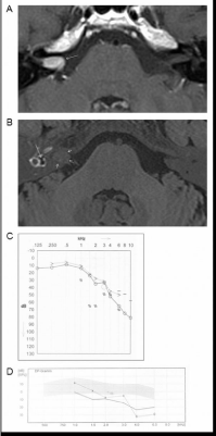
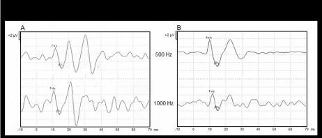
Nine years after the onset of symptoms and after the detection of the vestibular schwannoma in MR imaging, the follow-up MRI showed a stable tumor without evidence of growth (Figure 1A). By use of LEIM (Figure 1B), endolymphatic hydrops in the vestibulum could be visualized as an enlarged hypointense area within the contrast-enhanced perilymphatic space (greater than 40% endolymph/perilymph area ratio). There was no evidence of cochlear endolymphatic hydrops. In this case, clinical symptoms were mainly vestibular, which corresponds well to the LEIM that showed a vestibular endolymphatic hydrops. Caloric videonystagmography showed regular horizontal semicircular canal function. Cervikal Vestibular Evoked Myogenic Potential (cVEMP) testing revealed normal results. Ocular Vestibular Evoked Myogenic Potentials (oVEMP) revealed normal absolute amplitudes. However, oVEMP testing also revealed an ipsilaterally decreased 500/1000 Hz ratio of 0.87 (Figure 2); this shift in oVEMP frequency tuning has been shown to be indicative of ELH [2]. Audiometry merely showed a mild ipsilateral high-frequency hearing loss (Figure 1C). Otoacoustic emissions correlated well with the pure tone audiogram, with a loss of DPOAE above 4 kHz, indicating a cochlear rather than a retrocochlear damage (Figure 1D).

    Figure 1:  MR imaging and audiometric findings. A: A follow-up MRI nine
years after the detection of the vestibular schwannoma showed a stable tumor
(approximately 9 x 5 mm). An axialcontrast-enhanced T1-weighted sequence
is shown. B: Locally Enhanced Inner ear MRI (LEIM) with an axial 3D Real-IR
sequence of the right ear visualizing vestibular endolymphatic hydrops (long
arrow). Since the contrast agent primarily enters the perilymphatic space, the
endolymphatic space is visible as hypointense (dark) area bulging into the
contrast-enhanced (bright) perilymphatic space. The VS is also faintly visible
in this sequence (short arrows). C: Audiogram of the affected ear at the time
of the LEIM (nine years after the onset of symptoms). D: DPOAE-gram of the
affected ear. Thick line: noise floor. Thin line: DP level at f2.

    Figure 2:  oVEMP waveforms recorded in response to 500 Hz and 1000 Hz. The amplitude in response to 1000 Hz is larger than the amplitude in response to
500 Hz, so that the 500/1000 Hz ratiois decreased to 0.87. B: oVEMPs recorded in a healthy subject for comparison, with a normal 500/1000 Hz ratio above 1.2.
Discussion
Previously, several studies have detected endolymphatic hydrops in various inner ear syndromes like sudden sensorineural hearing loss and tinnitus by use of inner ear MRI. The potential presence of ELH in patients with vestibular schwannoma, however, has been sparsely investigated so far. By now, there is only one report of in vivo visualized ELH in patients with VS [5]. This retrospective study reported cochlear ELH in 1 of 13 and vestibular ELH in 4 of 13 patients with VS. Interestingly, ELH was found in 3 of 5 VS patients presenting with vertigo. No statistically significant correlation between vertigo and ELH could be detected with this small sample size, so that the authors concluded that vestibular ELH might not be the cause of vertigo in all patients. The lack of evidence of a significant correlation between vertigo and ELH, however, does not necessarily allow the conclusion that there is no relationship between ELH and vertigo. Even in Menière’s disease, there is no perfect correlation between the severity of ELH and a patient’s subjective symptoms, so that there might be so far unknown factors other than ELH which affect the patient’s complaints. Furthermore, the study by Naganawa et al. did not use LEIM but non-contrast-enhanced 3D-FLAIR images. In these MR images, the endolymphatic space is not demarcated from the perilymphatic space as clearly and distinctly as it is in Gadolinium contrast-enhanced inner ear MRI.
The origin of audio vestibular symptoms in vestibular schwannoma has been discussed in several previous studies, suggesting that vertigo and hearing loss in VS may not only be ascribed to a direct pressure of the tumor on the vestibulocochlear nerves which subsequently progressively lose their function, but also to a peripheral origin, i.e. to a dysfunction of the inner ear. Such a peripheral lesion may possibly be induced by tumor-related vascular compromise of the inner ear. Otoacoustic emissions have been reported to indicate a cochlear damage even in early stages of VS [6,7]. Furthermore, degeneration of the stria vascularis, hair cell degeneration and biochemical alterations in the inner ear fluids have been observed in histological studies [7,8].
Evidence that vestibular schwannomas may result in a dysfunction of the inner ear itself also arises from studies investigating the effect of gentamicin in patients with VS and vertigo. A recent study reported that 3 of 4 VS patients treated with gentamicin had no more intractable vertigo [9]. Gentamicin was therefore proposed as a treatment option particularly in patients with small, stable tumors but affected by vertigo attacks. These studies raised the question in which way an inner ear therapy could be effective on a retro labyrinthine disease. A possible explanation could be that gentamic in not only eliminates vestibular function by damaging vestibular hair cells but also reduces the production of endolymph by damaging cells of the stria vascularis [7].
The assumption that hearing loss and vertigo in VS are partly due to a dysfunction of the inner ear is therefore well supported by the literature. New inner ear imaging techniques now enable visualizing morphological changes in the cochlea and the vestibulum in vivo. Indeed, our present study shows signs of vestibular endolymphatic hydrops in a patient with VS. It is noteworthy that the patient in this study had small VS which did not increase in size over nine years and which caused mainly vestibular symptoms with long periods of freedom from complaints. Therefore, the present case study provides evidence in support of the notion of a peripheral origin of vertigo in patients with small vestibular schwannomas.
Conclusion
In vestibular schwannoma patients presenting with vestibular symptoms, locally contrast-enhanced inner ear MR imaging to detect endolymphatic hydrops may thus be of considerable diagnostic value. In case of confirmed ELH, a treatment directed against ELH may be indicated. Particularly in those patients with small tumors and preserved vestibular function, not only gentamicin but also betahistine or endolymphatic sac surgery might be treatment options for recurrent vestibular symptoms.
Acknowledgement
This study was supported by the Federal Germ
References
- Gürkov R, Berman A, Dietrich O, Flatz W, Jerin C, Krause E, et al. MR volumetric assessment of endolymphatic hydrops. Eur Radiol. 2015; 25: 585-595.
- Gurkov R, Flatz W, Louza J, Strupp M, Ertl-Wagner B, Krause E. In vivo visualized endolymphatic hydrops and inner ear functions in patients with electrocochleographically confirmed Ménière's disease. Otol Neurotol. 2012; 33: 1040-1045.
- Gürkov R, Flatz W, Ertl-Wagner B, Krause E. Endolymphatic hydrops in the horizontal semicircular canal: a morphologic correlate for canal paresis in Ménière's disease. Laryngoscope. 2013; 123: 503-506.
- Pyykkö I, Nakashima T, Yoshida T, Zou J, Naganawa S. Meniere's disease: a reappraisal supported by a variable latency of symptoms and the MRI visualisation of endolymphatic hydrops. BMJ Open. 2013; 3.
- Naganawa S, Kawai H, Sone M, Nakashima T, Ikeda M. Endolympathic hydrops in patients with vestibular schwannoma: visualization by non-contrast-enhanced 3D FLAIR. Neuroradiology. 2011; 53: 1009-1015.
- Prasher DK, Tun T, Brookes GB, Luxon LM. Mechanisms of hearing loss in acoustic neuroma: an otoacoustic emission study. Acta Otolaryngol. 1995; 115: 375-381.
- Gouveris HT, Victor A, Mann WJ. Cochlear origin of early hearing loss in vestibular schwannoma. Laryngoscope. 2007; 117: 680-683.
- Gleich O, Strutz J, Schmid K. [Endolymph homeostasis and Menière's disease: fundamentals, pathological changes, aminoglycosides]. HNO. 2008; 56: 1243-1252.
- Giannuzzi AL, Merkus P, Falcioni M. The use of intratympanic gentamicin in patients with vestibular schwannoma and disabling vertigo. Otol Neurotol. 2013; 34: 1096-1098.