Case Report
Austin J Radiol. 2017; 4(2): 1069.
Ureteral Triplication Associated with Ipsilateral Ureterocele and Ureterolithiasis - Uncommon Congenital Anomaly
Nayyar N¹* and Shukla A²
¹Department of Radiodiagnosis, India
²Department of Surgery, India
*Corresponding author: Nayyar N, Department of Radiodiagnosis, CH Palampur Himachal Pradesh 176061, India
Received: July 17, 2017; Accepted: August 01, 2017; Published: August 11, 2017
Abstract
Ureteral triplication is a rare congenital malformation of the urinary system and classified into four types. Ureteral triplication is a developmental abnormality of the ureteral bud originating from the Wolffian duct at the 5th week of embryological life. With ureteral triplication, three ureteral buds likely develop independently from the mesonephric duct, or from early fission of one or more of the ureteral buds to join the metanephros.
Keywords: Ureteral triplication; Wolffian duct; CT urography; Ureterocele
Case Presentation
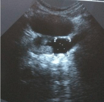
A 41 year old male adult from India presented with left sided flank pain off and on for last 1 year which was 2-3 times a month. He was investigated outside our hospital and diagnosed with left renal calculus and started treatment for the same. He reported to us after no clinical improvement, routine blood tests and urine analysis were normal. The patient underwent ultrasound examination of the kidneys that showed evidence of bilateral duplex urinary system. The upper and lower pole moieties of left kidney were dilated with extravesical ureterocele showing a calculus of size 14mm and having ectopic opening into prostatic urethra (Figure 1).

    Figure 1:  Ultrasound image showing left sided ureterocele with calculus with
ectopic opening into prostatic urethra.
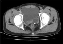
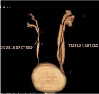
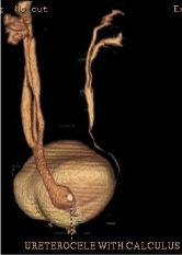
CT Urography was done for further work-up which showed Smith’s type III triplication on left side, all three left sided ureters are seen merging into a single ureter at S2 vertebral level (Figure 3). Abnormal dilatation of lower end of this common ureter seen measuring 2.7×2.4×3.2 cm with ectopic insertion into prostatic urethra (Figure 4). A calculus of size 14.9×7.8 mm seen in ureterocele (Figure 2). On right side duplication of pelvicalyceal system with partial duplication of ureters seen merging into single moiety at L4- L5 IV disc level with normal opening of right common ureter into urinary bladder (Figure 3).

Figure 2: Plain CT image showing left sided ureterocele with calculus.

    Figure 3:  CT Urography showing triplication of ureter on left side with right
sided duplex system.

Figure 4: CT Urography showing on left sided ureterocele with calculus.
Discussion
Ureteral triplication is a rare congenital malformation of the urinary system and classified into four types [1]. Ureteral duplication occurs in 0.3-0.8% of the population, but ureteral triplication has been very rarely described [2]. The most frequently encountered anomalies with ureteral triplication are contra lateral duplications (37%), renal ectopia (28%) and renal dysplasia (8%) [3]. Furthermore, ureteral triplication is occasionally an isolated anomaly [4]. The sporadic nature of this condition and its association with other anomalies makes its management difficult.
Smith classified triplication into four types: type I (35%), three separate ureters from the kidney with three separate draining orifices to the bladder; type II (21%), three ureters arising from the kidney with two of these joining and draining into two orifices; type III(31%), all three ureters joining together before reaching the bladder and draining through a single orifice; and type IV (9%), two ureters arising from the kidney with one becoming an inverse Y bifurcation and draining into three orifices [1].
Presentation of ureteral triplication is delayed due to paucity of symptoms and signs. Presenting symptoms are renal colic, recurrent UTI, and urinary incontinence [3,5]. Our case showed type III triplication on left side associated with ureterocele and ureterolithiasis with ectopic opening and contra lateral duplication which is very rare in itself.
Conclusion
Ureteral triplication is rare and difficult to diagnose because it occurs in four different forms, sometimes it may be misdiagnosed as duplex system. Due to lack of specific clinical signs, ultrasonography and urography remain the mainstay of diagnosis and can direct the best treatment strategy. Ureteral triplication should be included in differential diagnosis of congenital anomalies causing recurrent urinary tract infection.
References
- Smith I. Triplicate ureter. Br J Surg. 1946; 34: 182-185.
- Singh G, Murray K. Ureteral triplication, occasionally an isolated anomaly. Urol Int 1996; 56: 117-118.
- Sivrikaya A, Cay A, Imamoglu M, Sarihan H. A case of ureteral triplication associated with ureteropelvic junction obstruction. Int Urol Nephrol. 2007; 39: 755–757.
- Kudela G, Koszutski T, Mikosinski M, Utrata W. Ureteral triplication: report of four cases. Eur J Paediatr Surg. 2006; 16: 279–281.
- Fernbach SK, Poznanski AK. Pediatric case of the day. Complete ureteral triplication. Radiographics 1989; 9: 359-364.