
Research Article
Austin J Radiol. 2018; 5(1): 1079.
Emerging Trends Using Various Technological Advancements in Traumatic Brain Inury (TBI) in Western U.P
Mohd Arfat¹*, Gupta AK¹, Godara (Maj) SC2 and Yogesh CY3
¹Department of Radiology, Uttar Pradesh University of Medical Sciences, India
²Department of Radiology, Nims University, India
³Department of Pharmacology, Uttar Pradesh University of Medical Sciences India
*Corresponding author: Mohd Arfat, Department of Radiology, UPUMS Saifai, Guest Faculty (Paramedical College, Saifai), Senior Radiographer (MRI/CT Scan), Etawah, UP-206130, India
Received: May 01, 2017; Accepted: March 05, 2018; Published: March 20, 2018
Abstract
Objective: To study the various advance technology for the diagnosis of Traumatic Brain Injury (TBI) and find out Emerging trends occur in TBI patients.
Methods: The Present study was conducted with 80 patients, age between 02 year to 70 year mean age (36 Years) presenting to emergency department of Uttar Pradesh University of Medical Sciences, Saifai, Etawah, with a history of acute head trauma from October 2016 to March 2017. All patients were examined using 64 slices MDCT and 1.5T MRI Scanner also.
Results: Traumatic brain injury caused by various reasons like 65.0% Road Traffic Accidents (RTA) and 20.0% Fall From Height (FFH) being and 12.5% Assault/hit by hard object and 2.5% are Gunshot injury. Loss of consciousness was the most common complaint of the 58.75% TBI patients followed by 8.45% Vomiting and headache, 20.0% facial injury and 12.5% scalp injury. All TBI patients were diagnosed by MDCT 64 Slices Somatom Sensation Scanner who was observed 36.25% skull fractures, 12.5% Extra Dural hematoma, 13.75% Sub Dural hematoma, 13.75% Sub archnoid haemorrhage, 8.75% Intra cerebral hematoma, 25.0% brain contusions and 11.25% diffuse cerebral edema.
Conclusion: Road Traffic Accidents remain the leading cause of trauma in our country. MRI and MDCT is well characterized of the extent and various types of hemorrhages and skull fractures in TBI patients. The present study data is indicated 65.0% majority of TBI patients is suffered by Road traffic accidents mainly young males with alcoholism.
Keywords: Emerging trends; Extra-dural hematoma (EDH); Sub dural Hematoma (SDH); Diffuse axonal injury (DAI); Traumatic brain injury (TBI); UPUMS
Introduction
Traumatic Brain Injury (TBI) is a critical public health and socio-economic problem throughout the world. It is a major cause of death, especially among young adults [1] and lifelong disability is common in those who survive. Traumatic Brain Injury (TBI) has varied morbidity in serviving patients. The primary causes of TBI vary according to age of the peoples [2], Fall from height is the leading cause of Traumatic Brain Injury in children up to 4 years of age and persons more than 70 years or above. Traffic and vehicle injury is very common young and up to 50yrs of age, the cause behind is frequent and fast mobility for education and purpose of job & business. It is estimated that in the USA, around 5.3 million people are living with a TBI related disability [3]. Report shows one TBI every 15 seconds in the USA. TBI is the leading killer and disabler of young adults under the age of 35. In India 1.5 to 2 million people were injured every year. The Lancet reports that TBI projected become the third largest cause of disease burden in 2020.
Head injury requires immediate, quick diagnosis for the early management to show the incidence of mortality and morbidity can be minimized. CT Scan is the primary most important diagnostic modality for head trauma, it is superior to MRI for the diagnosis of bone injury and acute haemorrhage [4]. It has got limitation (i) Beam hardening effect there by not suitable for the posterior fossa of brain.
(ii) The age of the haemorrhage cannot be evaluated by the CTScan. (iii) The follow-up and complications are not visualized. (iv) Due to radiation effect cannot be utilized in pregnant women.
MRI has got some advantage (i) No radiation hazard.(ii) Age of the hematoma can be better evaluated.(iii) It is better modality of choice compare to CT scan to see the post TBI complications and follow up.(iv) Absence of beam hardening effect, multisectional imaging make it better modality for posterior fossa pathology. MRI is better than CT Scan in the detection of Non-hemorrhagic contusions and Diffuse Axonal Injury (DAI). A T2* Gradient Refocu ssed Echo (GRE) sequence is used to detect acute and chronic bleed. In this type of Sequence bleed appear black.
Material and Method
In the present study was done by diagnosis of 80 patients of acute head trauma and positive findings on head MDCT and MRI scanning between October 2016 and March 2017.
Including criteria
1- Patients of all ages, sexes and occupations were included.
2- Only patients with positive findings on brain MDCT and MRI scanning were included.
3- Taking complete history of all trauma patients.
4- General examination of the patients was done by the emergency department of U.P.UMS, Saifai, Etawah.
5- CT scan of head using MDCT scanner and MRI 1.5T Scanner without using intravenous contrast media.
Study area
The study will be carried out in the
• Department of Radiology, Uttar Pradesh University of Medical Sciences, Saifai, Etawah.
• Emergency department, Uttar Pradesh University of Medical Sciences, Saifai, Etawah.
Multidetector computed tomography technique
The diagnosis of TBI was performed using a 64 row Multi detector computed tomography scanner, Siemens somatom sensation. Axial section images (1.25 to 5 mm slice thickness and image interval of 5 mm), with a high standard frequency reconstruction algorithm. On 64 slice siemens somatom sensation scanner, CT head data sets were performed in the supine position. For adequate Multi planar reconstruction, scanning was performed to cover the area from orbito-meatal line to the vertex of head. Then makes the thin slice of whole data we acquired and load to MMWP work station, where MPR images were obtained in axial, coronal and sagittal planes whenever need. 3D technique including Shaded Surface Display (SSD), Volume Rendering Technique (VRT) is used to obtained three dimensional image according to the findings from the original image.
Magnetic Resonance Imaging Techniques
The experimental data has been generated by using PHILIPS 1.5T Achieva Nova machine. Fast Spin Echo (FSE) T1 and T2 weighted sequence are used in the evaluation of head trauma.
FLAIR (Fluid Attenuated Inversion Recovery) and FSE T2 weighted sequence are sensitive in the detection of non-hemorrhagic lesions such as contusions and Diffuse Axonal Injury (DAI) because of the sensitivity of these sequences to the presence of extracellular free water content. A T2 Gradient-Refocussed Echo (GRE) sequence with sensitivity to magnetic susceptibility effects will allow the detection of acute and chronic hemorrhagic lesions that may not be well visualized on FSE T2 weighted sequence. Acute hemorrhagic lesions are poorly seen on T1-weighted images because they are isointense or slightly hypointense. Diffusion–Weighted Image (DWI) sequences are also helpful in the evaluation of acute trauma. DWI has proven capable of detecting Diffuse Axonal Injury (DAI) that may not be seen on FLAIN and T2 GRE sequences. The multiplanar imaging capability and superior contrast resolution of MRI are advantages over CT Scan ,allowing more accurate localization and characterization of intracranial injuries.
Image parameters for T1- weighted images -Repetition Time (TR) = 500msec, Echo Time (TE) = 20msec, Number of Excitations (NEX)=2
For FLAIR images-TR=9000 msec, TE=155msec, inversion time (TI)=2200, NEX=1
For FSE T2-weighted images-TR=2000msec, TE= 80msec, NEX=1
For T2 GRE-TR=500msec, flip angle -20degrees
For DWI- TR=10,000msec, TE=95msec and NEX=1.
Results
In the present study, total 80 Traumatic Brain Injury patients were diagnosed in which 56 males and 24 females patients, with male and female ratio of 7:3. Their age ranges from 02 year to 70 year, with a mean age of 36 years. The peak age was the third decades including 24 patients with average of 30.0% from the total no. of patients (Table 1, Figure 1). The majority of the 80 patients studied, who have traumatic brain injury caused by Road Traffic Accidents (RTA) 65.0%, and Fall From Height (FFH) 20.0%, being hit by Assault/hard object 10.0% and Gun shot 2.5% (Table 2).
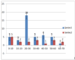
    Figure 1:  Age and sex distribution among the studied 80 patients with acute
Traumatic Brain Injury.
![]()
    S.N.
    Age in years
    Male [N(%)]
    Female [N (%)]
    Total [N (%)]


    1-
    0 to 10
    08 (10.0)
    07 (8.75)
    15 (18.75)


    2-
    10 to 20
    06 (7.5)
    02 (2.5)
    08 (10.0)


    3-
    20 to 30
    21 (26.25)
    03 (3.75)
    24 (30.0)


    4-
    30 to 40
    05 (6.25)
    04 (5.0)
    09 (11.25)


    5-
    40 to 50
    08 (10.0)
    02 (2.5)
    10 (12.5)


    6-
    50 to 60
    06 (7.5)
    03 (3.75)
    09 (11.25)


    7-
    60 to 70
    02 (2.5)
    03 (3.75)
    05 (6.25)


    Total
    56 (70.0)
    24 (30.0)
    80 (100)
Table 1: Age and sex distribution among the studied 80 patients with acute
Traumatic Brain Injury.
![]()
    S.NO.
    Causes of TBI
    Number of patients (%)


    1-
    Road traffic accidents
    52 (65.0)


    2-
    Fall from height
    16 (20.0)


    3-
    Hit by hard object/ Assault
    10 (12.5)


    4-
    Gun shot
    02 (2.5)
Table 2: Causes of Traumatic Brain Injury among the study population (80
patients).
The total no. of patients presented with different clinical presentation. 58.75% of TBI patients were complained Loss of consciousness, 8.45% Vomiting/headache, 20.0% facial injury and 12.5 % suffered scalp injury (Table 3).
![]()
    S.NO.
    Clinical    Presentation
    No.    Of Patients (%)


    1-
    Loss of    consciousness
    47 (58.75%)


    2-
    Vomiting/Headache
    07    (8.45%)


    3-
    Facial injury
    16 (20.0%)


    4-
    Scalp    injury
    10    (12.5%)
Table 3: Clinical representations among the studied (80 patients) with acute
Traumatic Brain Injury.
Most of the examined patients showed more than one lesion. Twenty nine (36.25%) of the eighty patients reported skull fractures. Ten patients had extra dural hematoma (12.5%), eleven patients had Sub dural hematoma (13.75%), eleven patients had Sub archnoid hemorrhage (13.75%), seven patients had Intra cerebellar hematoma (8.75%), twenty patients had brain contusions (25.0%) and nine patients had diffuse cerebral oedema (11.25%) (Table 4).
![]()
    S.NO.
    MDCT Findings
    No. of Patients (%)


    1-
    Skull fractures
    29 (36.25%)


    2-
    Extra    Dural Hematoma
    10    (12.5%)


    3-
    Sub Dural Hematoma
    11 (13.75%)


    4-
    Sub    archnoid haemorrhage
    11    (13.75%)


    5-
    Intra cerebral    Hematoma
    07 (8.75%)


    6-
    Brain    Contusions
    20    (25.0%)


    7-
    Diffuse cerebral    oedema
    09 (11.25%)
Table 4: MDCT findings among the 80 studied patients with acute Traumatic
Brain Injury.
Discussion
Injuries have been reported to be a neglected epidemic in developing countries and accounting for more than five million deaths per year, roughly equal to the number of deaths from HIV/ AIDS, malaria, and tuberculosis combined [5-7]. MDCT scanning is frequently used to diagnosis of all Traumatic Brain Injury patents those are admitted in hospital or their treatment. MDCT scanning is imaging technique used well assessment of the seriousness of injury and their image can be obtained by using multidetector high resolution scanners. The images could be observed using brain of bone contrast windows where as data can be obtained into 3D CT sets to indicated bony and intracranial injuries [8].
Axial CT scanning is used for evaluation of neurological injury in head trauma but it is limited in evaluation of the posterior fossa, the middle cranial fossa, and the inferior frontal lobes. Coronal and Sagittal CT reconstruction provide more informative of these areas [9].
Previously in study of acute traumatic brain injuries conducted earlier these are direct relation between the severities of clinical symptom and demonstration of abnormalities.
Youmans (1982) has been reported acute traumatic brain injuries are conducted on behalf of direct relation between the severities of clinical manifestation and relation with their abnormalities [10]. Whereas present study has seen intracranial sequelae on MDCT scanning.
Ashikaga et al (1997) reported use of MRI in skull fracture of TBI patients [11]. MDCT imaging was used for detecting fractures and depending on their location; type prompt surgical intervention can be done to prevent CSF leakage, infection, haemorrhage in which 36.25% of acute traumatic brain patient were suffered by open skull fractures and their pathological manifestation like depressed, crashes and abnormal thickened in skull in present studies of TBI patients. These results also supported to Ashikaga et al. (Figure 2).

Figure 2: Skull Fragment fracture.
Mittl et al., (1994) has been well demonstrated extra dural hematoma in mild head injury and normal head CT findings [12]. I present TBI patient cases were observed under MDCT imaging. 12.5% Extra Dural hematoma showed as biconvex hyper dense elliptical collection with sharp edge of acute traumatic brain patient. It was developed between the skull and dura associated skull fracture (75- 90%). It may be due to injured middle meningeal artery. Sub dural hematoma was found 13.75% case of present study of TBI patients. It was evaluated by using subdural CT window. It was showed as hyperdense and concave in shape on CT scan. Singh and Suryapratap (2009) also reported in an unusual case of a compound depressed skull fracture [13].
Subarachnoid hemorrhage was found 13.75% of acute traumatic brain injury patients (Figure 3). CT was showed more accurate in detecting acute sub arachnoid hematoma than routine MRI sequences. It may be due to the blood in acute sub arachnoid hematoma has a low deoxyhemoglobin and was appeared black. The present finding also supported to Bradley (1993) and Holme et al. (2008), studies about MR Appearance of hemorrhage in the brain [14,15].
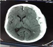
Figure 3: Sub-dural hematoma.
In the present study, it is revealed that 25.0% TBI patients suffered by brain contusions and 8.75% intra cerebral hematoma. The finding was confirmed due to scattered areas of bleeding seen at cortico- medullary junction or cortex of brain parenchyma and appeared as salt & pepper appearance. It could be hemorrhagic and non hemorrhagic. It was due to blunt head injury patients and in acceleration and deceleration trauma (Figure 4).
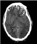
Figure 4: Multiple Brain Contusions.
Colorado et al (1997) also reported in Traumatic Brain Injury, 1990-1993 [16]. It was observed that the clot signal was similar to the brain parenchyma in MRI in acute stage of head trauma. It was observed that MDCT was more sensitive in detecting clots within 24 hours of injury than MRI. The present study of MDCT was well documented by Gutman et al. 1992, Intra cerebral hematoma was observed on CT [17]. It was well defined hyper dense area. ICH cause ruptured of blood vessels in Traumatic Brain Injury (TBI) patients. In previous studies of both ICH and contusions simultaneously present in same case [18].
In the present study, it is revealed that 11.25% diffuse cerebral edema of acute traumatic brain patients . Diffuse cerebral edema was made by loss of cerebral autoregulation due to significant increased blood flow and blood volume increased pressure on CSF leading to mildly increased density of white matter. The present finding was also well studied in various TBI patients [19,20].
In the present study, it is revealed that 2.5% patients of Traumatic Brain Injury due to Gun shot. MDCT is very useful in detecting of exact location of bullet with the help of Multi Planar Technique (MPR) and 3D reconstruction technique (Figure 5,6).
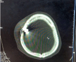
Figure 5: Bullet fracture the bone of skull.
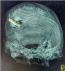
Figure 6: 3D (VRT) shows bullet site.
Magnetic Resonance Imaging (MRI) is playing an increasingly important role in the evaluation of Traumatic Brain Injury. MRI is more sensitive than CT Scan in the detection of Non-hemorrhagic contusions, Diffuse Axonal Injury (DAI). MRI is the modality of choice in assesment of age (time duration) of trauma. Development of MRI Compatible life support equipment such as Non-ferromagnetic ventilators, allows the severely injured comatose traumatic patients to be evaluated with MRI Scanner. FLAIR and FSE T2 weighted sequence are used in detection of non-hemorrhagic lesions such as -contusions, Diffuse Axonal Injury (DAI) (Figure 7). The Multiplanar Imaging capability and superior contrast resolution of MRI are the key advantage over CT Scan.
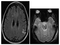
Figure 7: MRI shows small foci of bleed suggests Diffuse Axonal Injury (DAI).
Conclusion
Above all discussions we can say that, the fast modern life increasing incidents of road traffic accidents has increase the risk of Brain injury. Road Traffic Accidents (RTA) remain the leading cause of trauma. The present study indicated that 65.0% patients suffered with TBI by Road Traffic Accidents (RTA). The main cause of RTA occurs due to Alcoholism, lack of road side and driving precautionary majors. The role of two modern imaging CT Scan & MRI are able to diagnose the different brain injury, their outcome and follow up. In the modern medical science these two modalities are indispensible for neurosurgery and other medical field.
Acknowledgement
We gratefully acknowledge the Vice Chancellor of UPUMS , who supported well in this study and also emergency room doctors of UPUMS Saifai , Etawah.
Declaration of Interest
The present study was performed with support from Uttar Pradesh University of medical sciences Saifai Etawah U.P.
References
- Maas AI, Stocchetti N, Bullock R. Moderate and severe traumatic brain injury in adults. Lancet Neurol.2008; 7: 728–741.
- National Centre for Injury Prevention and Control. Report to Congress on Mild Traumatic Brain Injury in the United States: Steps to Prevent a Serious Public Health Problem. Atlanta, GA: Centre for Disease Control and Prevention. 2003.
- Langlois JA, Sattin RW. Traumatic brain injury in the United States: research and programs of the Centers for Disease Control and Prevention (CDC). J Head Trauma Rehabil. 2005; 20: 187–188.
- Ehimwenma Ogbeide, Alphonsus R Isara. Cranial computed tomography utilization in head trauma in a Southern Nigerian tertiary hospital. Sahel Medical Journal. 2015: 18: 27-30.
- Gosselin RA, Spiegel DA, Coughlin R, Zirkle LG. Injuries: The neglected burden in developing countries. Bull World Health Organ. 2009; 87: 246.
- Debas HT, Gosselin RA, McCord C, Thind A. Disease Control Priorities in Developing Countries. 2nd edition. New York: Oxford University Press. 2006.
- Lopez AD, Mathers CD, Ezzati M, Jamison DT, Murray CJ, editors. Global Burden of Disease and Risk Factors. New York: The World Bank, Oxford University Press. 2006.
- Coles JP. Imaging after brain injury. Br J Anaesth. 2007; 99: 49–60.
- Zacharia TT, Nguyen DT. Subtle pathology detection with multidetector row coronal and sagittal CT reformations in acute head trauma. Emerg Radiol. 2010; 17: 97-102.
- Youmans JR. Neurological surgery. Saunders, Philadelphia. 1982.
- Ashikaga R, Araki Y, Ishida O. MRI of head injury using FLAIR. Neuroradiol. 1997; 39: 239-242.
- Mittl RL, Grossman RI, Hiehle JF, Hurst RW, Kau- der DR, Gennarelli TA, et al. Prevalence of MR evidence of diffuse axonal injury in patients with mild head injury and normal head CT findings. Am J Neuroradiol.1994; 15: 1583- 1589.
- Singh Suryapratap. An unusual case of a compound depressed skull fracture. PaK J Med Sci. 2009; 14: 184-186.
- Bradley WG. MR Appearance of hemorrhage in the brain. Radiol. 1993; 189: 15-26.
- Holmes EJ, Forrest-Hay AC, Rakesh RM. Fundamentals of CT imaging. In: Holmes EJ, Forrest- Hay AC, Misra RR. Eds. Interpretation of Emer- gency Head CT: A Practical Handbook. Cambridge Uni- versity Press, Cambridge. 2008; 3-9.
- Traumatic brain injury--Colorado, Missouri, Okla- homa, and U. 1990-1993. Morbid Mortal W Repor. 1997; 46: 8-11.
- Gutman MB, Moulton RJ, Sullivan I, Hotz G, Tuc- ker, WS, Muller PJ. Risk factors predicting operable intracranial hematomas in head injury. J Neurosurg. 1992; 77: 9-14.
- Suryapratap ST, Bhargava A, Reddy N. Significance of computed tomography scans in head injury. OJCD. 2013; 3: 109-114.
- Hydel ML, Preston CA, Mills TJ, Luber S, Blau-dean E, Deblielux MC. Indications for com- puterized tomography in patients with minor head injury. N Engl J Med. 2000; 343: 100-105.
- Sosin DM, Sniezek JE, Thurman DJ. Incidence of mild and moderate brain injury in the United States. Brain Inj. 1996; 10: 47-54.