Special Article - Diagnostic Radiology
Austin J Radiol. 2018; 5(2): 1083.
Root Canal Morphology of Mandibular Incisors Using Cone-Beam Computed Tomography in Two Population Samples: A Cross-Sectional Study
Martínez I¹, Torres A2,3, Jacobs R3,4, Lepe A¹, Jara D¹, Ramírez V¹, Concha G¹, Briner A5 and Brizuela C1*
1Department of Endodontics, University of the Andes, Chile
2Department of Oral Health Sciences, University Hospitals Leuven, Belgium
3Department of Imaging and Pathology, KU Leuven, Belgium
4Department of Dental Medicine, Karolinska Institutet Stockholm, Sweden
5IMAX Centre of Radiology, Chile
*Corresponding author: Brizuela C, Department of Endodontics, Universidad de los Andes, Mons, Álvaro del Portillo 12.455,7620001 Las Condes, Santiago, Chile
Received: October 12, 2018; Accepted: November 12, 2018; Published: November 19, 2018
Abstract
Objective: To describe root canal morphology of mandibular incisors and their variability in Chilean and Belgian samples, using Cone-Beam Computed Tomography (CBCT).
Methods: A total of 698 mandibular incisors (353 from Chile and 345 from Belgium) were examined using CBCT images. The anatomical parameters analyzed were number of roots and canals, root canal configuration, presence of septum, tooth length, and radicular length. A descriptive analysis was performed. Differences between the two populations were explored using a generalized estimating equation for quantitative variables. A multilevel regression model was developed for nominal variables.
Results: The mandibular central incisor from both samples presented one root, as did the mandibular lateral incisor. One canal was present in most cases for both the mandibular central incisor and the lateral incisor. A type I root canal configuration was also predominant for the central incisor and the lateral incisor. The apical curvature was towards the vestibular direction in most cases, for both samples. A septum was present in 22% of Chilean central incisors and 33% of Belgian central incisors; for the lateral incisors the prevalence of a septum was 21% for the Chilean sample and 38% for the Belgian sample.
Conclusion: The anatomical parameters analyzed for the mandibular incisors in Chilean and Belgian samples had statistical differences. The proposed septum classification could be useful for future investigations to determine the length at which a practitioner could expect to find such anatomical variations.
Keywords: Anatomy; Cone-beam computed tomography; Dental pulp cavity; Incisor; Root canal
Introduction
Locating, cleaning and shaping the root canal system may be challenging [1]. Knowledge of its complexity is essential in order to understand the principles and problems of conformation [1]. Furthermore, this understanding may aid the comprehension of the cleaning process, and the determination of the apical limit and the dimensions of the instrumentation [1-6]. It has been established that one of the causes of failure of endodontic therapy is incomplete treatment, which leaves canals untreated [1]. Therefore, an understanding and knowledge of root canal anatomy is vital to ensuring thorough root canal treatment.
In earlier studies, the root canal anatomy of the mandibular incisor has been described as simple, with no associated complexities [7-9]. However, numerous studies have revealed the existence of variations in the root canal morphology, such as a second canal, an apical delta, the presence of a septum and bifurcations, among others [2,10-12]. These variations may be related to variables such as race, age, and gender [9,13].
Many studies have revealed variations in morphology among individuals of Asian race. A smaller amount of studies have analyzed African, Latin-American and Caucasian patients, but these also show morphological variations [14,15].
The techniques of clearing and staining, tooth extraction, observation of sectioned cuts, and radiographic evaluation have been used in studies evaluating the root canal morphology of mandibular incisors [2,16-18]. However, Cone-Beam Computed Tomography (CBTC) has been used to evaluate the anatomy of the root canal system [19,20] because it delivers high-resolution three-dimensional images in vivo [21] and, in comparison with micro CT scans, which provides precise quantitative and qualitative measurements of anatomical details, has a lower cost, requires less working time and can be applicable to larger sample, without being necessary to extract the tooth [12].
This research team is focused on characterizing the complexity and variability of root canal morphology of teeth in different populations as such to help creating clinical guidance notes assisting endodontists. Previously, a similar exercise had been done with regard to mandibular molars and mandibular premolars [22,23].
The purpose of this study was to describe root canal morphology of mandibular incisors and their variability in Chilean and Belgian samples, using CBCT, and to provide important information about the complexity of the root canal morphology to clinicians.
Materials and Methods
A cross-sectional study was conducted. All images analyzed were obtained from digital files. The methodology used was the same as previous studies in mandibular molars and mandibular premolars [22,23].
Patients
CBCT images were obtained from a non-probabilistic intentional sample from the database of a private dentomaxillo facial radiology practice in Santiago, Chile and from the database of the Dentomaxillo facial Imaging Center of the University Hospitals in Leuven, Belgium, between February 2011 and July 2013. The decision to undertake CBCT was made by the treating dentist as part of the dental treatment, with justifications such as preoperative implant planning, impacted mandibular third molars, complex periapical region and periodontal pathology, among others.
Ethical approval was obtained to carry out this study at the Leuven University Hospital (folio S57587), and an informed consent was obtained from the patients attending the hospital. For patients evaluated at the private dentomaxillo facial radiology practice in Santiago, Chile, authorization to use the information in the database for research purposes was obtained from the director of the center and ethical approval was obtained to carry out this study at the Universidad de los Andes, Santiago (folio CEC201756). All image data sets were anonymized to keep the identity of the patients confidential.
The following inclusion criteria were used: (1) the patient must present at least one mandibular incisor, either central incisor or lateral incisor; (2) the tooth to be evaluated should not present caries, nor occlusal or proximal restorations; (3) the tooth to be evaluated should not have had endodontic treatment; (4) the tooth to be evaluated must present apical closure compatible with complete root formation (Nolla stage 10) [24]; (5) absence of periapical lesions or periodontal ligament widening; (6) absence of radicular resorptions; (7) absence of root cysts or tumor lesions; (8) the patient must not have a condition that affects the normal development of teeth; and (9) a high-quality CBTC image must be available.
A random sample size was determined. The sample was selected by simple random sampling.
A total of 698 mandibular incisors (353 Chilean and 345 Belgian samples) from 220 patients (126 Chileans and 94 Belgians) met the required criteria and were analyzed.
Radiographic technique
The CBCT images were obtained using a 3D Accuitomo 170® Tomograph (Morita, Kyoto, Japan) operating at 90 kV and 8 mA, with different Fields of View (FOV), according to the needs of the case, ranging from 40 × 40 mm to 80 × 80 mm. The voxel size was 0.125 mm. All CBCT exposures were made with a low radiation dose by a trained medical imaging technologist and were reviewed by a specialist in oral and maxillofacial radiology.
Image analysis
All CBCT scans were analyzed using “i-Dixel One Volume Viewer” (version 2.0.0, Morita, Kyoto, Japan) under standardized conditions, in a quiet and dimmed room with a diagnostic display (Barco® MDRC-2120, Barco, Kortrijk, Belgium). Axial, coronal, and saggital two-dimensional sections were displayed on the monitor and the complete data set was evaluated. When necessary, the contrast and brightness of the image were adjusted using the program’s image processing tool to ensure optimal viewing.
Images from the Chilean sample were evaluated by a postgraduate endodontic student from Universidad de los Andes, who had been previously calibrated with another postgraduate endodontic student from the Katholieke Universiteit Leuven, who in turn analyzed the images from the Belgian sample. The Kappa index was used. Concordance between the observers was measured (variables were categorized into intervals with mutually exclusive categories). Cohen’s Kappa coefficient for interobserver and intraobserver agreement was 0.89.
Population, sex, age and type of teeth were recorded (31: left mandibular first incisor, 32: left mandibular second incisor, 41: right mandibular first incisor, 42: right mandibular second incisor).
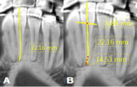
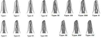
For all mandibular incisors, the observations included: (1) tooth length (Figure 1), (2) radicular length (Figure 1), (3) number of roots, (4) number of canals, (5) root canal configuration according to Vertucci’s classificationand Gulabivala classification (Figure 2) [25,26], (6) apical curvature, (7) type of septum (Figure 3), and (8) relationship of the septum to different anatomical structures (Figure 4).

    Figure 1:  CBCT images of a mandibular incisor.
A Tooth length measured from the incisal border to the root apex. B Root
length measured from the cement—enamel junction (represented by the
horizontal line) to the root apex.

    Figure 2:  Vertucci’s classification and Gulabivala’s classification of root canal morphology.
Type I: a single canal extends from the pulp chamber to the apex. Type II: two separate canals leave the pulp chamber and join short of the apex to form one canal.
Type III: one canal leaves the pulp chamber, divides into two within the root, and then merges to exit as one canal. Type IV: two separate and distinct canals extend
from the pulp chamber to the apex. Type V: one canal leaves the pulp chamber and divides short of the apex into two separate and distinct canals with separate
apical foramina. Type VI: two separate canals leave the pulp chamber, merge in the body of the root, and re-divide short of the apex to exit as two distinct canals.
Type VII: one canal leaves the pulp chamber, divides and then rejoins within the body of the root, and finally re-divides into two distinct canals short of the apex.
Type VIII: three separate and distinct canals extend from the pulp chamber to the apex. Type IX: three separate canals leave the pulp chamber and join short of the
apex to form one canal. Type X: two separate canals leave the pulp chamber, unite and then divide into two within the body of the root, and finally join short of the
apex to form one canal. Type XI: four canals leave the pulp chamber; both pairs unite within the root and emerge as two separate canals. Type XII: three canals
leave the pulp chamber, two of them unite within the root, and finally they emerge as two separate canals. Type XIII: two canals leave the pulp chamber, one of
them divides within the root and they emerge as three separate canals. Type XIV: four canals extend from the pulp chamber to the apex. Type XV: five canals leave
the pulp chamber; two of them join within the root, and finally they emerge as four separate canals.

    Figure 3:  Types of septum in mandibular incisors.
A and B Type I: septum with oval shape, defined and clear limits,
measurements can be made. C and D Type II: septum with elongated shape
and undefined limits, for which no measurements can be made because it is
difficult to establish stable references.

    Figure 4:  Measurements from the septum to different anatomical structures.
A Measurement from the incisal border to the coronal limit of the septum.
B Measurement from the cement—enamel junction (represented by the
horizontal line) to the coronal limit of the septum. C Measurement from the
apical limit of the septum to the root apex.
A descriptive analysis was performed for all variables. For quantitative variables (age, root length, bifurcation and unification of the canals), the mean, Standard Deviation (SD) and range were reported. Nominal variables (country of origin, evaluated incisor, number of roots, number of canals, root canal configuration, and type of septum) were tabulated according to absolute frequencies and percentages.
Differences between the two populations were explored using a generalized estimating equation for quantitative variables and a multilevel regression model for nominal variables, with a p-value of 0.05 or lower. The analysis was performed using Stata® v14 software (Stata Corp, Texas, USA).
Prevention of bias
To prevent bias, different measures were applied such as simple random sampling, calibration of the evaluators, the same instrument of measurement, management of the environmental variables that could affect imaging analysis, registration of the gender and age of the patients, strict selection criteria and compliance with ethical standards, approval of the ethics committee, obtaining informed consent, and declaration of no conflict of interest.
Results
A total of 220 CBCT scans were analyzed. In the Belgian sample, consisting of 94 patients with a total of 345 mandibular incisors (174 first mandibular incisors and 171 second mandibular incisors), 55% of the patients were female and 45% male. In the Chilean sample, of 126 patients with a total of 353 mandibular incisors (171 mandibular central incisors and 182 mandibular lateral incisors), 36% of the patients were female and 64% were male. The nominal variables are shown in Table 1. The quantitative variables are shown in Table 2. There were also significant differences between the groups in the number of canals, presence of a septum, and type of septum (Table 3). There were significant differences in root length and tooth length between the groups (Table 4). Age-related effects were found for tooth length in the Chilean sample, but no age-related effects were found for root length in either sample. Gender-related effects were found for tooth and root length in both samples. Age-related effects were also found for the distance between the intercanal septum to the incisal edge and for the distance between the intercanal septum and the apical foramen in mandibular lateral incisors, in both samples (Table 4).
    Mandibular central incisor
    Mandibular lateral incisor


    Belgium
    Chile
    Belgium
    Chile


    N
    %
    N
    %
    N
    %
    N
    %


    Number of roots


    1
    174
    100
    171
    100
    170
    99.42
    182
    100


    2
    0
    0
    0
    0
    1
    0.58
    0
    0


    Number of canals


    1
    107
    61.49
    134
    78.36
    102
    59.65
    144
    79.12


    2
    67
    38.51
    37
    21.64
    69
    40.35
    38
    20.88


    Root canal configuration


    Type I
    107
    61.49
    133
    77.78
    102
    59.65
    144
    79.12


    Type II
    1
    0.58
    5
    2.92
    1
    0.58
    2
    1.1


    Type III
    56
    32.18
    33
    19.3
    64
    37.44
    36
    19.78


    Type V
    7
    4.02
    0
    0
    3
    1.75
    0
    0


    Type VI
    1
    0.58
    0
    0
    0
    0
    0
    0


    Type X
    2
    1.15
    0
    0
    1
    0.58
    0
    0


    Isthmus


    Absent
    116
    66.67
    134
    78.36
    106
    61.97
    143
    78.57


    Type I
    25
    14.36
    24
    14.04
    21
    12.28
    31
    17.03


    Type II
    33
    18.97
    11
    7.6
    44
    25.73
    8
    4.4


    Curvature in the apical third of the canal


    Buccal
    130
    78.31
    137
    82.04
    108
    64.29
    122
    69.71


    Straight
    34
    20.48
    29
    17.37
    56
    33.33
    52
    29.71


    Lingual
    2
    1.21
    1
    0.59
    4
    2.38
    1
    0.58
Table 1: Descriptive analysis of nominal variables for mandibular central and lateral incisors, by population.
    Mandibular    central incisor
    Mandibular    lateral incisor


    N
    Mean(mm)
    SD
    Min (mm)
    Max (mm)
    N
    Mean(mm)
    SD
    Min (mm)
    Max (mm)


    Tooth length


    Belgium
    174
    20.9
    1.5
    17.3
    25
    171
    22.5
    1.6
    18.4
    26.3


    Chile
    171
    20.3
    1.8
    16
    24.7
    184
    21.9
    1.8
    18.8
    27.3


    Root length


    Belgium
    174
    12.9
    1.3
    9.8
    16.6
    171
    14.1
    1.4
    10.3
    17.8


    Chile
    171
    13.5
    1.3
    10.1
    16.6
    184
    14.7
    1.4
    11.3
    19.1


    IS to IB


    Belgium
    25
    11.8
    1.3
    8.2
    13.8
    21
    12.3
    1
    10.4
    14.4


    Chile
    24
    11.7
    2
    8.1
    14.7
    31
    11.7
    1.8
    14.2
    6.5


    IS to CRL


    Belgium
    25
    3.5
    1.1
    0
    4.9
    21
    3.9
    0.8
    2.7
    5.4


    Chile
    24
    3.2
    1.2
    0.9
    4.9
    31
    3.3
    1.1
    0
    4.9


    IS to AF


    Belgium
    25
    3.5
    1.5
    1.3
    8.3
    21
    4.4
    1.6
    1.4
    7.2


    Chile
    24
    4.1
    1.2
    2.1
    6
    31
    4.1
    1.4
    1.1
    6.7
Table 2: Descriptive analysis of quantitative variables for mandibular central and lateral incisors, by population (IS: Intercanal Septum; IB: Incisal Border; CRL:
Coronoradicular Limit; AF: Apical Foramen).
    Number    of canals
    Presence    of septum
    Type    of septum


    Odds Ratio
    Confidence interval 95%
    p value
    Odds Ratio
    Confidence Interval 95%
    p value
    Odds Ratio
    Confidence Interval 95%
    p value


    Central incisor


    Country of origin
    2.27
    1.41—3.65
    0.001*
    1.81
    1.12—2.93
    0.02*
    2.43
    1.03—5.74
    0.04*


    Gender


    Belgium
    1.84
    0.99—3.44
    0.05*
    1.69
    0.89—3.23
    0.11
    0.64
    0.21—1.91
    0.42


    Chile
    0.99
    0.47—2.10
    0.98
    0.99
    0.47—2.10
    0.98
    0.35
    0.08—1.65
    0.19


    Lateral incisor


    Country of origin
    2.6
    1.62—4.16
    <0.0001*
    2.28
    1.42—3.65
    0.001*
    8.12
    3.17—20.78
    <0.001*


    Gender


    Belgium
    1.58
    0.85—2.94
    0.17
    1.62
    0.86—3.03
    0.13
    1.59
    0.55—4.60
    0.39


    Chile
    1.38
    0.92—-3.92
    0.08
    2.04
    0.99—4.20
    0.05*
    0.27
    0.04—0.92
    0.15
Table 3: Comparative analysis of nominal variables for the mandibular central and latetal incisor, by country of origin and gender (multilevel regression model).
    Tooth length
    Root length
    IS to IB
    IS to CRL
    IS to AF


    Coef (mm)
    Confidence interval 95%
    p value
    Coef (mm)
    Confidence interval 95%
    p value
    Coef (mm)
    Confidence interval 95%
    p value
    Coef (mm)
    Confidence interval 95%
    p value
    Coef (mm)
    Confidence interval 95%
    p value


    Central incisor


    Country of origin
    0.6
    0.2—1.1
    0.007*
    (—)0.6
    (—)1.0— (—)0.3
    0.001*
    0.1
    (—)1.1—1.3
    0.84
    0.3
    (—)0.4—1.1
    0.42
    (—)0.6
    (—)1.4—0.3
    0.21


    Gender


    Belgium
    0.9
    0.3—1.5
    0.002*
    0.8
    0.3—1.3
    0.001*
    0.6
    (—)0.4—1.6
    0.26
    0.3
    (—)0.5—1.1
    0.51
    (—)0.8
    (—)2.0—0.3
    0.16


    Chile
    1.0
    0.4—1.7
    0.003*
    0.9
    0.4—1.4
    <0.001*
    0.8
    (—)1.3—2.9
    0.44
    0.5
    (—)0.6—1.6
    0.34
    (—)0.1
    (—)1.2—1.0
    0.83


    Age


    Belgium
    (—)0.01
    (—)0.03—0.01
    0.25
    0.01
    (—)0.003—0.03
    0.11
    (—)0.03
    (—)0.05—0.0003
    0.05*
    (—)0.01
    (—)0.04—0.02
    0.59
    0.03
    (—)0.01—0.06
    0.13


    Chile
    (—)0.03
    (—)0.04— (—)0.01
    <0.001*
    (—)0.01
    (—)0.02—0.002
    0.09
    (—)0.09
    (—)0.11— (—)0.07
    <0.001*
    (—)0.04
    (—)0.06— (—)0.02
    <0.001*
    0.02
    (—)0.01—0.05
    0.12


    Lateral    incisor


    Country    of origin
    0.7
    0.2—1.1
    0.004*
    (—)0.6
    (—)0.9— (—)0.2
    0.002*
    0.6
    (—)0.4—1.6
    0.22
    0.6
    (—)0.003—1.2
    0.05
    0.1
    (—)0.9—1.1
    0.82


    Gender


    Belgium
    0.9
    0.3—1.5
    0.006*
    0.5
    0.02—1.1
    0.04*
    0.2
    (—)0.7—1.1
    0.64
    0.02
    (—)0.7—0.7
    0.95
    (—)0.5
    (—)2.1—1.1
    0.55


    Chile
    0.9
    0.3—1.6
    0.005*
    0.8
    0.3—1.2
    0.003*
    1.1
    (—)0.5—2.8
    0.16
    0.9
    (—)0.1—1.9
    0.08
    (—)0.4
    (—)1.5—0.7
    0.49


    Age


    Belgium
    (—)0.004
    (—)0.02—0.02
    0.69
    0.01
    (—)0.003—0.03
    0.12
    (—)0.03
    (—)0.05— (—)0.007
    0.01*
    (—)0.02
    (—)0.03—0.001
    0.07
    0.07
    0.04—0.1
    <0.001*


    Chile
    (—)0.02
    (—)0.04— (—)0.01
    0.001*
    (—)0.005
    (—)0.02—0.006
    0.36
    (—)0.04
    (—)0.07— (—)0.00002
    0.05*
    (—)0.02
    (—)0.04—0.009
    0.22
    0.03
    0.01—0.06
    0.002*
Table 4: Comparative analysis of quantitative variables for the mandibular central and lateral incisor, by country of origin, gender and age (generalized estimating
equation).
Discussion
Periapical radiography is an essential tool for endodontic treatment. However, it has limitations such as a two-dimensional view of three-dimensional structure, revealing solely restricted aspects of the three-dimensional anatomy [27]. In distinction, CBCT is asophisticated diagnostic imaging technique that doesn’t present geometrical distortion or anatomical noise; it provides threedimensional images of structures and permits accurate measurements of the information acquired [28]. This makes CBCT a useful tool for endodontic assessment, because it permits an in depth analysis of the anatomy before treatment.
It is possible that studies using CBCT for the study of root canal anatomy are hampered by limited resolution and slice thickness, in comparison with micro-CT [12], but the use of CBCT allows a method that is feasible and reproducible in an in vivo scenario, that’s better than a periapical radiography. In an ex vivo study, micro-CT should be the chosen technique because it provides accuracy, high resolution and can be used for details measurement of the root canal anatomy [12].
Mandibular incisors can have sometimes complex root canal morphology. They can present multiple variations such as a second canal, apical delta, intercanal septum, and bifurcations, among others, which can become challenging during root canal treatment [2-6].
An inappropriate diagnosis of the anatomical configuration can lead to a series of complications and the failure of the endodontic treatment [2,29]. Therefore, it is critical to obtain a successful outcome, that professionals are informed about possible variations in these teeth before treating them.
In this study, all CBCT images were obtained using the same scanner, from patients who required a CBCT examination as an integral part of their diagnosis and treatment planning. Standard protocol for acquiring images was not obtained because it varied on a case-by-case basis to ensure the lowest possible radiation dose. These variations may have compromised the collection of standardized images and the accuracy of the assessment. In future research, a standard image acquisition protocol should be defined, as far as possible, to ensure an optimal comparison.
The ethnic background was not taken into consideration as an inclusion criterion. Assessing specific ethnic groups may be challenging. Therefore, the individuals analyzed represent only a sample of the population attending the radiological centers in each country.
The literature has shown that the mandibular central incisor in most cases presents one root and one canal [7,10,26,30,31], corresponding with the results obtained in the present study. However, Sertand Bayirli published a study reporting a prevalence of one canal of only 32.5%, which is not in line with the results obtained in this or any other published study [32]. That study used clearing and staining techniques to evaluate root canal morphology. The sample comprised 2800 maxillary and mandibular teeth from a Turkish population sample; 200 mandibular central incisors and 200 mandibular lateral incisors were evaluated.
The most common canal configurations in our study were Vertucci types I, III and II. The most prevalent in both samples was type I, followed by type III. In the Chilean simple, type I was present in 77.78% of the cases, while in the Belgian simple it was present in 61.49%. Vertucci described a prevalence of 70% of type I cases (2), Han et al. reported a prevalence of 84.29% (10), while Zhengyanet al. found 96.24% in a Chinese sample [33].
The mandibular lateral incisor presented in this study with one root and one canal in most cases for both samples. Only one case with two roots was reported, in the Belgian sample; this rare variation has been described elsewhere in the literature [33]. A similar prevalence has also been reported by other authors [2,4,32].
The prevalence of different root canal configurations of the mandibular lateral incisor was similar to that of the mandibular central incisor and is consistent with reports in the available literature, which classifies type I as the most prevalent [1,4,29,32].
The curvature of the apical third of the canal is not commonly described in the literature. In this study, it was found that a curvature to the vestibular side was more prevalent in both central and lateral mandibular incisors, for both samples. This corresponds with the studies by Willershausenet al. [33] and Miyashita et al. [4]. There is no description available in the literature for the classification of the intracanal septum present in mandibular incisors. It is vital for a dentist to know the parameter of length in relation to the presence of a septum, to be prepared for the anatomical variations that may be present. The classification proposed in this study could be useful for future investigations to determine at what length the practitioner could expect to find the bifurcation of the canal or the joining of it, which is vital for treatment planning.
This investigation has shown that the prevalence of two canals is high, up to 47.06%, therefore it is the responsibility of the dentist always to search for a second canal and be prepared for the possible configurations, of which type III is one of the most common.
Given that type III has a high prevalence in the mandibular incisor, the practitioner must be prepared for bifurcation and union of canals. The proposed classification of the intercanal septum and its relation to different anatomical structures tells the clinician when to expect bifurcation or unification. Furthermore, it indicates at what length to expect a second canal, giving critical information for root canal treatment. According to this investigation, professionals should expect bifurcation of the canals to occur between 11.69 mm and 12.25 mm, taking the incisal edge as a reference, and can expect the unification of the canals to occur between 3.51 mm and 4.4 mm from the apical foramen. If a type II septum is diagnosed, it could disappear during mechanical preparation owing to its lack of definition and density, causing unification of the two canals.
This investigation also compared the nominal variables by gender and country of origin. The results show that the Belgian sample showed a greater likelihood of having two canals, an intercanal septum and atype II septum. When compared by gender, men in the Belgian sample had a greater risk of having two canals and an intercanal septum. This relationship was not statistically significant in the Chilean sample, but when comparing the type of intercanal septum with gender in the Chilean sample, women presented a greater likelihood of having type II and men type I.
For the quantitative variables, comparison was made by gender, country of origin, and age. The Belgian sample presented longer teeth, but for roots the Chilean sample showed a greater length. In both populations men had longer teeth and roots than women. Furthermore, an age-related effect was found for tooth length in the Chilean sample and for the distance from the intercanal septum to the in cisal border in both samples. This implies that as age increases the length, for both parameters, decreases.
The root canal morphology of 698 mandibular incisors was analyzed using CBCT in Chilean and Belgian samples. The mandibular central and lateral incisors presented more frequently with one root and one canal, in both samples. Types I, III and II canal configurations were most commonly found.
When comparing the characteristics of the mandibular incisor in the two sample groups, statistically significant differences were found for tooth length, radicular length, number of canals and the presence and type of intercanal septum. Furthermore, an age-related effect was found for tooth length in the Chilean sample, for the mandibular lateral and central incisors.
Compliance with Ethical Standards
Human rights statements and informed consent
All procedures followed were in accordance with the ethical standards of the responsible committee on human experimentation (institutional and national) and with the Helsinki Declaration of 1975, as revised in 2008. Informed consent was obtained from all patients for being included in the study.
References
- Vertucci FJ. Root canal morphology and its relationship to endodontic procedures. Endod Top. 2005; 10: 3-29.
- Vertucci FJ. Root canal anatomy of the mandibular anterior teeth. J Am Dent Assoc. 1974; 89: 369-371.
- Funato A, Funato H, Matsumoto K. Mandibular central incisor with two root canals. Dent Traumatol. 1998; 14: 285-286.
- Miyashita M, Kasahara E, Yasuda E, Yamamoto A, Sekizawa T. Root canal system of the mandibular incisor. J Endod. 1997; 23: 479-484.
- Al-Qudah AA, Awawdeh LA. Root canal morphology of mandibular incisors in a Jordanian population. Int Endod J. 2006; 39: 873-877.
- Kabak YS, Abbott PV. Endodontic treatment of mandibular incisors with two root canals: Report of two cases. Aust Endod J. 2007; 33: 27-31.
- Madeira MC, Hetem S. Incidence of bifurcations in mandibular incisors. Oral Surg Oral Med Oral Pathol. 1973; 36: 589-591.
- Pineda F, Kuttler Y. Mesiodistal and buccolingual roentgenographic investigation of 7,275 root canals. Oral Surg Oral Med Oral Pathol. 1972; 33: 101-110.
- Benjamin KA, Dowson J. Incidence of two root canals in human mandibular incisor teeth. Oral Surg Oral Med Oral Pathol. 1974; 38: 122-126.
- Han T, Ma Y, Yang L, Chen X, Zhang X, Wang Y. A study of the root canal morphology of mandibular anterior teeth using cone-beam computed tomography in a Chinese subpopulation. J Endod. 2014; 40: 1309-1314.
- Martins JNR, Marques D, Mata A, Caramês J. Root and root canal morphology of the permanent dentition in a Caucasian population: a conebeam computed tomography study. Int Endod J. 2017.
- Ordinola-Zapata R, Bramante CM, Versiani MA, Moldauer BI, Topham G, Gutmann JL, et al. Comparative accuracy of the Clearing Technique, CBCT and Micro-CT methods in studying the mesial root canal configuration of mandibular first molars. Int Endod J. 2017; 50: 90-96.
- Rankine-Wilson RW, Henry P. The Bifurcated Root Canal in Lower Anterior Teeth. J Am Dent Assoc 1939. 1965; 70: 1162-1165.
- Walker RT. Root form and canal anatomy of maxillary first premolars in a southern Chinese population. Endod Dent Traumatol. 1987; 3: 130-134.
- Walker RT. Root form and canal anatomy of mandibular first molars in a southern Chinese population. Dent Traumatol. 1988; 4: 19-22.
- Bellizzi R, Hartwell G. Clinical investigation of in vivo endodontically treated mandibular anterior teeth. J Endod. 1983; 9: 246-248.
- Mauger MJ, Schindler WG, Walker WA. An evaluation of canal morphology at different levels of root resection in mandibular incisors. J Endod. 1998; 24: 607-609.
- Miyoshi S, Fujiwara J, Tsuji YH, Nakata T, Yamamoto K. Bifurcated Root Canals and Crown Diameter. J Dent Res. 1977; 56: 1425-1425.
- Zhang R, Wang H, Tian Y-Y, Yu X, Hu T, Dummer PMH. Use of cone-beam computed tomography to evaluate root and canal morphology of mandibular molars in Chinese individuals. Int Endod J. 2011; 44: 990-999.
- Zhang R, Yang H, Yu X, Wang H, Hu T, Dummer PMH. Use of CBCT to identify the morphology of maxillary permanent molar teeth in a Chinese subpopulation. Int Endod J. 2011; 44: 162-169.
- Cotton TP, Geisler TM, Holden DT, Schwartz SA, Schindler WG. Endodontic applications of cone-beam volumetric tomography. J Endod. 2007; 33: 1121- 1132.
- Pedemonte E, Cabrera C, Torres A, Jacobs R, Harnisch A, Ramírez V, et al. Root and canal morphology of mandibular premolars using cone-beam computed tomography in a Chilean and Belgian subpopulation: a crosssectional study. Oral Radiol. 2017; 3: 1-8.
- Torres A, Jacobs R, Lambrechts P, Brizuela C, Cabrera C, Concha G, et al. Characterization of mandibular molar root and canal morphology using cone beam computed tomography and its variability in Belgian and Chilean population samples. Imaging Sci Dent. 2015; 45: 95-101.
- Nolla C. The development of the permanent teeth. Journal of Dentistry for Children. 1960; 27: 254-266.
- Gulabivala K, Aung TH, Alavi A, Ng YL. Root and canal morphology of Burmese mandibular molars. Int Endod J. 2001; 34: 359-370.
- Vertucci FJ. Root canal anatomy of the human permanent teeth. Oral Surg Oral Med Oral Pathol. 1984; 58: 589-599.
- Patel S, Dawood A, Ford TP, Whaites E. The potential applications of cone beam computed tomography in the management of endodontic problems. Int Endod J. 2007; 40: 818-830.
- Scarfe WC, Farman AG, Sukovic P. Clinical applications of cone-beam computed tomography in dental practice. J Can Dent Assoc. 2006; 72: 75-80.
- Arslan H, Ertas H, Ertas ET, Kalabalik F, Saygili G, Capar ID. Evaluating root canal configuration of mandibular incisors with cone-beam computed tomography in a Turkish population. J Dent Sci. 2015; 10: 359-364.
- Lin Z, Hu Q, Wang T, Ge J, Liu S, Zhu M, et al. Use of CBCT to investigate the root canal morphology of mandibular incisors. Surg Radiol Anat SRA. 2014; 36: 877-882.
- Zhengyan Y, Keke L, Fei W, Yueheng L, Zhi Z. Cone-beam computed tomography study of the root and canal morphology of mandibular permanent anterior teeth in a Chongqing population. Ther Clin Risk Manag. 2015; 12: 19-25.
- Sert S, Bayirli GS. Evaluation of the Root Canal Configurations of the Mandibular and Maxillary Permanent Teeth by Gender in the Turkish Population. J Endod. 2004; 30: 391-398.
- Willershausen B, Kasaj A, Röhrig B, Marroquin BB. Radiographic Investigation of Frequency and Location of Root Canal Curvatures in Human Mandibular Anterior Incisors In Vitro. J Endod. 2008; 34: 152-156.
