Clinical Image
Austin J Radiol. 2018; 5(2): 1086.
A Late Revelation of a Right Bochdalek Hernia with Hepatic and Colic Content
Amarir M* and Dafiri R
Department of Radiopediatrics, Mohammed V University, Morocco
*Corresponding author: Amarir M, Departement of Radiopediatrics, Children’s Hospital, Faculty of Medicine and Pharmacy, Mohammed V University, Rabat, Morocco
Received: December 10, 2018; Accepted: December 27, 2018; Published: December 31, 2018
Clinical Image
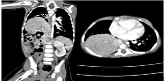
A12month old female infant with no significant history is admitted to emergency for respiratory distress without fever; the chest x-ray shows a heterogeneous hydric opacity with aeric structures occupying the 2/3 inferiors of the right hemithorax. Thoracic CT shows an intra-thoracic ascent of the liver and part of the ascending colon through a right posterior lateral diaphragmatic defect (Figures 1&2). The diagnosis retained is a late revelation of a posterolateral Bochdalek hernia. A drain was inserted in the chest cavity to decompress it for better lung expansion. The infant was operated with favorable postoperative evolution. Bochdalek’s hernia is a congenital diaphragmatic malformation, its occurs when the diaphragm muscle fails to close during fetal development, the defect is more common on the left, only 3% of cases in the right, exceptional on both sides. The diagnosis is made at the ante or neonatal period, in 5 to 25% of the cases the hernia can be revelated 4 weeks later, after months as our case or many years later, respiratory distress is the most frequent revealing symptom.

    Figure 1:  Chest x-ray: A heterogeneous hydric opacity with aeric structures
occupying the 2/3 inferiors of the right hemithorax.

    Figure 2:  Thoracic CT in coronal and axial sections shows an intra-thoracic
ascent of the liver and part of the ascending colon through a right posterior
lateral diaphragmatic defect.