Research Article
Austin J Radiol. 2019; 6(1): 1089.
Symphalangism on Radiographs of Foot - A Cross Sectional Study
Venkatraman Indiran*, Naorem Vinod Singh and Prabakaran Maduraimuthu
Department of Radiodiagnosis, Sree Balaji Medical College and Hospital, Chennai, India
*Corresponding author: Venkatraman Indiran, Department of Radiodiagnosis, Sree Balaji Medical College and Hospital, Chromepet, Chennai, India
Received: January 25, 2019; Accepted: February 27, 2019; Published: March 06, 2019
Abstract
Aims and Objectives: To analyze the number of phalanges and prevalence of symphalangism in the second to fifth toes, with respect to age and gender in the Indian Population.
Materials and Methods: Analysis of 431 radiographs of the foot (anteroposterior and oblique views) in patients presenting to the Radiology department of Sree Balaji Medical college and Hospital was carried out. Number of phalanges in the second to fifth toes were counted on all the radiographs and were assessed for fusion of phalanges (symphalangism)
Results: Symphalangism of fifth toe was seen in 170 radiographs with isolated fifth toe involvement on 156 radiographs. The rest of the 14 cases have associated symphalangism in second, third or fourth toe. Similar prevalence rate was seen in male and female populations. Maximum prevalence symphalangism was found in the age group of 21 to 30 years (males were more affected than the female).
Conclusion: The presence of two phalanges is a common anatomical variant in the Indian population. It was also observed that the second, third and fourth toe symphalangism was never seen in the absence of fifth toe symphalangism.
Keywords: Phalanges; Fusion; Biphalangeal; Symphalangism
Introduction
The phalanges are long bones in the foot located distal to the metatarsals. In a normal anatomy of human feet, each toe consists of three phalanges, which are named the proximal, middle and distal phalanges. However, the great toe only has two phalanges, a proximal and a distal one. Symphalangism of feet refers to the fusion of the two phalanges of the same toe. There is a lack of attention regarding symphalangism due to its asymptomatic nature. The study is not common in the international scientific literature. This is the second case study done in the Indian population and first case study purely based on the radiography. Some authors have considered relation to ethnicity, since symphalangism is less common among the African, American and English while it is extremely common among the Japanese and Korean [1,2].
Materials and Methods
Four hundred and fifty consecutive foot radiographs taken in the department of Radio-diagnosis between December 2016 to May 2017 were considered for analysis in this cross sectional study, irrespective of the clinical indications for radiography. Nineteen patients who had destruction of phalanges of 2nd to 5th toe were excluded from the study. Patients from different states of India were included in the study. Plain radiographs were performed 600 mA x ray machine (Allengers 625, India). Antero - posterior and oblique views of foot were obtained from all the patients. For anterior-posterior view, the patients were made to lie in a supine position on the x ray table with the knee in flexion and plantar surface of foot placed over the cassette. The x ray tube is placed 100 cm away from the source. For oblique view, the patients were made to lie in a supine position in the x ray table with knee in flexion and foot externally rotated until the plantar surface is at 45 angle to the cassette. The x ray tube was placed 100 cm away from the source. The radiographic parameters used were 50-55 kV power, 100 mA current and 0.08 seconds exposure time. Informed consent was obtained from the patients for the radiography procedure. Ethical committee approval was obtained for this study.
Results
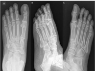
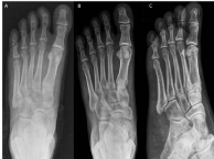
Of the 431 cases, 162 were female (37.6%) and 269 were male (62.4%). Age of the patients included in our study ranged from 9 to 90 years with mean age of 36.7 years. This study showed the presence of 3 phalanges in 261 cases (60.6%) and 2 phalanges in 170 cases (39.4%) in the fifth toe (Figure 1). Among the 269 males, 167 (62.1%) had triphalangeal 5th toe and 102 (37.9%) had biphalangeal 5th toe. On the other hand, among the 162 females, 94 (58.1%) had triphalangeal in 5th toe and 68 feet (41.9%) had biphalangeal 5th toe (Figure 2). The proportion between females and males was almost similar (Table 1). The biphalangeal fourth toe was never seen in the in the absence of biphalangeal fifth toe. Likewise, the third toe symphalangism was never seen in the absence of fourth and fifth toe symphalangism. Similarly, the second toe symphalangism also was never seen in the absence of third, fourth and fifth toe symphalangism in this study population. The presence of two phalanges were observed in fourth, third and second toes in 14 cases (3.2%), 4 cases (0.92%) and 3 cases (0.69%) respectively (Table 2). It was also observed that the second, third and fourth toe symphalangism was never seen in the absence of fifth toe symphalangism.
    AGE AND GENDER


    FUSED PHALANGES
    TOE
    1-10    yrs
    11-20    yrs
    21-30    yrs
    31-40    yrs
    41-50    yrs
    51-60    yrs
    61-70    yrs
    71-80    yrs
    81-90    yrs
    TOTAL


    M
    F
    M
    F
    M
    F
    M
    F
    M
    F
    M
    F
    M
    F
    M
    F
    M
    F
    M
    F
    COM


    5THTOE
    1
    1
    14
    5
    34
    9
    21
    14
    16
    19
    8
    14
    5
    5
    3
    1
    -
    -
    102
    68
    170


    4THTOE
    -
    -
    -
    1
    4
    1
    1
    2
    1
    2
    -
    1
    -
    1
    -
    -
    -
    -
    6
    8
    14


    3RDTOE
    -
    -
    -
    -
    2
    -
    -
    1
    -
    -
    -
    -
    -
    1
    -
    -
    -
    -
    2
    2
    4


    2NDTOE
    -
    -
    -
    -
    1
    -
    -
    1
    -
    -
    -
    -
    -
    1
    -
    -
    -
    -
    1
    2
    3
Table 1: Age and gender wise distribution in patients with symphalangism.

    Figure 1:  Normal foot radiographs anterior-posterior view (A) and oblique
view (B) showing triphalangeal fifth toe. Oblique view of another foot showing
biphalangeal fifth toe (C).

    Figure 2:  Anterior-posterior view of foot showing biphalangeal fourth & fifth
toe (A) and biphalangeal third, fourth and fifth toe (B). Oblique radiograph foot
showing biphalangeal second, third, fourth and fifth toe (C).
    FUSED    PHALANGES
    FEMALE
    MALE
    TOTAL
    STUDY    POPULATION
    PERCENTAGE    (F+M)


    5TH TOE
    68
    102
    170
    431
    39%


    4TH toe associated with 5th toe
    8
    6
    14
    431
    3.20%


    3RD toe associated with 4th&5th toe
    2
    2
    4
    431
    0.92%


    2ND toe associated with 3rd, 4th& 5th toe
    2
    1
    3
    431
    0.69%
Table 2: Percentage of fused phalanges.
Discussion
The presence of 2 phalanges in the 5th toe was first described by Leonardo da Vinci in 1492 (O’Malley & Saunders, 1952) and is recognized as a normal anatomical variant [3,4]. Studies on symphalangism have been based either on cadaveric studies or radiographic methods. Studies on symphalangism have revealed varying prevalence in different ethnic populations (Table 3). Biphalangeal fifth toe is probably a true anatomical variant, resulting from incomplete segmentation rather than the result of phalangeal fusion [5,6]. This variant is exclusively a human phenomenon, suggesting that it is a response to bipedalism and that it would result primarily from the failure of the distal interphalangeal joint to develop [7].
    AUTHORS
    YEAR
    SAMPLE    POPULATION
    NO. OF    FEET
    COUNTING    METHOD
    STUDY    TYPE
    SYMPHALANGISM OF 5TH TOE (%)


    Nakaishi
    1942
    Japanese    adults
    500
    Feet
    Radiograph
    72.2


    Venning
    1956
    European    children and adults
    4632
    Feet
    Radiograph
    42.5


    Asin
    1966
    American    adults
    417
    Individual    feet
    Radiograph
    42.5


    Ellis    et al.
    1968
    American    adults
    390
    Individual    feet
    Radiograph
    47.5


    Sandstorm    and Hedman
    1971
    Swedish    children and adults
    496
    Feet
    Radiograph
    34.5


    Winiecki
    1978
    American    adults
    974
    Feet
    Radiograph
    42.1


    Carroll    et al.
    1978
    American    adults
    1324
    Individual feet
    Radiograph
    33.8


    Le    Minor
    1995
    French    adults
    2550
    Individual feet
    Radiograph
    41


    Nakashima    et al.
    1995
    Japanese    children and adults
    488
    Feet
    Radiograph
    72.5


    Park    and Sohn
    1998
    Korean    adults
    1187
    Feet
    Radiograph
    74


    George
    2001
    English    old and young adults
    204
    Feet
    Radiograph
    37.7


    Chae    et al.
    2002
    Koreans    adults
    1290
    Feet
    Radiograph
    72.4


    Rabi    et al.
    2005
    South    Indian fetuses children and adults
    24
      112
      263
    Feet
    Radiograph
    87.5
      9.8
      11.8


    Sohn    et al.
    2006
    Korean    adults
    175
    Feet
    Radiograph
    74.2


    Moultron    et al.
    2012
    English    adults
    606
    Feet
    Radiograph
    44.4


    Gallart    et al
    2014
    Spanish    adults
    2494
    Feet
    Radiograph
    46.3
Table 3: Prevalence of symphalangism in different parts of the world.
In comparison with others studies conducted over the years, the percentage of symphalangism (39.4%) in this study appears lower than in the American [4,8], English [9], Korean [10], Japanese population [2] and higher than the Swedish population [11]. Study conducted by M.George in 2001 revealed biphalangeal fifth toe in 38.5% of the 204 patients, which was similar to this study (39.3%) [12].
In a study of symphalangism in the digits of Japanese feet, reported overall incidence of symphalangism in the 5th toe was 72.5%, which was significantly higher than that in the European population [2] as well as in this study population.
In their study, Gallart et al found that the risk of suffering from hammer toe of 5th toe was almost 4 times more in triphalangeal toe than the biphalangeal toe. It did not find any significant differences regarding the need for surgery of the fifth toe of the biphalangeal (39.1%) versus triphalangeal toes (60. 9%). They postulated that there may be an association between pathologic deviations and bigger mobility of the triphalangeal fifth toes. However, biphalangeal fifth toes show more rigidity leading to smaller accommodation inside the shoe, which may lead to less painful feet and decreased proportion of surgery [13].
Turan et al, in their case report showed that, the presence of a biphalangeal fifth toe delayed the diagnosis of a fracture, although proper radiographic examination had been performed [14]. The fracture line was at the same level of the joint line and transverse in configuration. This type of fracture pattern may cause the confusion and misdiagnosis. The fracture line and joint line can be differentiated with careful observation. Knowledge of pedal symphalangism with the presence of ecchymosis, swelling, and severe tenderness on clinical examination with a history of traumatic event, helps in the correct diagnosis. In a case of trauma, biphalangeal toes may pose a diagnostic challenge and fractures may be interpreted as normal, which can lead to misdiagnosis and under treatment [14].
Case et al suggested that additional genetic or developmental factors may play a role in the expression of pedal symphalangism in each of the toes as they never observed fourth toe symphalangism in the absence of fifth toe involvement [1]. Similarly, in the present study the second, third and fourth toe symphalangism was never seen in the absence of fifth toe symphalangism.
In this study population, all the cases were included with regardless of complaints of the patient or pathology of the toes. This could be a limitation of the study. Non-inclusion of the opposite foot in all the patients limited this study’s ability to exactly assess the laterality of symphalangism.
Conclusion
The presence of two phalanges in the fifth toe is a common anatomical variant in the Indian population. It is also observed that the second, third and fourth toe symphalangism is never seen in the absence of fifth toe symphalangism. Further prospective study may be done in patients with biphalangeal toes and toe complaints to assess the clinical impact of symphalangism.
Compliance with Ethical Standards
Ethical approval (animals): This article does not contain any studies with animals performed by any of the author(s).
Ethical approval: All procedures performed in studies involving human participants were in accordance with the ethical standards of the institutional and/or national research committee and with the 1964 Helsinki declaration and its later amendments or comparable ethical standards.
Informed consent: Informed consent was obtained from individual participant included in the study.
References
- Case DT, Heilman J. Pedal symphalangism in modern American and Japanese skeletons. Homo. 2005; 55: 251-262.
- Nakashima T, Hojo T, Suzuki K, Ijichi M. Symphalangism (two phalanges) in the digits of the Japanese foot. Ann Anat. 1995; 177: 275-278.
- O' Malley CD, Saunders JB, De CM. Leonardo da Vinci on the Human Body: The Anatomical, Physiological, and Embryological Drawings of Leonardo da Vinci with Translations, Emendations, and a Biological Introduction, New York: Henry Schuman. 1952; 62-65.
- Ellis R, Short JG, Knepley DW. The two-phalanged ?fth toe. J Am Med Assoc. 1968; 206: 2526.
- Venning P. Variation of the digital skeleton of the foot. Clinical Orthopaedics and Related Research. 1960; 16: 26-40.
- Thompson FM, Chang VK. The two-boned fifth toe: clinical implications. Foot Ankle Int. 1995; 16: 34-36.
- Le Minor JM. Biphalangeal and triphalangeal toes in the evolution of the human foot. Acta Anat. 1995; 154: 236-241.
- Asin HM. Symphalangia of the ?fth toes. J Am Podiatry Assoc. 1966; 56: 411-413.
- Moulton LS, Prasad S, Lamb RG, Sirikonda SP. How many joints does the 5th toe have? A review of 606 patients of 655 foot radiographs. Foot Ankle Surg. 2012; 18: 263-265.
- Chae WY, Park SB, Lee SG. Biphalangeal Toes in the Korean Foot. J Korean Acad Rehabil Med. 2002; 26: 193-197.
- Sandström B, Hedman G. Biphalangea of the lateral toes. A study on the incidence in a Swedish population together with some observations on digital sesamoid bones in the foot. Am J Phys Antropol. 1971; 34: 37-41.
- George M. Biphalangeal fifth toe: an increasingly common variant? J Anat. 2001; 198: 251.
- Gallart J, González D, Valero J, Deus J, Serrano P, Lahoz M. Biphalangeal/triphalangeal fifth toe and impact in the pathology of the fifth ray. BMC Musculoskeletal Disord. 2014; 15: 295.
- Turan A, Kose O, Guler F, Ozyurek S. Fracture of biphalangeal fifth toe: A diagnostic pitfall in the emergency department. Journal of Emergency Medicine, Trauma & Acute Care. 2016: 11.