
Clinical Image
Austin J Radiol. 2019; 6(1): 1090.
Spontaneous Ruptured Intracranial Dermoid Cyst
Choudhary G1*, Chokr J2 and Jagpal A3
1Department to Radiology, University of Alabama at Birmingham, USA
2Department of Radiology, Clemenceau Medical Center affiliated with Johns Hopkins International Clemenceau Street. Beirut, Lebanon
3Division of Clinical Immunology and Rheumatology, University of Alabama at Birmingham, USA
*Corresponding author: Gagandeep Choudhary, Department to Radiology, University of Alabama at Birmingham, 619 19th Street South, Birmingham, AL 35249-6835, USA
Received: March 09, 2019; Accepted: March 13, 2019; Published: March 20, 2019
Spontaneous Ruptured Intracranial Dermoid Cyst
A 31-year-old man with history of chronic headache for years, presented with acute worsening of the headache. Emergent CT scan of the head demonstrated a 4 cm low attenuation (fat density) mass within the frontal horn of right lateral ventricle. There was dilatation of both lateral and 3rd ventricles with normal 4th ventricle consistent with obstructive hydrocephalus at the level of cerebral aqueduct. Multiple fat density foci were seen within the subarachnoid sulcal spaces and cistern along with fat fluid level within the ventricles, consistent with intracranial dermoid cyst rupture. Figure 1,2 Patient was taken to the OR for resection and shunt placement for the hydrocephalus.
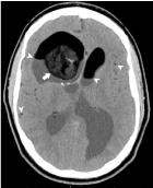
    Figure 1: Non-contrast axial CT image of the brain show low density mass
in the right lateral ventricle (arrow) with small foci of calcification. Fat fluid
levels and multiple fat droplets are seen in the ventricles and sulcal spaces
(arrow heads).
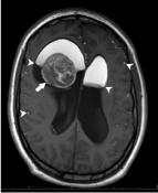
    Figure 2: T1 weighted non-contrast axial image of the brain show
heterogeneously hyperintense mass in the right lateral ventricle (arrow).
Fat fluid levels and multiple fat droplets are seen in the ventricles and sulcal
spaces (arrow heads).
Intracranial dermoid cysts are extremely rare congenital tumors of ectodermal origin, accounting for 0.5% of all primary intracranial tumors. Cysts are lined by stratified squamous epithelium and may contain epidermal appendages like hair follicles, sweet glands, sebaceous glands and even teeth. Cysts enlarge slowly over time due to accumulation of secretion from these glands. They are often asymptomatic unless they rupture or cause mass effect. The rupture can be spontaneous, traumatic, or iatrogenic. Complications such as chemical meningitis, hydrocephalus, vasospasm, infraction and even death may occur. Imaging findings of ruptured dermoid cysts are characteristic. CT findings include, non-enhancing low-density lesion with multiple fat density droplets in the subarachnoid spaces and ventricles with fat fluid levels. MRI is superior in evaluating the extent of lipid dissemination [1,2].
References