Case Report
Austin J Radiol. 2019; 6(1): 1091.
Multi-Detector CT Urethrography Diagnosis of an Urethro-Cutaneous Fistula and Associated Urethral Stricture: An Un-Conventional Imaging Style
Bhatt S1*, Swarup MS1, Tandon A1, Khullar T1 and Dangwal S2
1Department of Radiology, University College of Medical Sciences and GTB Hospital, India
2Himalyan Institute of Medical Sciences, Swami Rama Himalyan University, India
*Corresponding author: Bhatt S, Department of Radiology, University College of Medical Sciences and GTB Hospital, University of Delhi, Dilshad Garden, Delhi-110095, India
Received: February 02, 2019; Accepted: March 14, 2019; Published: March 21, 2019
Abstract
We report a case of a young male with Urethro-Cutaneous Fistula (UCF) diagnosed on Multi-Detector CT Urethrography (MDCTU), an un-conventional imaging modality to evaluate the urethra. This highlights the clinical utility of MDCTU to accurately delineate the UCF and the associated urethral stricture, when Conventional Urethrography (CU) failed to do so. Though sparingly used, MDCTU is a promising method for evaluation of the urethral pathologies and can be utilized as a reserve modality when routine techniques are not feasible or fail to provide the required diagnostic information. MDCTU has several advantages over CU including better patient compliance. It also provides additional diagnostic information in complicated urethral conditions like periurethral abscess, fistula, diverticula and urethral trauma. Thus, in a select group of patients the superior diagnostic capability of MDCTU can prove useful for appropriate management decisions.
Keywords: CT; Urethrography; Urethro-cutaneous fistula; Urethral stricture; Male urethra
Abbreviations
CT: Computed Tomography; UCF: Urethro-Cutaneous Fistula; MDCTU: Multi-Detector CT Urethrography; CU: Conventional Urethrography; RGU: Retrograde Urethrography; VCUG: Voiding Cysto-Urethrography; CTU CT Urethrography
Case Presentation
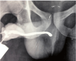
A 23 year old gentleman presented with passage of urine from an abnormal opening since one month. There was past history of pelvic trauma, associated with hematuria and urinary retention for which urethral catheterization was done. He remained asymptomatic for six months following catheter removal but gradually developed perineal pain and thinning of the urinary stream. These symptoms were present for the past eight months but recently he started passing urine from an abnormal opening in the perineum. Clinical diagnosis of urethral stricture with urethra-cutaneous fistula was made. RGU revealed a narrowing at the peno-bulbar junction and only little contrast passed beyond it (Figure 1). Inadequate contrast opacification failed to demonstrate the complete stricture. Even the smallest size (5F) infant feeding tube could not be negotiated beyond the stricture and therefore the VCUG study could not be conducted. Attempt to directly opacify the fistulous tract also failed due to regurgitation of contrast and poor patient compliance.

    Figure 1: Conventional retrograde urethrography image showing a normal
penile urethra with narrowing at the peno-bulbar junction and only little
passage of contrast beyond the stricture.
The referring surgeon agreed for a MDCTU as suggested by the radiologist. A written informed consent was obtained from the patient and the study conducted using a 64 slice MDCT scanner. As direct filling of bladder was not possible due to inability to pass the urethral catheter beyond the stricture; bladder filling was achieved by renal excretion of intravenously administered contrast (30 ml of 350 mg % Iohexol) as the serum creatinine was within normal limits (1.2 mg/dl). Thus, a technical difficulty of bladder filling encountered during the conventional study could be overcome with the use of MDCTU.
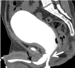
Thin axial CT sections were obtained as the patient voided into a condom catheter in a lying position. MDCTU clearly demonstrated the entire lower urinary tract- bladder, posterior and anterior urethra as well as the urethro-cutaneous fistula with a 9 mm tight stricture in the distal bulbar urethra with proximal urethral dilatation and irregularity. A thick fistulous tract of 6.4 cm length was present from prostato-membranous junction to perineal skin (Figure 2). Bladder wall was thickened and irregular suggestive of bladder outflow obstruction. The final imaging diagnosis was possible with MDCTU and was a tight short-segment peno-bulbar stricture with urethracutaneous fistula and urethritis.

    Figure 2: Sagittal reformatted CT urethrographic image shows short segment
stricture (black arrow) at distal bulbar urethra with dilatation and irregularity
of proximal urethra. A contrast filled fistulous tract is seen with its ostium
at prostato-membranous junction and extending to perineal region (white
arrow). The prevesical fat is obliterated and anterior bladder wall adhered
to abdominal wall indicating site of Previous Suprapubic Catheter (SPC)
insertion. Voiding during the CT acquisition allowed filling up of the urethral
lumen as well as the abnormal fistulous tract, ensuring its visualization and,
thus, its evaluation.
Discussion
Urethro-Cutaneous Fistula (UCF) is an uncommon entity and rarely encountered in clinical practice. It may occur due to a congenital defect or may be acquired after trauma (accidental or surgical) or following a urethral infection [1]. For best surgical results, delineation of the length and course of the fistula and its relation to the underlying urethra is required. Conventional Urethrography (CU) ie. Retrograde Urethrography (RGU) and Voiding Cysto- Urethrography (VCUG) is used to demonstrate the fistula in the anterior and posterior urethra respectively [2,3]. MRI can be considered in cases where CU fails to demonstrate the fistula [4]; but availability and affordability is always an issue with MRI. Though CT is not routinely used, CT Urethrography (CTU) can be utilized to evaluate the distended urethra as the patient voids a contrast-filled bladder. It is called Multi-detector CT Urethrography (MDCTU) when a multi-detector CT is used to acquire the scan. In our case MDCTU successfully demonstrated both the anterior and posterior urethra, UCF and the urethral stricture in a single setting. The sagittal reconstruction displayed the entire diagnostic information in a single image, thus providing a vivid road-map for surgical correction as well.
By definition, UCF is a tract connecting the urethra to the skin, with perineum being the most common site. It clinically presents with perineal infection and dribbling of urine through the abnormal external skin opening. Conventional studies like VCUG, RGU and fistulography can demonstrate fistulous details and use of CT is usually limited to detection of complications like peri-urethral or perineal abscess [5,6]. MDCTU involves acquiring thin axial scans after retrograde or antegrade contrast filling of the urethra via a catheter or during voiding of the contrast filled bladder, respectively. In this patient, the normal urethra as well as the stricture and the UCF were adequately demonstrated when CU miserably failed to do so. Various procedure and patient related limitations of CU are well known, but still it remains the corner stone imaging for the urethral pathologies.
CT cysto-urethrography was introduced way back in 2003 [7] and simultaneously depicts both the urinary bladder and the entire urethra and has definite advantages over CU [8-10]. However, lack of knowledge of this effective imaging both among the radiologists and urologists has resulted in under-utilization of this modality.
MDCTU is capable of overcoming various limitations of CU due to greater sensitivity to detect contrast, option of indirect filling of urinary bladder for a voiding study via intravenous injection of contrast, flexibility of patient position during micturition and adequate delineation of entire urethra by a single CT acquisition. Post-processing to depict contrast filled urethral lumen after bone removal is a great advantage, especially for demonstrating the posterior urethra. Moreover, radiation exposure of the operator can be avoided unlike in CU [9]. Compared to conventional imaging, it has better compliance as it allows indirect bladder filling without the need of catheterization and the patient to assume a comfortable position during the micturition phase [9]. MDCTU may prove better in un-cooperative patients when a technical difficulty is expected, where overlapping pelvic bones or extravasated contrast material may make interpretation difficult or when CU fails to diagnose or rule out a suspected urethral pathology.
Thin isotropic axial sections can be represented into high quality coronal, sagittal and oblique Multi-Planar Reconstruction (MPR) images to provide a clear perspective of the patho-anatomy of the entire lower urinary tract (bladder, both anterior and posterior urethra as well as the surrounding peri-urethral soft tissues). Other benefits of CTU include accurate measurement of lesion especially the stricture length, without magnification or distortion, better delineation of associated fistula, diverticula and false passages [8,9]. Precise location of the fistulous opening in the urethra and the exact length of the stricture, are important to guide surgery [10]. Being more accurate, it ensures better treatment decision and better surgical planning [7,9].
Use of special software allows endoluminal views of the urethra presenting an endoscopic perspective of the pathology which is especially useful if an endoscopic treatment is being contemplated. These 3D virtual cysto-urethroscopic images obtained from axial CT data have definite advantages over conventional cysto-urethroscopy with higher detection rate of urethro-cutaneous fistula [10]. In MDCTU, all the information in the axial datasets can be represented in a single composite CTU image with a comprehensive view of the urethral and peri-urethral pathology; as was seen in this patient.
Management decisions regarding surgical correction become easy with imaging support as it provides accurate information of the altered morphology. The only disadvantage is a higher radiation dose to the patient, which can be reduced by low dose techniques, mAs modulation and limited Z-axis coverage during scanning [8,9].
MDCTU is a useful technique which can be tried in a difficult to diagnose patient of urethral pathology to increase the diagnostic yield or in situations where conventional imaging fails.
References
- Yucel M, Kabay S, Sahin L, KoplayM, Yalcinkaya S, Cucioglu T, et al. Posttraumatic ventral urethral fistula: a case report. Cases Journal. 2009; 2: 8644.
- Hagedorn JC, Voelzke BB. Pelvic-fracture urethral injury in children. Arab J Urol. 2015; 13: 37-42.
- Martínez-Piñeiro L, Djakovic N, Plas E, Mor Y, Santucci RA, Serafetinidis E, et al. EAU Guidelines on Urethral Trauma. Eur Urol. 2010; 57: 791-803.
- Pichler R, Fritsch H, Skradski V, Horninger W, Schlenck B, Rehder P, et al. Diagnosis and management of pediatric urethral injuries. Urol Int. 2012; 89: 136-142.
- Burivong W, Leelasithorn V, Varavithya V. Common lower urinary tract fistulas: A review of clinical presentations, causes and radiographic imaging. International Journal of Case Reports and Images. 2011; 2: 1-7.
- Yu NC, Raman SS, Patel M, Barbaric Z. Fistulas of the genitourinary tract: a radiologic review. Radiographics. 2004; 24: 1331-1352.
- El-Kassaby AW, Osman T, Abdel-Aal A, Sadek M, Nayef N. Dynamic threedimensional spiral computed tomographic cysto-urethrography: a nove technique for evaluating post-traumatic posterior urethral defects. BJU Int. 2003; 92: 993-996.
- Chou CP, Huang JS, Wu MT, Pan HB, Huang FD, Yu CC, et al. CT Voiding Urethrography and Virtual Urethroscopy: Preliminary Study with 16-MDCT. AJR Am J Roentgenol. 2005; 184: 1882-1888.
- Zhang XM, Hu WL, He HX, Lv J, Nie HB, Yao HQ, et al. Diagnosis of male posterior urethral stricture: comparison of 64-MDCT urethrography vs. standard urethrography. Abdom Imaging. 2011; 36: 771-775.
- Lv XG, Peng XF, Feng C, Xu YM, Shen YL. The application of CT voiding urethrography in the evaluation of urethral stricture associated with fistula: a preliminary report. Int Urol Nephrol. 2016; 48: 1267-1273.