Clinical Image
Austin J Radiol. 2019; 6(2): 1093.
A Rare Case of Emphysematous Cystitis
Chaoui S*, Hamri H, Laamrani FZ and Jroundi L
Imaging Department, Mohammed V University, Morocco
*Corresponding author: Chaoui S, Imaging Department, Ibn Sina Hospital, Faculty of Medicine and Pharmacy, Mohammed V University, Rabat, Morocco
Received: May 31, 2019;Accepted: June 10, 2019; Published: June 17, 2019
Clinical Image
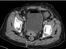
A 68 years old woman with history of uncontrolled diabetes presented to the emergency with abdominal pain and fever. The blood level of C-reactive protein and glucose was elevated. An abdominopelvic CT scan was performed showing intraluminal and intramural gas in the bladder (Figure 1). The diagnosis of emphysematous cystitis was made based on the CT. Glycemic control was established, a Foley catheter was placed, and the patient underwent antibiotherapy subsequently adapted to the results of urine culture that was positive for Escherichia coli. Emphysematous cystitis is a rare form of complicated urinary tract infection, commonly caused by Escherichia coli and Klebsiella pneumoniae, characterized by the presence of gas in the bladder wall and/or lumen. The primary risk factor is diabetes mellitus. Clinical manifestations are variable ranging from asymptomatic to severe sepsis. Abdominopelvic CT is the best diagnostic tool, contributing also to accurate assessment of the severity of this condition.
