Clinical Image
Austin J Radiol. 2019; 6(4): 1102.
A Rare Case of Abdominal Pain and Hematuria from Retroaortic Left Renal Vein
Pérez J* and Pozo D
Department of Radiology, Medical Science University of Havana, Cuba
*Corresponding author: Pérez J, Radiology Department, Hospital Clínico Quirúrgico “Hermanos Ameijeiras” Calle San Lázaro # 701 esq. a Belascoaín, Piso 5, Centro Habana, Havana, Cuba
Received: October 16, 2019; Accepted: October 22, 2019; Published: October 29, 2019
Clinical Image
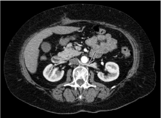
A 62-year-old female with a history of arterial hypertension, attended the emergency department due to pain in the left flank. On physical examination no showed signs of peritoneal irritation. Urinalysis was indicated, that reported microscopic hematuria and negative abdominal ultrasound. Then urotomography was performed, which confirmed the presence of retroaortic renal vein with a slight tortuous path near to renal pelvis (Figure 1).

    Figure 1: Urotomography in venous phase- Retroaortic renal vein with a
slight tortuous path near to renal pelvis (arrow).
Conservative treatment with analgesics and outpatient control by urology is treated. Four days later, the pain improved spontaneously as did hematuria.
The formation of this rare variant occurs in the ventral portion of the sub supra and intercardinal anastomosis or in its dorsal part or the presence of a persistent cardinal vein. This vascular variant can generate compression between the aorta and the anterior aspect of the vertebral body and can cause abdominal pain in the left flank with or without hematuria. The CT contributes to its diagnosis.